A general understanding of the anatomy of a canine body is very important for any dog owner: a kennel owner, a breeder or a simple fancier. Anatomy studies the external and internal structure of the dog's body. The internal structure consists of the skeletal system and internal organs. It is this knowledge, combined with physiology, that can help, for example, provide first aid to a pet in time or correctly assess the exterior of a dog.
Anatomical parts of the dog's body
Features of the location of various parts of the body, physique and general appearance of the dog in accordance with their breed characteristics are called exterior. To assess the exterior Anatomically, several sections of the dog's body are distinguished:
- Head. The skull and muzzle, eyes, ears, dental system are evaluated.
- Torso. Along the top line look at the withers, back, loin, croup and tail. On the bottom line evaluate thoracic region and belly.
- Limbs. Presented front and rear.
Knowledge of the features of the exterior is especially necessary for the owners breed dogs. It helps to control, preserve and develop dog breeds.
Skeletal system
The study of anatomy is necessary start by looking at the skeletal system. The skeleton is the bone base of the dog's body. The development and productivity of the canine organism as a whole depends on its condition. The skeletal system together with joints, ligaments, muscles and tendons make up the musculoskeletal system. There are axial and peripheral parts of the skeletal system.
Axial Division System
The structure of the axial skeleton includes:
- Scull.
- Vertebral column.
- Thorax with ribs.
 Skulls are dolichocephalic (long) and brachycephalic (short). The first ones are typical for breeds of shepherds, dobermans, collies, the second type of skull - for Pekingese, pugs, bulldogs. Dog skull has cranial and facial (muzzle) parts. The bones of the skull, with the exception of the lower jaw, are fixedly connected. The mobility of the lower jaw is due to the need to grasp, hold and chew food. The dental system is actively involved in this process. Adult dogs have 42 teeth, puppies have 28. There are incisors, canines, premolars and molars. Puppies are missing molars and one premolar.
Depending on breed characteristics the closing of the front teeth (incisors) form a certain bite. The most preferred and most often obligatory for most breeds is the scissor-shaped, in which the upper incisors lie tightly behind the lower ones. In a level bite, which is acceptable for some breed groups, the surfaces of the incisors are closed together by the cutting edges. Undershot bite is manifested by a strong protrusion of the upper jaw in front of the lower, so that a large gap is formed between them. Undershot bite is characterized by protrusion of the lower jaw, resulting in the lower incisors protruding in front of the upper ones and is a characteristic feature of breeds with a short muzzle.
The dog's vertebral column consists of seven cervical, thirteen thoracic, seven lumbar, three sacral and several caudal vertebrae.
The cervical region is made up of seven cervical vertebrae, which begin with the first - the atlas and the second - the epistrophy. The skull is attached to them. and they allow the dog's head to be movable in different directions.
The thoracic region is represented by thirteen vertebrae, to which curved ribs of various lengths are attached. The first four pairs of ribs are closed in the costal arch, the remaining nine pairs are shortened in the direction lumbar, bend freely. The ribs serve as protection for the internal organs of the dog and are involved in the breathing process.
 The lumbar region is made up of seven segments. The loin should not be long - this is considered a big disadvantage. Ideally, it is desirable to be short, convex and wide, reliably connecting the thoracic and pelvic spine and capable of acting as a spring. The long loin is very strongly reflected in the movements of the dog, the gait becomes lax, the back begins to wag.
Typically, dogs are characterized by the presence of 20-23 tail vertebrae. There are also smaller numbers. To meet the standard in some breeds, the tail vertebrae are cut off (stopped), leaving a few segments.
Peripheral skeletal system
The department is represented by the front and hind limbs of the dog.
The forelimb consists of a scapula, preferably obliquely set, to which the humerus is attached by means of the humeroscapular joint. The shoulder through the elbow joint is connected to the bones of the forearm, consisting of two bones - the ulna and the radius. For most breeds, it is highly desirable that the lowest point of the costal arch reach or be below the elbow joint. . chest depth- one of the important parameters of the exterior. A rather deep chest, with its moderate width, creates the basis for the good development of the internal chest organs: heart, lungs, blood vessels.
The wrist joint is made up of seven bones that connect the bones of the forearm to the five bones of the metacarpus. The forelimb ends with fingers, each of them is equipped with a hard claw at the end, which cannot be retracted. Four fingers have three phalanges, and one has only two.
The forelimb is attached to the vertebral skeleton very strong muscles shoulder. The protrusion of the obliquely set shoulder blade, rising above the thoracic vertebrae, creates a prominent withers. Measurements from the highest point The withers to the ground of a calmly standing dog is a very important conformation parameter and is called "height at the withers" for evaluation. Depending on the accepted breed standard, the height at the withers has a different meaning. The fluctuation of the height at the withers in different breeds is sometimes simply amazing with the wonders of the selection work of breeders and breeders. So great is the difference in height between a miniature room pocket dog and the giants of the dog world, Great Danes and Wolfhounds - from 6.5 cm to 111.8 cm in height at the withers.
 The hind limb girdle begins at the hip joint, which articulates the entire hind limb with the pelvic bone of the dog's spine. The hind limb consists of the femur, which, through the knee joint, is connected to two bones of the lower leg: the tibia and the tibia.
The usually inconspicuous knee joint plays an important role in the dog's musculoskeletal system. . Straightening, he gives rise to a push y, which produces the hind limb. This push ends with the extension of the hock (tarsus), which connects the bones of the lower leg to the metatarsus. The large heel bone clearly stands out on the hock joint. Four bones of the metacarpus, occasionally five pass into three phalangeal fingers, which end in strong claws.
Puppies are sometimes born with a fifth toe on their hind limbs. These dewclaws are often injured, so they are removed, which is prescribed by the breed standards. At rare breeds the dewclaws are left. Beaucerons(French Shepherd) they must be double, their absence leads to disqualification of the dog. In the Tibetan Mastiff and the Italian Hound, dewclaws are left at the request of the breeder or owner.
The internal structure of the dog's body
The internal organ system consists of the digestive, respiratory, excretory and reproductive organs.
digestive
Its main purpose in consumption, promotion, digestion, assimilation of food and water. Starting in the mouth with teeth, it passes into the esophagus, which is adjacent to the stomach. In the stomach, food and water mix and with the help of the secreted of hydrochloric acid are broken down into nutrients (the process of digestion). Moving further, the food lump enters the duodenum of the intestine.
The intestine is the main organ for further digestion and absorption of split particles - nutrients. They open their ducts into it and secrete the substances necessary for digestion, pancreatic secretion and bile, the pancreas and liver with gallbladder respectively. The intestinal section is very long, its length is from two and a half to seven meters. The intestine is divided into the small intestine and the large intestine, which ends at the anus.
Respiratory
 The respiratory system is designed for gas exchange in the lungs. Oxygen enters the blood from the air and is removed back carbon dioxide. By contracting and relaxing, the rib muscles cause the lungs to contract to remove carbon dioxide and inflate to suck in oxygen. The respiratory system consists from the nasal and oral cavities, larynx, trachea and lungs.
excretory
The system consists of two kidneys with a ureter, bladder and urethra. The end products of metabolism from the blood in the kidneys are filtered into urine, which is collected in the bladder through the ureters and periodically removed from the body through the urethra.
reproductive system
The organs of the reproductive system serve to procreate. Their structure is different in different sexes. In males, it includes the testes located in the scrotum, the vas deferens, the penis covered by the prepuce . In females, the reproductive organ system has an internal location in the body and consists of the ovaries, fallopian tubes, uterus, vagina and external genitalia.
Whole body management
All body systems are controlled by the nervous, circulatory, immune, lymphatic, hormonal, skin and sensory systems.
nervous
The system is divided into central and vegetative. It is made up of nerve fibers. Due to its high development in dogs, such sense organs as smell, sight and hearing are exacerbated. The central nervous system, together with the cerebral cortex, through congenital and acquired reflexes during life, regulates all systems of the dog's body.
circulatory
The cardiovascular system includes the heart and blood vessels: arterial, coming from the heart, and venous, coming to this organ. The main arterial vessel is called the aorta. The cardiovascular system is designed to supply all organs and cells of the body with oxygen and nutrients and to remove the end products of metabolism. The location of the heart is the chest. It is located on the left side of it.
Sense organs and skin
 External and internal influences are perceived and analyzed by the sense organs. A dog has five senses: visual, auditory, olfactory, gustatory and tactile. The visual consists of the eye with the pupil, eye muscles and nerves.
auditory analyzer includes the ear, the structure of which is such that it not only perceives vibrations sound waves, turning them into sound, but also has the function of correct orientation in space - balance. The sense of smell in dogs is highly developed, its sharpness depends on individual characteristics and fitness. Taste buds are located on the dog's tongue and serve to analyze the composition and quality of substances that enter the mouth.
The skin organ of touch is primarily a barrier between the external environment and the internal system of the dog's body. The tactile function protects the organs from adverse effects. The composition of the skin:
- Subcutaneous tissue.
- Epidermis.
- Wool is a derivative of leather.
Knowledge of canine anatomy but allows you to better understand the reasons that prompt our pets to behave in one way or another.
In the article I will consider the structural features of the internal skeleton of a dog, how it differs from the anatomy of other animals. I will tell you in detail about each department of the skeleton. I will indicate how many bones the pet has.
The anatomical structure must be studied by all dog owners, as these are quite mobile animals. And the structure of the skeleton plays an important role and is of great importance.
The skeleton is the base upon which all soft tissues are attached. This is not just a set of muscles and joints, here everything is thought out by nature so subtly that it is the skeleton that is responsible for the variety of movements.
The structure of the skeleton of a dogHow is the internal skeleton of a dog
Upper spine (neck). It consists of seven vertebral bones. The very first is called "Atlas" (translated from Latin "Atlant"). It differs from the others in its annular shape and provides vertical mobility of the head. The second vertebra is called "Epistrophy" ("Epistropheus"), it is responsible for the horizontal movements of the animal's head.
The dog's head can rotate 350 degrees.
Thoracic department.
It mainly consists of 13 vertebrae, but there are individuals with twelve vertebrae.
Ribs are attached to the transverse processes of the vertebrae of this department. The spinous processes from 1 to 10 vertebrae are directed towards the tail, but the eleventh is called diaphragmatic. Its spinous process is directed upwards. The same processes from the 12th to 13th vertebrae are directed towards the head of the animal.
 Comparison of human and dog skeleton
 Loin or lumbar region. These vertebrae are oval. Their processes are long, flat, ribbon-like transversely costal articular processes are superbly developed.
Basically, there are seven vertebrae in this department, but there are representatives with six.
The spinous processes of the lumbar at the vertebrae are directed towards the head. The length of each (up to the fifth) gradually increases, after which it immediately decreases.
The sacrum is the fusion of three or four sacral vertebrae into one bone. The main function of this section of the spine is the strong bonding of the spinal column with the hind limbs.
Finally, the bones of the sacrum grow together at the age of 2 - 2.5 years.
In females, the sacrum is longer and wider than in males. Such sizes are due to the reproductive function of females. In this section of the spine, the spinal processes merge into the crest of the same name.
In most cases in dogs, the spinal process of the first sacral vertebrae remains separate.
 The bones of the tail hold the muscles due to which the dog wags its tail.
 Tail. The first four vertebrae are well developed. They are endowed with all the relevant characteristics, like ordinary vertebrae. Further, the vertebrae of the tail section serve only for attaching the muscles that allow you to move the tail.
At different breeds meets different amount vertebral bones in the tail. Basically, their number is from 20 to 23, in more rare cases from 15 to 25.
In case of spinal cord injury or congenital pathologies and prescribe treatment.
The shoulder girdle includes the scapular bones and the rudiments of the clavicle. The scapular bone is attached to the body of the dog near the first pair of ribs. Thanks to this belt, the forelimbs are attached to the skeleton.
 Comparison of animal limb bones
 Limbs. Dogs have only four legs.
These pets have thoracic and pelvic limbs.
The thoracic limb belt consists of:
- The shoulder, which consists of the humerus.
- Forearm, includes the ulna and radius.
- Brush. It consists of seven carpal bones, five bones of the metacarpus and phalanges of the fingers. The dog has five fingers, which consist of three phalanges.
The hanging finger is the first finger and has only two phalanges. Some breeds of dogs may not have it at all.
The pelvic limb belt includes:
- Pelvic bones (iliac, pubic, ischial).
- The hips are made up of the femur and the patella.
- The lower leg includes the tibia and fibula.
- Stop. It consists of seven tarsal bones and five metatarsal bones. The phalanges of the fingers and their structure are the same as the thoracic region.
 Pelvic bone of a dog
 Dog skull anatomy
Skull and teeth. The connection of the bones of the skull is movable. It is this that gives the pet the ability to chew, gnaw, and so on.
Adult dogs have forty-two teeth, puppies have twenty-eight milk teeth.
The teeth formula includes: canines, incisors, molars and premolars.
The bite is influenced by the breed and breed standards.
 bite patterns in dogs
 - Scissors. Here, the lower ones, as it were, are under the upper incisors, and also have a tight connection with each other.
- Pincer-shaped - this form of bite occurs when the incisors are closed to each other.
Dogs differ from each other in bite.
- Straight. The incisors are stacked on top of each other.
- Snack. The lower jaw protrudes forward and the teeth do not match.
 The structure of the skull
 The structure of the skull is directly related to the breed of the dog and its age. Now many are able to distinguish by the shape of the skull, to which breed this or that individual belongs.
There are two types into which all watchdogs are divided:

In the structure of the skull there are paired and unpaired bones.
The unpaired bones include the "pterygoid", "occipital", hyoid bones, as well as the "vomer". Additionally, the skeleton includes and does not have a pair of ethmoid bone and sphenoid with interparietal.
Paired bones include two bones of the upper jaw, bones of the cheekbone, lacrimal, nasal, palatine, and two more incisive bones, bones of the lower jaw, frontal, vertex and temples.
Structural features
 timing of bone growth
 The skeleton of any breed performs the most important function. It is not just a foundation for the whole organism, it is a lever that provides movement, it also performs a supporting function for all organs, muscles and systems of the animal.
The skeleton is involved in almost all biological processes in the animal body.
Bone tissue is strong and light compared to other systems in the pet's body.
How many bones
In total, the dog's skeleton consists of 247 bones and 262 joints.
In the human, there are only from 205 to 207 bones, while there are about two hundred joints. the same number of bones is about 244 pieces.
The dog's skeleton is unique in its composition and functions. Thanks to him, these animals are mobile and active. They are well coordinated and can be very hardy.
Axial and peripheral skeleton of a dog. Purpose, components.
The skeleton plays an important role in the life of the body. It serves as a lever of movement, support for the soft parts of the body, protection, a place for the development of hematopoietic organs, and also participates in metabolic and biochemical processes in the body. The skeleton is unique in its structure. The skeleton is a rigid structure, consisting of individual bones, connected to each other motionlessly or by joints. Muscles are attached to the skeleton, which set in motion its individual sections, which makes it possible for the animal to move in space. Distinctive features of the skeletal system are strength and lightness compared to other tissues. In young animals, the bones are more elastic than in old ones. As we age, bones become more brittle.
The musculoskeletal system consists of bones of the skeleton, joints with ligaments and muscles with tendons. The movement manifests itself in the form of a change in the position of the joints under the influence of contraction of the skeletal muscles, which serve as engines for each joint, or are carried out without the participation of the osteoarticular apparatus by the same muscles (closing and opening the eyelids, the work of mimic muscles, etc.). Bones, muscles, tendons have special nerve endings - receptors that send impulses to cells of various levels of the central nervous system. They are abundantly supplied with blood and lymphatic vessels. In this regard, the lack of sufficient physical activity reduces the amount of mechanical energy, in connection with which innervation and blood circulation are disturbed in the body, delivery of impulses to the brain worsens, the outflow of metabolic products from all organs of the body slows down, and metabolism in them is disturbed.
Recent studies have shown that the state of the skeleton can be used to judge the health of animals: the skeleton is called a mirror reflecting the state of the body.
- The degree of development of the skeleton is of great importance in the life of the animal. It is not only a rigid supporting structure, it also bleeds, its part - the red bone marrow - produces blood cells, including erythrocytes that carry out gas exchange, and stem cells, which, developing, further form protective immune cells that ensure the viability of the body.
- The bone marrow, in addition to the formation of blood elements (erythrocytes and leukocytes), produces protective immune cells that ensure the vitality of the body.
- They act as a depot of minerals, maintain reserve blood alkalinity and electrolyte balance in the body.
- Under the influence of a sharp decrease in motor activity, muscle atrophy occurs, the structure of bones changes, the amount of adipose tissue increases, metabolic processes are disrupted, the structure and state of the central nervous system changes. The skeleton suffers greatly during hypodynamia, which is the first to experience the effect of physical stress that occurs during movement.
- The skeleton provides a certain ratio of Ca and P in the blood and, finally, the skeleton maintains the electrolytic balance in the body. Throughout life, the skeleton is rebuilt, destroyed and restored, and, as it turned out, all these functions of the skeleton developed in connection with the movement of the animal and turned out to be dependent on it.
- Studies have shown that the lack of the necessary physical activity leads to a violation of the processes of hematopoiesis, bone metabolism, which leads to animal disease, loosening of bones, their softening - demineralization, and a decrease in bone strength. The animal loses the ability to move. Elastic deformations of bones that occur during movement lead to tension of collagen fibers, without which bone mineralization does not occur. And from this it follows that if the bone does not experience the action of the necessary, at least the minimum dose of mechanical energy, normal processes of bone formation, hematopoiesis, metabolism and electrolyte balance will not be able to proceed in it.
- About the nature of mineral metabolic processes in the body of a dog, they are judged by the degree of bone development in the metacarpus, metatarsus, the severity of the carpal and hock joints, and the condition of the teeth.
- Curvature of the bones of the forearm, knotty carpal joints - a sign of rickets.
- Disproportions in the development of bones and other organs or parts of the body indicate dysfunctions in the hormonal system.
- The underdevelopment of the facial bones of the skull, the weak severity of the tubercles on the bones indicate a deeper violation of the mineral and general metabolism in the body. This is also evidenced by the absence of individual teeth, the destruction of enamel, small or not located on the same line of incisors, all deviations from the normal bite.
- The listed shortcomings and defects can be hereditary.
The skeleton of a dog consists of 289 - 292 bones (fluctuations in the number associated with the tail vertebrae and 262 joints. The bones of the various shapes, interconnected by ligaments, cartilage or bone tissue into such large sections as the spinal column, skull and skeleton of the limbs.
The skeleton is divided into:
Rice. 1.Dog skeleton: 1 - upper jaw; 2 - lower jaw; 3 - skull; 4 - parietal bone; 5 - occipital protuberance; 6 - cervical vertebrae; 7 - thoracic vertebrae; 8 - lumbar vertebrae; 9 - tail vertebrae; 10 - scapula; 11 - humerus; 12 - bones of the forearm; 13 - bones of the wrist; 14 - bones of the metacarpus; 15 - phalanges of fingers; 16 - ribs; 17 - costal cartilages; 18 - sternum; 19 - pelvic bone; 20 - hip joint; 21- femur; 22 - knee joint; 23 - tibia; 24 - fibula; 25- calcaneus; 26 - hock; 27 - tarsus; 28 - metatarsus; 29 - fingers
The axial skeleton includes:
1 . Skeleton of the head ( skull), consisting of the bones of the brain and facial skull. The skull is formed in the greater part of the plane by the bones connected immovably in young animals with the help of cartilage or connective tissue (in weak puppies, the joints between the bones do not ossify for a long time, they are palpable in the form of soft sutures). In older dogs, all the bones of the skull are fused. Only the lower jaw is connected to the temporal bone by a very mobile joint, thanks to which the dog grabs and "cuts" food. The work of this jaw joint is provided by the strongest - masticatory muscles. On the posterior edge of the skull, a triangular-shaped occipital crest is clearly palpable, the more pronounced the stronger the muscles of the neck are attached to it. Below the occipital crest, on the border with the first cervical vertebra, there is a large occipital foramen of the skull, through which it exits from the brain spinal cord, heading into the spinal canal of the spinal column. At the back of the skull, the cranial cavity is formed, where the brain is located. Anterior to the cranial cavity is nasal cavity, which in dogs is very complex. It can be entered through the nostrils located on the always moist, hairless skin of the top (lobe) of the nose. The nasal cavity is divided in the middle by a cartilaginous nasal septum and in each of its 2 halves are located, attached to its side wall, thin bone plates wrapped in tubules. These plates are called shells. The shells fill both halves of the nasal cavity, leaving only narrow gaps ( passages) between them, through which air passes through the nasal cavity, heading to the lungs. Below the nasal cavity, the bones of the skull form the oral cavity, framed from below by the movable lower jaw. Teeth are located on the incisor bone, upper and lower jaws.
Paired and unpaired bones of the skull:
Paired: temporal bone, parietal, frontal bone, lower jaw, incisor bone, palatine, lacrimal, nasal, zygomatic bones and upper jaw;
Unpaired: sphenoid, interparietal, ethmoid, vomer, hyoid, occipital and sphenoid.

Rice. 2. Dog skull: 1 - incisor bone; 2 - nasal bone; 3 - maxillary bone; 4 - lacrimal bone; 5 - zygomatic bone; 6 - frontal bone;7 - parietal bone; 8 - temporal bone; 9 - occipital bone; 10 - lower jaw
2 . The bones of the spinal column, including the cervical, thoracic, lumbar, sacral and caudal vertebrae. The spinal column is a series of vertebrae connected by intervertebral cartilage and joints. Above the supporting part of the spinal column, in its canal, lies the spinal cord, from which nerves go to all parts of the body through the intervertebral foramina.
7 cervical vertebrae. The cervical spine of the dog is the most mobile, regardless of the size of the animal.
13 inactive thoracic vertebrae (but often there can be 12 and in rare cases 14).
7 firmly connected lumbar vertebrae (in isolated cases 6). Below the vertebrae are the kidneys, in females behind them lie the ovaries.
3 sacral fused vertebrae, to which the ilium of the pelvis is attached with a tight joint.
up to 20 - 23 tail vertebrae (the number of vertebrae is determined by the standard)
Table 1. Sections of the spine and the number of vertebrae in a dog.

The sacrum, the first tail vertebrae and the pelvic bones - ilium (above), pubic and ischium (at the bottom of the pelvis) - form the pelvic cavity. Outside, along with the muscles, this area is called the croup. The bones of the pelvis are firmly connected to the sacrum and the first caudal vertebrae by strong ligaments, and along the bottom of the pelvis, the right and left bones are connected in young animals by cartilage, forming the so-called pelvic suture. Before whelping, the connection between the bones relaxes, which contributes to the best passage fetus through the pelvic cavity. After childbirth, the connection between the bones becomes rigid again.
3 . Thirteen pairs of ribs - 26
9 pairs are true, because connected to the sternum by their own costal cartilages
4 pairs are false, because the costal cartilages of these ribs are first combined with each other, and only then connected to the sternum. The last pair of ribs with their free cartilaginous end can end in the muscles, so this pair of ribs is called hanging ribs.
4 . Breast bone.
The thoracic vertebrae, ribs and sternum together form the ribcage. The movement of its wall ensures breathing - the expansion of the chest wall, together with the contraction of the muscles of the diaphragm, provides inspiration; constriction of the chest wall, relaxation of the diaphragm and pressure on it of the internal organs, while simultaneously contracting the muscles of the abdominal wall, ensures exhalation. The posterior edge of the chest, formed by the edges of the last ribs and costal cartilages, is called the costal arch.
peripheral skeleton.
The thoracic limb begins with the scapula, then the humerus, forearm, wrist (7 carpal bones), metacarpus (5 metacarpus bones).
The fingers at the end are equipped with strong non-retractable claws. The thoracic limb is connected to the spine by muscles. It is attached with the help of the scapula and muscles to chest and back of the neck. Withers are formed above the shoulder blade. The pelvic ( rear) limb begins with the femur, which, in turn, passes into the lower leg (large and small tibia), then into the tarsus (consists of 7 bones).
This is followed by the metatarsus (from 4-5 metatarsal bones), then 4 phalangeal fingers ending in claws.
Sometimes a rudimentary ( profit) finger grows from the inside. At a young age, it is usually amputated. The pelvic limb has an articular connection with the pelvis and is fixed by the muscles of the hip group. Paired thoracic and pelvic limbs have a similar structural plan - they are composed of 3 links:
- 1st link - shoulder (on the chest) or thigh (on the pelvic), which are based on long tubular bones - the humerus and femur.
- 2nd link - forearm or lower leg. The basis of this link is 2 bones: the radius and ulna with a large olecranon on the forearm, and the tibia and fibula - on the lower leg, and the ulna and fibula are much thinner and less pronounced than the radius and tibia - the main bones on which the weight of the body falls .
- 3rd link of the limbs - hand or foot. These are the hardest parts. The hand and foot each have 3 links of bones: the 1st link has 2 or 3 rows of short bones of the wrist (on the hand) and tarsus (on the foot). 2nd - long, thin 4 or 5 bones of the metacarpus (on the hand) or metatarsus (on the foot), interconnected by short ligaments. Fingers are attached to each of the bones of the metacarpus or metatarsus, each finger consists of 3 phalanges.
The dog is a digitigrade animal, it relies only on the finger. The longest middle fingers (3rd and 4th), the shorter ones are the 2nd and 5th, and the 1st finger is hanging and may be absent altogether. In dogs, the calcaneus of the tarsus is raised high off the ground, while in plantigrades, the heel rests on the ground.
All links of the limbs are interconnected by movable joints - hermetically sealed capsules and reinforced ligaments. There is a clear, viscous synovial fluid inside the joint, so the first sign of a joint puncture will be the release of a yellowish transparent synovium through the puncture. Groups of muscles act on each joint, connected by means of nerves with certain centers of the spinal cord. The muscular-ligamentous apparatus of the limbs is a powerful shock-absorbing apparatus that softens the shock load on the skeleton. For the possibility of faster movement, the lower parts of the limb are facilitated - mainly only muscle tendons go along the hand and foot. Most of the muscle mass is concentrated in the scapula or pelvis, shoulder and thigh. All skeletal muscles, contracting, not only cause the movement of the animal, but also contribute to the formation of thermal energy. This should be remembered and when working with a dog, take into account the ambient temperature so as not to cause heat stroke.
One of the most difficult areas of the body is the head area. It contains: nasal and oral cavities, pharynx and larynx, brain, organs of vision and hearing.
In the nasal cavity, an upper narrow passage is distinguished between the shells and the nasal bone, which falls directly into the labyrinth of the ethmoid bone - the organ of smell, therefore it is called olfactory. In order for the air to get into it, the dog "holds" its breath and draws in the air more strongly - it sniffs. The shells, between which narrow passages are formed in the nasal cavity, form a kind of filter, passing through which the inhaled air is cleaned, heated and checked for smell.
The cavities of the frontal and maxillary bones of the skull, called sinuses, communicate with the nasal cavity. Because of this, inflammation of the membrane of the nasal cavity can cause inflammation not only of the membrane of the sinuses, but, worse, of the olfactory region, as a result of which the dog's sense of smell can be disturbed.
In front of the nasal cavity of the dog there are small holes through which you can get into the cavity of the eye, where the lacrimal canal leads.
From the nasal cavity, the exit leads to the pharyngeal cavity, where the respiratory and digestive tracts cross. It is located under the base of the skull. On its side walls there are holes going into the auditory tubes, and therefore there is a danger of infection from the pharynx into the middle ear.
The entrance to the oral cavity is formed by teeth. The gap between the teeth and gums on one side and the cheeks on the other is called the vestibule of the oral cavity. In the middle part of the buccal mucosa, at the level between the arcades of closed teeth, ducts of very small parotid salivary glands located at the base of the auricles open. By opening the jaws, you can get into the oral cavity. At the bottom of it, under the tongue, two more salivary glands open - the submandibular gland, which lies behind and under the lower jaw next to the parotid gland, and the sublingual gland, which lies on the side of the base of the tongue. Both glands open at the bottom of the mouth.
Dog teeth are located along the edges of the incisor, maxillary bone and lower jaw. Ahead they are covered with skin folds - lips, and from the sides - cheeks. The dog's mouth is very large. It almost reaches the angle between the upper and lower jaws, the dog does not chew, but "chops" the food. Her teeth and jaws are not adapted for chewing food, she can capture and swallow large pieces of food. In front of the dog there are 6 upper and 6 lower incisor teeth, on the sides of them there are 2 fangs, behind which there are molars: on each side 6 on the upper and 7 on the lower jaws. However, it must be borne in mind that all incisors, canines and front 4 molars ( premolars) on each side of each jaw change. The back molars - molars grow later and do not change (there are 2 molars on the upper jaw on each side, 3 on the lower jaw).
Puppies are born without teeth on the surface of the gums, which erupt only on the 18th - 25th day after birth. Delayed teething indicates a developmental delay in the puppy.
At the bottom of the mouth is the tongue. In a dog, it is thin and very mobile, on top (along the back) it is covered with delicate filiform papillae, among which taste buds are scattered.
On top of the oral cavity, rollers of the hard palate are visible, passing at the entrance to the pharynx into the palatine curtain. The gums and hard palate may be unevenly pigmented, that is, have a spotted color. At the exit from the oral cavity into the pharynx, on the sides of the pharynx, there are tonsils, lymphoid formations that perform a protective function - the neutralization of microflora that enters the oral cavity from the external environment.
In the special recesses of the skull - the orbits are the organs of vision of the dog. In dogs, the orbit forms an incomplete bony ring. Here, in special fat pads, the eyeballs lie, covered in front by the upper and lower eyelids. Eyelashes grow along the edges of the eyelids. From the inside, the eyelids are covered with a mucous membrane of a pale pink color, which passes to the surface of the eyeball and is called the conjunctiva, its inflammation is called conjunctivitis. To the back of the inner surface upper eyelid the ducts of the lacrimal gland, which lies above the eyeball, open. The tear constantly washes the mucous membrane of the eyelids and eyes and flows into the region of the inner corner of the eye, where small pinholes of the lacrimal canaliculi are visible on the edges of the upper and lower eyelids, through which the tear enters the lacrimal canal and flows into the anterior part of the nasal cavity. If the openings of the lacrimal ducts are inflamed, or “clogged”, the eyes begin to “water”, as tears no longer flow into the nasal cavity, but onto the front surface (this is sometimes observed in older dogs).
The eyeball itself, which perceives light irritation, is a three-layer bubble. The outer layer has a transparent part - the cornea and a dense white shell - the sclera. Under the cornea, a second membrane is visible - the vascular. In the region of the cornea, it has a color and is therefore called the iris. In the center of it you can see a hole - the pupil, through which a beam of light penetrates into the eyeball. The pupil with the help of muscles can narrow or expand. Behind the pupil lies a transparent lens - the lens, held by special ligaments with muscles. Muscles, contracting, act on the curvature of the surface of the lens. Behind the lens, the eyeball is filled with a gelatinous, transparent mass - the vitreous body. The third layer of the eyeball is the retina, on which nerve cells are located, their processes are connected with the nerve cells of the brain through a special optic nerve.
The dog's hearing organ is divided into the outer, middle and inner ear. The outer ear is the auricles, which in dogs have the most diverse shape characteristic of each breed. Under the skin of the auricle there is a cartilaginous plate that provides the position of the auricle - dense cartilage lies at the base of the standing ear, thin - forms the basis of the hanging. From the auricle comes the external auditory canal, which at the entrance to the middle ear is tightened by the tympanic membrane. The middle and inner ear are located in a special bone of the skull - the petrous bone.
The middle ear is a bony cavity containing the auditory ossicles, the malleus, anvil, lenticular bone, and stirrup. They transmit sound waves from the outer ear to the inner ear. Two openings lead from the middle ear to the inner ear, also covered by the eardrums. The ossicles of the middle ear are connected by the malleus to the outer tympanic membrane, and by the stirrup to the inner. The middle ear has an opening through the auditory tube into the pharynx. Directly the organ of hearing and balance is located in inner ear, from the sensitive cells of which processes go to the centers of the brain located in the cranial cavity.
Thus, the dog's head is a very complex and important area of the body.
The neck region is characterized by the fact that under its vertebrae are located: the esophagus, which runs along the trachea, very large vessels and nerve trunks. In young puppies, the central organ of the lymphoid system, the thymus, or thymus, is located along the trachea.
The chest area is the location of a very important organs: lungs and heart. They lie in hermetically sealed separate cavities formed by a special transparent serous membrane, which secretes a serous fluid that “moisturizes” the surface of organs. Thus, the cavity of the right lung does not communicate with the cavity of the left, and both of them do not communicate with the cavity in which the heart is located. The esophagus, large trunks of two nerves innervating the diaphragm, all the internal organs of the chest and abdominal cavity. Under the spinal column lies the aorta coming from the heart, which passes through the opening of the diaphragm into the abdominal cavity. Behind the chest is fenced off from the abdominal cavity by the diaphragm, or, as it is called, the abdominal barrier. The nerve that innervates the diaphragm (without it, the diaphragm becomes paralyzed) comes from the lower neck area, so injuries to the lower neck can hit this nerve and cause diaphragmatic malfunction, which in turn can lead to severe breathing problems.
Under the lumbar region behind the chest and diaphragm is the abdominal cavity. Its roof is the lower back, from behind it freely passes into the pelvic cavity, and its side walls are formed by muscles located in 4 layers. Below, along the midline of the abdomen, these muscles of the left and right sides are “sewn together”, forming the so-called physiological suture line, or white line. In males, in the back of the abdominal wall, somewhat retreating from the white line, you can feel narrow gaps, they are called inguinal rings, through which you can get into the inguinal canals (right and left), where the spermatic cords lie - the right and left strands, consisting of vessels , nerves and vas deferens. In females, the inguinal canal is not pronounced.
Located in the abdominal cavity most of digestive organs. Immediately behind the diaphragm, slightly to the left of the midline, lies the stomach, into which the esophagus flows and the spleen is attached. The duodenum comes out of the stomach, the ducts of large glands open into it - the liver and pancreas. The liver is attached to the right to the diaphragm and runs with it during inhalation and exhalation.
In the abdominal cavity under the lower back are the kidneys, from which urine is discharged through the ureters into bladder- a reservoir where urine accumulates and is periodically excreted from the dog's body through the urethra.
In the pelvic cavity, located under the sacral region and the first caudal vertebrae of the spinal column, lies the rectum. In females, the internal genital organs are located under it: the uterus, vagina, urogenital vestibule, which ends under the anus with the external labia. In the lower corner of the genital slit is the clitoris (a rudiment of the male penis). At the bottom of the pelvis under the uterus and vagina lies the bladder and urethra, which opens into the lower wall between the vagina and its vestibule. In males, in the pelvic cavity under the rectum lies the bladder and the pelvic part of the urogenital canal. The urogenital canal goes from the neck of the bladder and here the male has a large and only accessory sex gland - the prostate, which secretes a fluid in which the male germ cells - spermatozoa are located. The urogenital canal emerges from the pelvic cavity and runs along the underside of the penis, opening on its head with the urogenital process.
All organs located in the pelvic cavity, like the anus, are connected by nerves with the sacral centers of the spinal cord. The defeat of the centers of the sacral spinal cord can lead to a violation of not only the act of defecation, but also urination and sexual functions.
Breathing apparatus. Structure, functions.
The breathing apparatus ensures the supply of oxygen to the body and the removal of carbon dioxide, that is, the exchange of gases between atmospheric air and blood. In domestic animals, gas exchange occurs in the lungs, which are located in the chest. Alternate contraction of the muscles of the inhalers and expirators leads to the expansion and narrowing of the chest, and with it the lungs. This ensures that air is sucked in through the airways into the lungs and pushed back out. The contractions of the respiratory muscles are controlled by the nervous system.
During passage through the airways, the inhaled air is humidified, warmed, cleaned of dust, and also examined for odors using the olfactory organ. With exhaled air, part of the water (in the form of steam), excess heat, and some gases are removed from the body. In the airways ( larynx) sounds are reproduced.
The respiratory organs are represented by the nose and nasal cavity, larynx, trachea and lungs.
NOSE AND NOSE CAVITY
The nose together with the mouth make up the anterior part of the head in animals - the muzzle. The nose contains a paired nasal cavity, which is the initial section of the airways. In the nasal cavity, the inhaled air is examined for odors, heated, moistened, and cleaned of contaminants. The nasal cavity communicates with the external environment through the nostrils, with the pharynx through the choanae, with the conjunctival sac through the lacrimal canal, and also with the paranasal sinuses. On the nose, the top, back, side parts and root are distinguished. At the top there are two holes - nostrils. The nasal cavity is divided by the nasal septum into right and left parts. The basis of this partition is hyaline cartilage.
The paranasal glands communicate with the nasal cavity paranasal sinuses. The paranasal sinuses are cavities filled with air and lined with mucous membrane between the outer and inner plates of some flat bones of the skull (for example, the frontal bone). Because of this message, inflammatory processes from the mucous membrane of the nasal cavity can easily spread to the sinuses, which complicates the course of the disease.
LARYNX
The larynx is the section of the breathing tube that is located between the pharynx and the trachea. In a dog, it is short and wide. The peculiar structure of the larynx allows it to perform, in addition to conducting air, other functions. It isolates the airway when food is swallowed, is a support for the trachea, pharynx and the beginning of the esophagus, and serves as a vocal organ. The skeleton of the larynx is formed by five movably interconnected cartilages, on which the muscles of the larynx and pharynx are attached. This is an annular cartilage, in front of and below it is the thyroid cartilage, in front and above it are two arytenoid cartilages, and below it is the epiglottic cartilage. The cavity of the larynx is lined with a mucous membrane. Between the vocal process of the arytenoid cartilage and the body of the thyroid cartilage, a transverse fold passes on the right and left - the so-called vocal lip, which divides the cavity of the larynx into two parts. It contains the vocal cord and vocal muscle. The space between the right and left vocal lips is called the glottis. The tension of the vocal lips during exhalation creates and regulates sounds. Dogs have large vocal lips, which makes it possible for your four-legged pet to make various sounds.
TRACHEA
The trachea serves to carry air to and from the lungs. This is a tube with a constantly gaping lumen, which is ensured by the rings of hyaline cartilage that are not closed from above in its wall. The inside of the trachea is lined with a mucous membrane. It extends from the larynx to the base of the heart, where it divides into two bronchi, which form the basis of the roots of the lungs. This place, which occurs at the level of the 4th rib, is called the tracheal bifurcation. The length of the trachea depends on the length of the neck, and therefore the number of cartilages in dogs ranges from 42 to 46.
LUNGS
These are the main respiratory organs, directly in which gas exchange occurs between the inhaled air and blood through a thin wall separating them. To ensure gas exchange, it is necessary big square contact between air and blood channels. In accordance with this, the airways of the lungs - bronchi - like a tree branch many times to bronchioles (small bronchi) and end with numerous small pulmonary vesicles - alveoli, which form the parenchyma of the lungs (parenchyma is a specific part of the organ that performs its main function). The blood vessels branch parallel to the bronchi and encircle the alveoli with a dense capillary network, where gas exchange takes place. Thus, the main components of the lungs are the airways and blood vessels.
The connective tissue combines them into a paired compact organ - the right and left lungs. The right lung is somewhat larger than the left, since the heart located between the lungs is displaced to the left. The relative weight of the lungs is 1.7% relative to body weight.
The lungs are located in the chest cavity, adjacent to its walls. As a result, they have the shape of a truncated cone, somewhat compressed from the sides. Each lung is divided into lobes by deep interlobar fissures: the left one into three, and the right one into four.
The frequency of respiratory movements in dogs depends on the load on the body, age, state of health, temperature and humidity of the environment.
Normally, the number of inhalations and exhalations (breaths) in healthy dog fluctuates widely: from 14 to 25-30 per minute. This breadth of range depends on a number of factors. So, puppies breathe more often than adult dogs, because they have a more active metabolism. Bitches breathe faster than males. Pregnant or lactating dogs breathe more frequently than non-pregnant dogs. The breed of the dog, its emotional state, and the size of the dog also affect the respiratory rate. Small breed dogs breathe more often than large breeds: miniature pinscher, the Japanese Chin breathes 20-25 times per minute, and the Airedale Terrier breathes 10-14 times. This is due to the different speed of the metabolic process, and, as a result, a greater loss of heat.
Breathing largely depends on the position of the dog's body. Animals breathe easier when they are standing. In diseases accompanied by damage to the heart and respiratory organs, animals take a sitting position, which helps to facilitate breathing.

Topography of the dog's lungs, right side view: 1 - trachea; 2,3,4 - cranial middle lobe of the lung; 5 - heart; 6 - diaphragm; 7 - dorsal edge of the lung; 8 - basal edge of the lung; 9 - stomach; 10 - ventral edge of the lung
The breathing process is also affected by the time of day and season. At night, at rest, the dog breathes less frequently. In summer, in hot weather, as well as in stuffy rooms with high humidity, breathing quickens. In winter, breathing in dogs at rest is even and imperceptible.
Muscular work sharply speeds up the dog's breathing. The excitability factor of the animal also has a certain value. The appearance of a stranger, a new environment can cause rapid breathing.
Physiology of reproduction.
GENITAL ORGANS OF THE FEMALE
In the female, internal and external genital organs are distinguished.
The internal reproductive organs include the ovaries, fallopian tubes, uterus, and vagina.
The ovaries (Ovaria, Oophoron) are the primary paired sex gland that performs reproductive and hormonal functions. The ovaries are ovoid in shape, somewhat flattened laterally. During sexual hunting, the luteal phase of the sexual cycle and during pregnancy, their shape can be grape-like. The size of the ovaries in dogs varies greatly depending on the morphofunctional state of the organ and the size of the animal. For example, in large breed dogs during the luteal phase of the sexual cycle and during pregnancy, the ovaries can reach 2-2.5 cm in length and 1-1.5 cm in width.
The ovaries are located in the abdominal cavity behind and below the kidneys in an open ovarian bursa. The walls of the ovarian bursa are formed by the mesentery of the ovaries and fallopian tubes. The abdominal opening of the ovarian bursa is small - does not exceed 1-1.5 cm in length. With the help of its own ligament, the ovary is connected to the top of the corresponding uterine horn, and attached to the lumbar vertebrae by means of an additional ligament. Accessory ovarian ligaments in dogs are short, contain a lot of fat and blood vessels. Specified anatomical features restrict access to the ovaries and complicate their surgical removal.
Outside, the ovary is covered with a single-layer cubic epithelium, under which there is a fibrous ( albuginea) membrane. The parenchyma of the ovary is represented by the medulla and cortex. The medulla is composed of connective tissue, blood vessels, and nerves. The follicular apparatus (primary, secondary and tertiary follicles) and corpus luteum are located in the connective tissue basis of the cortical substance.
Primary, or primordial, resting, follicles, which are first-order oocytes surrounded by a single layer of follicular cells, are formed in dogs in fetal (fetal) ovaries. At birth, there are 700,000 follicles in the ovaries, at the onset of puberty - 250,000, at the age of 5 years - 33,000, at the age of 10 years - 500 primary follicles.
Secondary, or growing, follicles are first-order oocytes surrounded by two or more layers of follicular cells. At this stage of folliculogenesis, the egg is actively growing and covered with a transparent membrane.
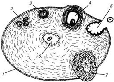
Rice. 2. Ovarian bursa:
A - side view, medial surface; B - top view. the dorsal wall of the bursa is opened; 1 - abdominal opening of the ovarian bursa; 2 - ovary; 3 - fallopian tube; 4 - funnel of the fallopian tube

Tertiary, or bubble, cavity, Graafian, follicles (the last stage of folliculogenesis) contain a micro- or macroscopic cavity filled with follicular fluid. Their wall is lined from the inside with stratified follicular epithelium, from the outside - by the inner and outer layers of the connective tissue membrane. The cells of the follicular epithelium form an oviparous tubercle, in the center of which there is a first-order oocyte. Tertiary follicles produce estrogenic hormones. The hormonal activity of Graaffian follicles depends on their degree of maturity. The preovulatory follicles, which have entered the final stage of their development, are the most active in the endocrine respect. Shortly before ovulation, they reach 6 - 8 mm in diameter, the number can vary from 1 to 14. Ovulation in dogs occurs spontaneously.
The corpus luteum, which forms at the site of the ovulated follicle, is an endocrine gland of temporary secretion. Cells of the corpus luteum ( luteocytes) produce progesterone, a hormone necessary to maintain pregnancy. There are yellow bodies of the sexual cycle and pregnancy. In dogs, the corpus luteum of the sexual cycle functions for the same amount of time as the corpus luteum of pregnancy.
The fallopian tubes (Tuba uterina, salpinx), or oviducts, fallopian tubes, are a paired organ in the form of a convoluted tube extending from each horn of the uterus. The fallopian tubes are located in their own mesentery, formed by the inner leaf of the broad uterine ligament. Their opposite end opens into the cavity of the ovarian bursa; the wall consists of mucous, muscular and serous membranes. The mucous membrane is folded, its single-layer cylindrical epithelium is represented by secretory and ciliated cells. In the fallopian tubes, sperm mature, the egg is fertilized, and the embryo develops to the stage of a 16-cell blastomere. Sex cells and the embryo are transported to the uterus due to fluctuations in the cilia of epithelial cells and the contraction of smooth muscle fibers of the organ wall. The contractile activity of the muscular wall of the fallopian tubes is stimulated by estrogens and inhibited by progesterone.
The uterus (Uterus, histera, metra) in dogs is bicornuate, consists of a neck, body and horns. The cervix and body of the uterus are short, the horns are long and serve as a fruit-bearing place. The horns diverge at an acute angle, which gives the uterus the shape of a slingshot. The size of the uterine horns in dogs varies greatly and depends on the size of the animal and the physiological state of the body - the stage of the sexual cycle and the timing of pregnancy. The wall of the uterus is built of three membranes: outer - serous ( perimetrium), middle - muscular ( myometrium) and internal - mucous ( endometrium). The muscular layer is represented by longitudinal and circular layers, between which there is a layer rich in vessels and nerves. The contractile activity of the myometrium of the body and the horns of the uterus is stimulated by estrogens and inhibited by progesterone. The structure of the mucous membrane of the body and the horns of the uterus is quite complex: it is covered with a single-layer cylindrical epithelium, in its thickness there are numerous tubular glands, the ducts of which open into the uterine cavity. The glands produce the so-called royal jelly, necessary for the nutrition of the embryo. The endometrium, like the myometrium, serves as a target tissue for sex hormones. Estrogens increase the vascularization of the endometrium, stimulate the growth of endometrial glands. Excessive vascularization of the endometrium leads to leakage (diapedesis) of blood cells into the lumen of the uterus and the appearance of hemorrhagic discharge from the genital slit in the proestrus stage. Progesterone causes branching of the tubular glands and stimulates the production of royal jelly.
During pregnancy in dogs, as well as in other placental animals, the placenta is formed from the mucous membrane of the uterus and the choroid of the fetus, which, according to the microscopic structure, belongs to the endotheliochorial type, and according to the macroscopic structure, to the zonal type. During childbirth, only the baby's part of the placenta falls off.
The cervix (Cervix uteri) has a narrow canal, a thick wall with a well-developed muscular layer. In dogs, the cervix reaches a length of 1-1.5 cm and is characterized by the absence of clear boundaries with the body of the uterus and the vagina. The entrance to the cervical canal from the side of the vagina is covered by the postcervical vaginal fold and is not accessible for vaginal examination. The cervix acts as a sphincter of the uterus. Full disclosure of its canal and postcervical vaginal fold (false cervix) is noted during childbirth, partial - during estrus, estrus and in the postpartum period. The opening of the cervix during childbirth is stimulated by estrogens and relaxin, during estrus and sexual hunting - only estrogenic hormones. The epithelium of the mucous membrane of the cervix is single-layer cylindrical and is represented mainly by secretory cells that produce a mucous secretion with bactericidal and bacteriostatic properties.
The uterus is located in the abdominal cavity, it is supported by wide and round uterine ligaments. The broad ligaments of the uterus are double layers of the peritoneum running from the lesser curvature of the horns, the lateral surface of the body, the cervix and the cranial part of the vagina to the side walls of the pelvis. Round ligaments of the uterus in the form of cords begin at the top of the uterine horns and end at the internal opening of the inguinal canal.
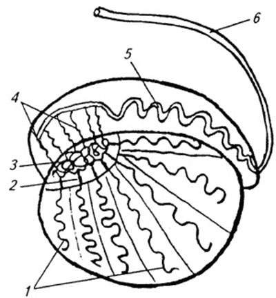
Figure 3. Schematic representation of the ovary, sagittal section:
1 - integumentary epithelium; 2 - primary follicles; 3 - secondary follicle; 4 - tertiary follicle; 5- follicle atresia; 6 - ovulated follicle; 7- corpus luteum
The vagina (Vagina), or vagina, is located in the pelvic cavity between the cervix and the opening of the urethra (urethra channel). It is a thin-walled elastic tube and serves as an organ of copulation and a birth canal. From the inside, the wall of the vagina is lined with a mucous membrane, devoid of glands and covered with stratified squamous epithelium. Under the influence of estrogenic hormones during proestrus and especially estrus (sexual hunting), the number of layers of epithelial cells increases, surface cells become keratinized, lose their nucleus, and keratin accumulates in their cytoplasm. Under the mucous membrane there are two layers of muscles: longitudinal and circular ( transverse). The cranial part of the vaginal tube is covered on the outside with a serous ( peritoneal) membrane, while the rest of it is loose connective tissue, which, together with pararectal connective tissue, provides fixation of the vagina and rectum in the pelvic cavity.
The external genitalia include the vestibule, labia, and clitoris.
The vestibule of the vagina (Vestibulum vaginae) serves as the urogenital canal. Its mucous membrane does not contain vestibular glands, is covered with stratified squamous epithelium and performs only a protective function. The muscular membrane is well developed and forms the sphincter of the vestibule of the vagina, which ensures the adhesion of the genital organs of the female and male during intercourse. The boundary between the vagina and its vestibule is the opening of the urethra. The hymen (Hymen) in dogs is poorly developed or absent. The vestibule of the vagina caudally passes into the genital gap (Rima pudendi), bounded by the labia (Labia vulvae), or the vulva, the genital loop. The upper corner of the vulva is rounded, the lower one is pointed. In the lower corner of the genital slit is the clitoris (Clitoris) - a homologue of the penis that does not contain the genital bone. The clitoris is composed of fibrous, adipose, and erectile tissues and is rich in sensory nerve endings.
The genital organs of females are supplied with blood vessels extending from the ovarian, or ovarian, artery (Arteria ovaricd) and branches of the internal pudendal artery (A. pudenda inlerna).
The ovarian artery branches directly from the aorta behind the renal artery and divides into two branches - tubal (Ramus tubarius) and uterine (R. uterinus), which vascularize the ovaries, fallopian tubes and the cranial part of the uterine horns.
The internal pudendal artery originates from the internal iliac artery (A. iliaca intema) and is divided into several branches. In the blood supply to the genital organs of females, two of them are of primary importance - the vaginal (A. vaginalis) and the ventral perineal (A. perinealis ventralis) arteries. The vaginal artery feeds the vaginal wall and at the level of the cervix passes into the uterine artery (A. uterina), which vascularizes the walls of the cervix, body and ⅔ of the uterine horns. Branches of the ventral perineal artery supply the external genital organs and perineal tissues.
Ovarian veins (Venae ovaricae) serve as the main trunk through which venous blood is drained from the genital organs. In this case, the right ovarian vein (Vena ovarica dextra) flows into the posterior vena cava (V. cava caudalis), the left (V. ovarica sinistra) into the renal vein (V. renalis).
The lymphatic system of the genital organs of females is very well developed. Lymph is collected in regional The lymph nodes- pelvic, sacral and inguinal, performing filtration-barrier and immune functions.
The most important functions of the genital organs of females:
|
Organ |
Function |
|
ovaries |
1. Reproductive - formation and isolation of oocytes 2. Hormonal - production of estrogen, progesterone and inhibin |
|
The fallopian tubes |
1. Transport of germ cells 2. Place of sperm maturation 3. Place of fertilization of the egg and development of the embryo to the morula stage |
|
Uterus |
1. Sperm storage 2. Organ of the fetus-place 3. Heat |
|
Cervix |
1. Sphincter of the uterus 2. Birth canal 3. Mucus production |
|
Vagina |
1. Organ of copulation 2. Birth canal |
|
Vaginal vestibule |
1. Urogenital canal 2. Clutch of male and female genital organs during coitus |
|
Clitoris |
Sexual organ |
|
Labia |
Closure of the genital gap |
The sympathetic and parasympathetic systems participate in the innervation of the reproductive organs of females. Sympathetic fibers depart from the pelvic plexus (Plexus pelvinus), parasympathetic - from the sacral nerves (Nervi sacrales). The external genitalia and vagina are also well supplied with sensory nerve fibers.
GENITAL ORGANS OF A MALE.
The reproductive organs of the male consist of the testes, their excretory ducts (adnexa of the testes, sperm ducts, and urogenital canal), prostate, penis, prepuce, and scrotum (Fig. 4).
Rice. 4. Male genitals, side view:
1 - scrotum; 2 - testis; 3 - appendage of the testis; 4 - penis; 5 - urogenital canal; 6 - prostate gland; 7 - sperm duct ampulla; 8 - sperm pipeline; 9 - bladder; 10 - sexual bone; 11 - prepuce; 12 - bulb of the head of the genital

The testicles ( Testis, orchis, didymis), or testicles, are the primary paired sexual organ that performs reproductive and hormonal functions: it produces male sperm cells and the male sex hormone testosterone. The testicles are oval in shape, densely elastic in consistency and reach a length of 2 ... 4 cm. On the testis, the capitate and caudate ends, free and adnexal edges, lateral and medial surfaces are distinguished.
Outside, the testis is covered with its own vaginal ( serous) membrane, under which the protein membrane is located. Its radial strands divide the parenchyma of the organ into numerous pyramidal lobules and form the connective tissue mediastinum of the testis. The top of the pyramidal lobules faces the mediastinum of the testis, the base - to the albuginea.
Each lobule contains several convoluted tubules surrounded by loose connective tissue with a large number of blood vessels. In the connective tissue basis of the pyramidal lobules are Leydig cells that produce the androgenic hormone testosterone. The convoluted tubules begin as a blind sac and, merging at the top of the pyramidal lobule, flow into the straight tubules of the testes, the ducts of which open into the network of the testis. Sperm are formed in the convoluted tubules of the testes, the function of the direct tubules and the network of the testis is the transport of germ cells. The wall of the convoluted tubules consists of two layers: connective tissue and epithelial, separated from each other by a basement membrane, which serves as a hematotesticular barrier.
Rice. 5. Schematic representation of the testis and its appendage, sagittal section:
1 - convoluted tubules; 2 - straight tubules; 3 - testis network; 4 - sperm-carrying tubules; 5 - canal of the epididymis; 6 - sperm pipeline
Rice. 6. Microstructure of the wall of the convoluted tubule of the testis:
1 - spermatogonium; 2 - spermatocyte of the first order; 3 - spermatocyte of the second order; 4 - spermatids; 5 - sperm; 6 - Sertoli cell; 7 - fibrocytes

The process of sperm formation is characterized by a clear time cycle and continues throughout the reproductive life of the male. The spermatogenic epithelium of sexually mature dogs is multilayered and consists of spermatogonia, spermatocytes of the first and second orders, spermatids and spermatozoa. All these cells are interconnected by syncytial processes of Sertoli cells, which perform nutritional and secretory functions: produce testicular fluid, produce testosterone-binding protein, and the hormone inhibin, which inhibits the secretion of follicle-stimulating hormone (FSH).
Scrotum (Scrotum) - a special formation of the abdominal wall in which the testes are located. Performs protective and thermoregulatory functions. In dogs, the scrotum is located between the thighs and is a musculoskeletal sac, divided by a septum into the right and left chambers, which communicate with the abdominal cavity through the corresponding inguinal canals. The skin of the scrotum in dogs - with a sparse hairline, contains a large number of sebaceous and sweat glands. Due to the sweat glands, the scrotum is able to actively maintain the optimal temperature for spermatogenesis in the testes - several degrees Celsius below the animal's body temperature. Secret sebaceous glands reduces heat transfer and protects the skin of the scrotum from adverse environmental factors. The skin is closely fused with the muscular-elastic membrane that forms the scrotal septum. Behind the musculo-elastic membrane is the general vaginal membrane of the testis, which is a parietal sheet of the peritoneum. Muscular-elastic and general vaginal membranes are loosely interconnected, they are easy to separate from each other. The general vaginal membrane through the vaginal ( testicular) ligament, passing to the tail end of the testis, is connected to the own vaginal membrane of the testis. The testis lifter (M. cremaster) is attached to the outer surface of the vaginal membrane from the side and back, which, with the musculo-elastic membrane, participates in temperature regulation in the testes and its appendages, changing the volume of the scrotum and the distance between the testes and inguinal canals.
The testicles in dogs are located in the cavity of the scrotum in an almost horizontal position. They are suspended in front on the spermatic cord, behind - on their own ligament of the testis.
The spermatic cord (Funiculus spermaticus) is a cord extending from the capitate end of the testis to the internal inguinal ring. It consists of the testis levator, highly convoluted testicular vessels, nerves and sperm duct. A dense network of venous vessels, providing a decrease in the temperature of arterial blood in the testes, forms a venous plexus.
Testicular appendages (Epididymis) - a paired organ closely adjacent to the surface of the testes. In the epididymis, the head, body and tail are distinguished. The head consists of 12-18 spermatic tubules connecting the testicular network with a highly convoluted canal of the epididymis, from which the spermatic duct begins. In the epididymis, sperm mature and concentrate. The functions of the organ also include the storage and transport of sperm. As they move along the canal of the epididymis, the spermatozoa are released from the cytoplasmic drop (the remnants of the cytoplasm of the spermatids), are covered with a protective sheath, acquire a negative electric charge, the ability to rectilinearly progressive movement and fertilization. In an acidic anoxic environment at a temperature below the animal's body temperature by several degrees Celsius, they retain their fertilizing ability for several months.
Sperm ducts (Ductus deferens) - a paired tubular organ consisting of mucous, muscular and serous membranes; provides transport of sperm from the canal of the tail of the epididymis to the urogenital canal. Four parts are distinguished in the spermatic duct: testis, corresponding to the length of the testis; cord, passing as part of the spermatic cord to the superficial inguinal ring; inguinal - in the inguinal canal; pelvic part - the area from the deep inguinal ring to the place where it flows into the urinary canal. Near the neck of the bladder, the end parts of the sperm ducts expand, become spindle-shaped and form ampoules. The wall of the ampulla contains secretory active tubular glands.
The urogenital canal (Canalis urogenitalis), which provides the transport of urine and sperm, begins at the confluence of the sperm ducts into the urinary canal. It distinguishes between the pelvic (up to the ischial notch) and the penis part. The mucous membrane of the urogenital canal in dogs does not contain urethral glands and is represented by stratified squamous non-keratinized epithelium. Behind the mucosa is a layer of smooth muscle fibers. The penis part of the urogenital canal is surrounded by spongy tissue and is located in a special groove of the genital bone. The urogenital canal ends at the glans penis with the urogenital opening.
The prostate gland (Prostata) in dogs is bilobular, tubular-alveolar in structure. Located in the pelvic cavity above the neck of the bladder, the ducts open into the pelvic part of the urogenital canal. The prostate gland produces a secret that is part of the semen. Vesicular and bulbous glands are absent in dogs.
The penis (Penis), or penis, is the organ of copulation and urinary excretion. In dogs, it is of a vascular type with a sex bone (Os penis), which gives it elasticity. The penis is divided into root, body and head. The root consists of two legs, originating from the ischial tuberosities. The legs, surrounded by a developed bulbous-cavernous muscle (M. bulbospongiosus), are connected above the urogenital canal and together with it form the body of the penis, ending with the head. The genital bone, located in the head of the penis, fills the urogenital canal by ⅔, narrowing its opening. In dogs of large breeds, the genital bone reaches 8-10 cm in length. The basis of the penis is two cavernous bodies and one spongy, surrounding the urogenital canal and forming the bulb of the penis in dogs. These bodies are covered with protein membranes and contain numerous interconnected cavities ( cavities), capable of accumulating blood during contraction of the bulbous-cavernous ( erector ) muscle during sexual arousal and causing an erection of the penis.
Sperm from the penis is released due to peristaltic contractions of the wall of the urogenital canal and rhythmic contractions of the bulbocavernosus muscle located at the base of the penis.
The coanial part of the penis is located in the preputial sac on the ventral surface of the abdomen. Outside, the prepuce is covered with skin, from the inside it is lined with stratified squamous non-keratinized epithelium (parietal sheet), which also covers the glans penis (visceral sheet). The parietal leaf of the poeputium in dogs does not contain preputial glands. In the preputial sac, the penis is held by a special retractor muscle (M. retractor penis), consisting of smooth muscle fibers. The muscle originates at the first tail vertebrae and ends at the base of the head of the penis. When erect, the penis increases in size and extends beyond the preputial sac. The bulb of the penis swells strongly, which contributes to the adhesion of the genital organs of the male and female during intercourse.
The genital organs of males are supplied with blood by the seminal artery (A. testicularis) and branches of the internal pudendal artery. The seminal artery departs from the aorta and feeds the testis and its appendages. The internal pudendal artery originates from the internal iliac artery and gives three main branches involved in the blood supply to the genital organs of males: the prostatic (A. prostatica), ventral perineal and penis artery (A. penis). The prostate artery vascularizes the prostate and bladder. Ventral perineum - tissues of the perineum and scrotum. The artery of the penis is divided into three branches - the dorsal artery of the penis (A. dorsalis penis), the artery of the bulb of the penis (A. buibi penis) and the deep artery of the penis (A. profunda penis).
The outflow of blood from the genital organs is provided by the veins of the same name. Lymph from the genital organs is collected in the regional lymph nodes.
The autonomic and somatic nervous systems also take part in the innervation of the genital organs of males. The external genital organs - the scrotum, prepuce, and especially the cranial part of the penis - are well supplied with sensory nerve endings. Irritation during sexual intercourse of the thermo- and baro-receptors of the glans penis initiates ejaculation (semen release). Baroreceptors play a leading role in the manifestation of the ejaculation reflex.
The most important functions of the male genital organs are summarized below.
|
Organ |
Function |
|
testicles |
1. Reproductive - formation and transport of testicular sperm 2. Hormonal - secretion of testosterone and inhibin |
|
Testicular appendages |
1. Transport of sperm 2. Place of sperm maturation 3. Concentration and storage of spermatozoa |
|
spermatic cord |
1. Supporting apparatus of the testes and their appendages 2. Thermoregulation |
|
sperm ducts |
Sperm transport |
|
Sperm tube ampoules |
1. Development of a secret 2. Short-term storage of sperm |
|
urogenital canal |
Excretion of urine and semen |
|
Prostate |
1. Secretion of sperm plasma 2. Cleansing the urogenital canal |
|
Penis |
Coitus organ |
|
Prepuce |
1. Receptacle of the penis 2. Protective |
|
Scrotum |
1. Receptacle of the testes and their appendages 2. Protective 3. Thermoregulation |
DEVELOPMENT OF THE GENERAL ORGANS AND FEATURES OF OVO- AND SPERMATOGENESIS
In the process of embryonic development, the male and female genital organs are simultaneously laid in the individual. The indifferent reproductive system consists of primary gonads, mesonephric ( wolf) and paramesonephric ( müllerian) ducts, urogenital sinus, genital tubercle and genital folds. Features of the differentiation of the genital organs of fetuses in dogs are shown in Table 1 and Figure 7.
Features of intrauterine development of the genital organs in dogs
|
indifferent sexual organs |
Differentiation of indifferent genital organs |
|
|
males |
females |
|
|
Primary gonads: cortical layer medulla |
regresses testicles |
ovaries regresses |
|
Müllerducts |
Rudiment |
Fallopian tubes, uterus, cranial part of the vagina |
|
wolfovsducts |
Testicular appendages, sperm ducts |
Rudiment |
|
Genitourinary sinus |
Urethra, prostate |
Urethra, caudal part of the vagina, vaginal vestibule |
|
Genital tubercle |
Penis |
Clitoris |
|
Sexual folds |
Scrotum |
Labia |
The sex glands are laid on the inner surface of the primary kidney. The primary gonad consists of cells of the coelomic epithelium (outer cortical layer), mesenchyme (inner medulla) and primary germ cells of extragonadal origin - gonocytes that migrate to the indifferent gonad from the endoderm of the yolk sac.
Sexual differentiation of the gonads is induced by a set of sex chromosomes that are formed in the zygote by the fusion of the sperm with the egg. Sex cells, unlike somatic cells, contain a haploid set of chromosomes. Sperm can carry either an X or Y chromosome, while an egg can only carry an X chromosome. The set of sex chromosomes XY induces differentiation of the gonads according to the male type, the set XX - according to the female type.
With the development of the gonads according to the male type, gonocytes are localized in the inner medulla of the gonad. They are introduced into the seminal cords formed by the cells of the coelomic epithelium. The seminiferous cords are differentiated into the network of the testis, straight and convoluted tubules of the testes. In the convoluted tubules, gonocytes are transformed into spermatogonia, cells of the coelomic epithelium into Sertoli cells. At the same time, Leydig cells are formed from mesenchymal cells. Fetal testicles are hormonally active. Sertoli cells produce an anti-Mullerian factor that causes regression of the paramesonephric channels, Leydig cells produce testosterone, which ensures the development of the male secondary genital organs from the embryonic anlage: testicular appendages, sperm ducts, prostate gland, penis, prepuce and scrotum.
Rice. 7. Differentiation of the genital organs:
A - indifferent stage: 1 - gonad; 2 - primary kidney (mesonephros); 3 - mesonephric ( wolfian) ducts; four -paramesonephric ( Mullerian) ducts; 5 - inguinal cord; 6 - bladder; 7 - urogenital sinus; 8 - sexual tubercle;
B- formation of the male genital organs: 1 - testis; 2 - appendage of the testis; 3 - sperm pipeline; 4 - testicular ligament (inguinal cord); 5 - bladder; 6 - prostate gland; 7 - penis;
B - formation of the female genital organs: 1 - ovary; 2 - remains of the primary kidney (para - and epoforon); 3 - fallopian tubes; 4 - round uterine ligament (inguinal cord); 5 - bladder; 6 - clitoris

At the end of the fetal period of development, the testicles are in the inguinal canal and on the 10th ... 14th day after the birth of the puppy descend into the scrotum due to the differentiated growth of the supporting ligaments of the testis and, above all, the inguinal cord of the testicular ligament. The absence of testicles in the scrotum may be due to malformations of the gonads - cryptorchidism, anorchism and ectopia.
In the postnatal period, the sexual and hypothalamic-pituitary systems mature, the interaction of their hormones is established, secondary sexual characteristics (the time of puberty) develop.
Spermatogenesis - the process of formation and maturation of male germ cells, precedes the onset of puberty and continues throughout the reproductive life of the male.
The average duration of spermatogenesis in dogs is 56.4 days. Sperm are produced in the convoluted tubules of the testes. During spermatogenesis, diploid spermatogonia are transformed into differentiated haploid male cells- sperm. Parental sex cells are divided according to the type of mitosis and meiosis. Spermatogonia reproduce by mitosis. During each mitotic division, spermatogonia differentiate into active, intermediate, and inactive variants. First-order spermatocytes, derived from active spermatogonia, grow and enter the first meiotic division, in which two second-order spermatocytes are formed from one first-order spermatocyte. During the first meiotic division, crossing over occurs - the exchange of blocks of genes within one chromosome and between homologous chromosomes, which creates the possibility of hereditary variation in the offspring. After a short period of rest, spermatocytes of the second order enter into the second meiotic division, which results in the formation of four spermatids with a haploid set of chromosomes. Spermatids no longer divide, but are modified, which leads to the formation of sperm.
Oogenesis is the process of formation of female germ cells. Intrauterine development of the reproductive system in female fetuses begins later than in male fetuses. Gonocytes localized in the outer cortical layer of the gonads turn into oogonia, which, like spermatogonia, contain a diploid set of chromosomes. Oogonia reproduce intensively by mitotic division. After completing the last mitotic division, the oogonia enter the first stage of meiosis and turn into a first-order oocyte. The maturation of the first order oocyte stops at the dictyoten stage of the prophase of the first meiosis. The blockade of meiosis coincides in time with the process of formation of the primary follicle - the formation of a single layer of follicular cells around the oocyte of the first order. By the time of the birth of the fetus, the ovaries are morphologically formed. In the cortical layer are multiple primary follicles. The medulla (the remnants of the mesenchymal layer of the indifferent gonad) consists of connective tissue, blood vessels and nerves.
Unlike the fetal testes, the hormonal activity of the fetal ovaries is negligible. Ovarian hormones do not have a decisive influence on the sexual differentiation of the reproductive system of females during prenatal development. At congenital absence gonads or as a result of their removal at the stage of the indifferent gonad, only the female sex organs develop. In hermaphroditism (anomalies of sexual development, when the gonads differentiate in two directions at once - ovarian and testicular), the development of the internal and external genitalia depends on the presence and activity of testosterone-secreting cells in the mixed-type gonad.
In the postnatal period during puberty, a daily cycle is formed and the release of gonadotropins increases, under the influence of which the secretion of estrogen hormones of the ovaries increases, an interaction is established between the ovaries and the hypothalamic-pituitary system. Folliculogenesis is incomplete. Follicles degenerate at different stages of their development. The process of degeneration of tertiary follicles is called atresia.
The ovaries begin to show generative activity at the onset of puberty. The maturation of germ cells is cyclical: during each sexual cycle, several tertiary follicles enter the final stage of their development, mature and ovulate. The ovum ovulates at the first-order oocyte stage (does not contain polar bodies). It is a regular spherical shape, its diameter is 1.2. 10 -5 mm, and together with a transparent shell 1.56. 10 -5 mm (Hoist P. A. et al., 1971). The oocyte matures in upper third oviducts undergoing two meiotic divisions. The first meiotic division is initiated by a preovulatory release of luteinizing hormone (LH) and leads to the formation of a second-order oocyte and the separation of the first polar body into the perivitelline space, which contains a small amount of cytoplasm and extra chromosomes. When a sperm cell enters the egg, the second-order oocyte undergoes the second division of meiosis, which results in the formation of one mature egg with a haploid set of chromosomes, capable of fertilization, and the second polar body.
NEUROENDOCRINE REGULATION OF SEXUAL PROCESSES
The most important function of the nervous system is to control the activity of the whole organism with the help of nervous and humoral signals based on the collection, analysis and integration of information coming from separate parts organism and from the environment.
According to the topographic feature, the nervous system is divided into central and peripheral. The central nervous system ( CNS) includes the spinal cord and brain, peripheral - spinal and cranial nerves, their branches and plexuses.

Rice. 8. Scheme of neuroendocrine regulation of sexual processes in females
[Solid lines show direct and positive feedback (stimulation), broken lines - negative (blocking)]:the hypothalamic hormone G-RG stimulates the release of FSH and LH from the adenohypophysis; FSH activates the growth and development of follicles and their production of estrogens and inhibin - inhibin selectively blocks the secretion of FSH; estrogens, acting on target organs (CNS, secondary genital organs), induce the manifestation of estrus, sexual arousal and hunting; at the end of proestrus and at the beginning of estrus, when the progesterone concentration is low, the preovulatory peak of Prestradiol initiates a cyclic release of G-RH, FSH and LH; preovulatory LH peak induces the maturation of preovulatory follicles, their ovulation, basal LH secretion - the formation of corpus luteum and their production of progesterone; progesterone, through negative feedback mechanisms, controls the secretion of G-RH, FSH and LH: its high concentrations block, and low ones stimulate the release of these hormones
On a functional basis, the somatic and autonomic nervous systems are distinguished.
The somatic system innervates the organs of the body ( soma) and connects the body with the external environment with the help of sensory organs, skin sensitivity and movement. The centers of the somatic system are located in the central nervous system, the highest section of which - the cerebral cortex - controls higher nervous activity.
A vegetative, or autonomous, system that provides innervation to the organs and systems of the body, which include smooth muscle fibers and glandular epithelium (organs of digestion, respiration, blood supply, excretion, reproduction and internal secretion), includes the sympathetic and parasympathetic parts. The centers of the sympathetic nervous system are located in the thoracolumbar spinal cord, the parasympathetic - in the brain stem and sacral spinal cord. The nerve impulse in the peripheral synapses of the sympathetic system is transmitted using norepinephrine, parasympathetic - acetylcholine. The sympathetic and parasympathetic parts coordinate the work of the internal organs, having the opposite effect on them, for example, in females, the excitation of the b-adrenergic receptors of the sympathetic system helps to relax the uterus, their blockade or the excitation of the cholinergic receptors of the parasympathetic system, on the contrary, stimulates contraction of the organ. In males, the sympathetic part stimulates the ejaculation reflex, while the parasympathetic part stimulates erections.
The manifestation of the sexual cycle and sexual reflexes depends on the interaction of the nervous and endocrine systems.
Hypothalamus - epithelium diencephalon, is both a nerve formation and an endocrine gland. It produces oxytocin, vasopressin and 10 more hypophysiotropic neurohormones, seven of which have a stimulating effect on the anterior pituitary gland (liberins), three - inhibitory (statins). Oxytocin, prolactostatin, gonadoliberin and corticoliberin are involved in the regulation of the reproductive function.
Rice. 9. Scheme of neuroendocrine regulation of sexual processes in males
[Solid lines show a direct relationship (stimulation), broken lines - negative feedback (blocking)]: the hypothalamic hormone G-RH stimulates the release of FSH and LH from the adenohypophysis; LH stimulates the production of the testosterone hormone by Leydig cells; testosterone supports spermatogenesis, sexual desire and, through negative feedback mechanisms, controls the secretion of G-RH, FSH and LH: its high concentrations block, and low ones stimulate the release of these hormones; FSH stimulates the production of the hormone inhibin by Sertoli cells; inhibin inhibits FSH secretion through feedback mechanisms

Oxytocin is a nanopeptide; produced by the hypothalamus and accumulated in the posterior pituitary gland (neurohypophysis). Oxytocin stimulates the contractile activity of the uterus, takes part in the act of childbirth and contributes to the return of the secret of the mammary glands during the act of sucking. In males, oxytocin appears to induce peristaltic contractions in the sperm ducts. The secretion of the hormone in the body is regulated by the neuroreflex pathway.
Prolactostatin, or prolactin inhibitory factor ( PIF), blocks the secretion of prolactin by the anterior pituitary (adenohypophysis). The secretion of prolactostatin is stimulated by dopamine. The suppression of lactation in veterinary and medical practice is based on the use of dopamine agonists.
Gonadoliberin, or luliberin, gonadotropin-releasing factor, gonadotropin-releasing hormone G-RH, LH-RH, FSH / LH-RH, is a decapeptide that regulates the synthesis and secretion of pituitary gonadotropic hormones - follitropin (follicle-stimulating hormone, or FSH) and lutropin (luteinizing hormone, or LH). FSH and LH are chemically glucoproteins. FSH stimulates the growth and development of follicles in females, and spermatogenesis in males; LH - maturation of preovulatory follicles, their ovulation, the formation of the corpus luteum and the production of progesterone. Together with FSH, lutropin also initiates the secretion of estrogens by tertiary follicles. In males, LH stimulates the production of the male sex hormone testosterone by Leydig cells.
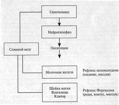
Rice. 10. Neuroreflex pathways for the release of oxytocin by the hypothalamic-pituitary system
Prolactin (PRL), lactogenic or luteotropic hormone - a polypeptide produced by the adenohypophysis; in females, it stimulates the process of milk formation, supports lactation, and exhibits luteotropic properties in the second half of pregnancy. In males, its effect is unknown. Feature PRL - target organs of prolactin (mammary gland, corpus luteum) do not synthesize hormones that inhibit its secretion (lack of feedback).
There are two types of gonadotropin secretion: tonic and cyclic. Tonic secretion is continuous, it is recorded in males and females throughout life. Cyclic secretion of FSH and LH precedes ovulation and is recorded in mature females. Preovulatory release of LH in spontaneously ovulating animals is initiated by a preovulatory peak of estradiol, in reflex ovulating animals (cats, rabbits, camels) - by sexual intercourse.
The pineal gland, or pineal gland, is an upper cerebral appendage related to the structures of the diencephalon. It produces the neurosecrete melatonin, which is characterized (depending on the type of animal) by anti- and progonadal activity: the ability to inhibit or, conversely, stimulate the secretion of GnRH. A distinctive feature of the epiphysis is the dependence of its secretory activity on illumination (photoperiodism). The activity of the gland increases in the dark. The neurohormone of the pineal gland controls the daily hormonal rhythm in the body. In many mono- (wolves, coyotes, jackals, wild dogs Dingo, etc.) and polycyclic animals (felines, sheep, horses, etc.), the light factor serves as the main climatic regulator of breeding season. The role of melatonin in dogs remains unclear, as dogs are sexually active regardless of the season.
The physiological role of sex hormones in the body of females and males is extremely diverse. The endocrine parts of the ovaries produce estrogens, progesterone and inhibin, while the testes produce testosterone and inhibin.
Estrogens - sex hormones of a steroid nature, consist of 18 carbon atoms (C 18). Produced by growing and maturing tertiary follicles and placenta. There are three fractions of estrogen - estradiol, estrone and estriol. Estradiol, the most active of these, is the primary estrogen that can be converted to estrone and estriol. In the body of females, estrogens are responsible for the development of secondary sexual characteristics and excretory ducts of the mammary glands. Estrogens induce estrus, sexual arousal and hunting, cause proliferation of the endometrium, myometrium, epithelium of the vaginal mucosa and its vestibule, increase the blood supply to the genital organs, promote the opening of the cervical canal, stimulate the contractile activity of the uterus, fallopian tubes and take an active part in labor activity, through positive feedback mechanisms induce preovulatory LH surge in spontaneously ovulating animals.
Progesterone is a steroid hormone (C 21). It is produced by the corpus luteum of the sexual cycle and pregnancy, as well as by the placenta. In dogs, the corpus luteum is the main producer of progesterone throughout the entire gestation period. Ovariectomy leads to termination of pregnancy at any stage. Progesterone transfers the endometrium to a secretory active state, prepares it for the attachment of the embryo, maintains the conditions in the uterus necessary for the development of the embryo and fetus, blocks the contractile activity of the uterus, causes the closure of the cervical canal, inhibits the maturation of follicles, the manifestation of estrus, sexual arousal and hunting, stimulates the development mammary alveoli and inhibits LH secretion.
Involvement of hormones in the regulation of reproductive function in dogs
|
Hormone |
Hormone function |
||
|
title |
place of production |
Chemical nature |
|
|
Gonadotropin-releasing hormone(G-RG) |
Hypothalamus |
Peptide |
Stimulation of FSH and LH secretion |
|
prolactin inhibitoryfactor (PIF) |
Hypothalamus |
Peptide |
Inhibition of the secretion of the hormone prolactin |
|
Corticotropin-releasing hormone(K-RG) |
Hypothalamus |
Peptide |
Stimulation of ACTH secretion |
|
Follicle stimulating hormone (FSH) |
adiohypophysis |
Glucoprotein |
1. Growth of follicles 2. Secretion of estrogen hormones 3. Spermatogenesis |
|
luteinizinghormone (LH) |
adiohypophysis |
Glucoprotein |
1. Ovulation 2. Formation of the corpus luteum and secretion of the hormone progesterone 3. Secretion of testosterone hormone |
|
Prolactin |
adiohypophysis |
Protein |
1. Lactation 2. Luteotropic factor |
|
Adrenocorticotropic hormone (ACTH) |
adiohypophysis |
Polypeptide |
Secretion of glucocorticoid hormones |
|
Oxytocin |
Hypothalamus |
Peptide |
1. Childbirth 2. Milk yield |
|
Estrogens |
ovaries Placenta |
Steroid |
1. Estrus, sexual arousal and sexual desire 2. Floor shaping 3. Childbirth 4. Growth of mammary glands |
|
Progesterone |
ovaries Placenta |
Steroid |
1. Preservation of pregnancy 2. Growth of mammary glands |
|
Relaxin |
ovaries |
Polypeptide |
1. Relaxation of the ligaments of the pelvis 2. Opening the cervix |
|
Testosterone |
testicles |
Steroid |
1. Sex drive 2. Spermatogenesis 3. Stimulation of the growth and development of the excretory ducts of the testes, accessory sex glands and penis |
|
Inhibin |
ovaries testicles |
Protein |
Inhibition of FSH secretion |
|
Cortisol |
adrenal glands |
Steroid |
1. Childbirth 2. Lactation |
Inhibin is a peptide hormone produced by the follicular epithelium of tertiary follicles and Sertoli cells. It inhibits the secretion of FSH in the body of females and males.
Relaxin is a peptide hormone produced by the corpus luteum at the end of pregnancy. Prepares the mother's body for childbirth, causing relaxation of the ligaments of the pelvis, muscles of the cervix, vagina and its vestibule.
Testosterone is a male sex hormone of a steroid nature (C19) produced in the testes, ovaries, and adrenal cortex. In the testes, testosterone is synthesized in Leydig cells, in the ovaries - in tertiary follicles, being only an intermediate stage of biosynthesis. The physiological role of testosterone in the embryonic period is participation in the sexual differentiation of the organism. Testosterone is essential for the development of secondary sexual characteristics, maintenance of spermatogenesis, stimulation of sexual desire, and regulation of LH secretion in males.
Maternal and fetal glucocorticosteroids and prostaglandin Fc also take part in the regulation of sexual processes.
Glucocorticoids are steroid hormones produced by the adrenal glands of the mother and fetus. Cortisol (stress hormone) is the most active and fundamental glucocorticoid hormone involved in the regulation of the birth process and lactogenesis. In the initiation of the birth act, fetal cortisol appears to play a leading role.
The secretion of glucocorticoids is controlled by corticoliberin and adrenocorticotropic hormone, or ACTH. Corticoliberin - a peptide hormone of the hypothalamus, stimulates the secretion of the polypeptide hormone of the adenohypophysis ACTH, characterized by corticotropic activity.
Prostaglandin F 2 a (PgF 2 a) is a biologically active compound with hormonal properties, a derivative of polyunsaturated fatty acids. It is produced by many cells and tissues and takes part in various processes and reactions of the body. In many domestic animals, PgP 2 a serves as a uterine luteolytic factor (causes regression of the corpus luteum of the sexual cycle and pregnancy) and takes an active part in the initiation and maintenance of the birth process. The yellow bodies of the sexual cycle and pregnancy in dogs and cats are not very sensitive to it. Abortion in animals of these species can usually be induced only after repeated application of PgF 2 a starting from the 5th day of diestrus.
The neuroendocrine regulation of the sexual function of dogs is significantly influenced by communication with the opposite sex (this is especially important for reflex ovulating animals - rabbits, camels, llamas, and felines). Coordination of sexual behavior of females and males is provided by sense organs, skin sensitivity and movement.
Important external factor that affects all the functions of the body of dogs, is food. Insufficient, excessive and (or) inadequate feeding not only reduces the reproductive ability of animals, but is often the cause of their infertility.
The most important reproductive functions of the hormones of the hypothalamus, pituitary, gonads, placenta and adrenal glands are shown in Table 2.
SEXUAL AND PHYSIOLOGICAL MATURITY
Puberty- this is the age from which males and females are able to participate in the process of sexual reproduction: to produce the corresponding germ cells (sperm, oocytes) and mate. Sexual activity in females is cyclical. They demonstrate readiness for mating only in a strictly fixed period of the sexual cycle - during sexual hunting. Ovulation in dogs is spontaneous and is tied to the onset of estrus. The selection of sperm (sperm and sperm plasma) from the genital organs of males and its introduction into the genitals of females occur during intercourse.
The timing of the onset of maturity in females is determined by the date of manifestation of the first sexual cycle. Usually the first sexual cycle is recorded at the age of 6-12 months. In males, puberty occurs approximately 1-1.5 months later than in females. Dogs of medium and dwarf breeds become sexually mature earlier than large breed dogs. The timing of the onset of puberty is significantly influenced by the state of health of the animal, the conditions of feeding and keeping, communication with the opposite sex.
Puberty usually occurs before the end of the main growth, structural and physiological development of the animal, ensuring its high fertility, the normal functioning of the body during pregnancy, childbirth and lactation, the birth of a healthy offspring.
Physiological maturity - the age at which it is advisable to use males and females to produce offspring. Physiological maturity in dogs coincides in time, as a rule, with the achievement of body sizes characteristic of an adult animal, and the manifestation of 2-3 sexual cycles in females. Females of most breeds become physiologically mature, usually at the age of 1.5 years, males - 2 years.
SEXUAL ACT
Sexual intercourse, or copulation, intercourse, copulation, is a set of sexual reflexes that ensure the removal of sperm from the male genital apparatus and its introduction into the female genital tract. In sexual intercourse, the following reflexes are distinguished: approximation, erection, cuddling, copulation and ejaculation.
The approach reflex is a set of behavioral responses that coordinate the sexual behavior of individuals of different sexes during mating.
The erection reflex is a strong filling with blood and an increase in the size of the penis, ensuring its removal from the prepuce and the possibility of insertion into the female genital tract. This reflex-vascular act is manifested in females by swelling of the clitoris and hyperemia of the mucous membrane of the vagina and its vestibule.
The cuddling reflex is the adoption by the female and male of the mating position. Simultaneously with the embracing, the copulatory reflex is also manifested.
The cumulative reflex is the insertion and friction of the male's penis into the female's vagina, leading to irritation of the erogenous zones of their genitals and the onset of ejaculation.
Ejaculation reflex - removal of sperm (ejaculate) from the male reproductive apparatus. Ejaculation, like an erection, is a neuroreflex act.
All sexual reflexes are inherently innate ( unconditioned). In the process of their formation and the acquisition of sexual experience by animals without conditioned reflexes conditions are layered. Some of them contribute to the development of full-fledged sexual reflexes, while others, on the contrary, by strengthening or weakening their manifestation, lead to the occurrence of abnormal sexual behavior.
The sexual behavior of animals is coordinated by the sense organs, skin sensitivity and movement.
During proestrus and estrus, females secrete sex pheromones and, in particular, methyl p-hydroxybenzoate, which stimulate male sex reflexes. At the beginning of proestrus, females behave passively or demonstrate aggressive behavior towards males; at the end of proestrus, they begin to show sexual interest in them. Males follow the female with signs of estrus and sexual arousal, lining up in a hierarchical rank. If the female allows, then the male favorite begins to actively sniff her, lick the external genitalia and other parts of her body, puts his head, one or two front paws on her back, and attempts to have sexual intercourse. At the onset of sexual hunting, the female demonstrates readiness for mating. It sniffs the genitals of the male, takes a pose for mating: it stands motionless, takes the tail to the side, pulls the genital loop up. The male jumps onto the female, clasps her sides with his front paws and makes pushing movements with his pelvis, ensuring the introduction of the penis into the vagina.

Rice. 11. Dog Mating Postures:
A - at the beginning of sexual intercourse; B - during the period of adhesion of the genital organs
With the full introduction of the penis, the sphincter of the vestibule of the vagina is reflexively compressed, which leads to a strong swelling of the bulb of the penis and adhesion ( gluing) of the genital organs of animals. The male jumps off the female, and the animals assume a tail-to-tail posture. Coupling of the genital organs (genital lock, mating) lasts 5-45 minutes. The release of sperm is accompanied by rhythmic movements of the root of the tail and wave-like contractions of the wall of the urogenital canal. Sperm is released into the vagina in the form of three fractions: the first fraction acts as a lubricant, the second contains sperm, and the third ensures the pushing of sperm into the uterine cavity.
Muscles of the head.
The muscles of the head are divided into mimic and chewable. The first differ in that they begin on the bones or fascia and end in the skin. Part of the muscles grouped around natural openings form sphincters (contribute to the narrowing of the opening) or dilators (contribute to the expansion of the opening). Function facial muscles heads - ensuring the mobility of the lips, corners of the mouth, nostrils, eyelids, skin of the muzzle, chin, cheeks, forehead, etc. In addition to the utilitarian significance of these movements, important for nutrition, breathing, vision, etc., facial muscles provide communication links between animals, since the expression of the eyes, mouth, the position of the lips, ears, the relief of the back of the nose play a signal value in the communication of animals among themselves. The facial expressions of the dog are extremely diverse and convey the various mental states of the animal in a way understandable to most animals. Some facial expressions (in this context, you can’t call it otherwise) in a dog are similar to human facial expressions, others are understandable only to an observant dog breeder from the experience of communicating with a particular dog. Work disruptions mimic muscles can introduce very significant difficulties in the collective actions of animals, sometimes in wild nature it can cost the animal its life. Great role mimic muscles and in the conformation assessment of a thoroughbred dog. It is not for nothing that in the standard of many breeds the description often begins with a characteristic expression of the eyes, the muzzle of the dog. Deviations in the direction of strengthening or weakening the breed characteristics in the exterior of the dog may be associated with features of the work of facial muscles. So, insufficient tone subcutaneous muscles of the mouth, incisor and canine muscles contribute to the appearance of saggy wet lips in the Slovak kuvach. Weakness these same muscles zygomatic muscle leads to drooping lips with drooping jowls, drooping ears, which is a serious defect in the Rottweiler's conformation. Flaccidity of the external buccal and orbicularis muscles The mouth droops the upper lip, simulating the depth of the muzzle - this is beneficial to the conformation of the English Bulldog and St. Bernard, but may be the reason for the culling of the Great Dane. Weakness of the zygomatic muscle for german shepherd or the Pomeranian may cause a loss of chances for a show career, as it leads to the formation of floppy ears. Hanging ears, also associated with weakness of the zygomatic muscle, is a vicious sign for dogs of a number of breeds - husky, Scotch, Doberman. Narrow nostrils, associated with weakness of the own levator labrum and transverse nasal muscles, is a vice of the English bulldog, but the dignity of a greyhound. Chewing muscles in connection with their more significant work than mimic ones, they are much more powerful. They start from various bones of the skull and are attached mainly to the lower jaw. Their reduction provides a variety of movements of the jaws to capture, bite and grind solid food. In case of violation of the act of chewing (for example, due to a painful focus in the gums), insufficient training of the masticatory muscles (for example, when feeding pasty food) or due to a traumatic nature, phenomena of asymmetric or general atrophy and weakness of these muscles may occur. Weakness, spastic contraction of individual muscles can distort the appearance of the dog.
Functionally important muscles of the dog's head.

A - facial muscles: 1 - subcutaneous muscle of the face, 2 - circular muscle of the mouth, 3 - zygomatic muscle, 4 - nasolabial levator, 5 - circular muscle of the eye.
B - chewing muscles: 1 - a large chewing muscle (superficial and deep layers), 2 - temporalis muscle, 3 - digastric muscle.
Musculature of the dog's head and its location.
ROUND MUSCLE OF THE MOUTH - in the lower lip forms faint bundles; in the upper lip they are somewhat more strongly developed, but do not form a continuous ring. Part of them at the middle sagittal line bends upward and is fixed on the cartilaginous framework of the nose.
INCITOR MUSCLES - poorly developed.
CHIN MUSCLE - poorly developed.
Zygomatic muscle - departs from the thyroid ear cartilage, stretches in the form of a thin ribbon under the skin along the lateral surface of the head to the corner of the lips and ends under the skin muscle of the lips in the upper lip. The original, in comparison with other animals, should be considered primitive. In other animals, it apparently differentiated into two parts - ear and zygomatic, with the first from the thyroid cartilage going to the area of the zygomatic arch, being included in the general ear muscles.
NOSO - LIP LIFT - wide, separated by a weak border from the lower eyelid lowerer and originates from the lamellar tendon in the medial angle of the eyes from the frontal fascia and on the upper jaw. It breaks up into a deep and superficial part and ends in the upper lip. The superficial part extends back into the buccal muscle, and the deep part gives off bundles to the lateral wall of the nasal cartilaginous tube.
FANGING MUSCLE - begins on the lateral surface of the upper jaw near the infraorbital foramen and, gradually expanding, ends in the upper lip; only a very small beam passes in the region of the outer wing of the nose.
SPECIAL UPPER LIP LIFT - starts along with the previous one, goes under the nasolabial lifter, gradually expands, and sometimes even bifurcates, and is fixed with thin tendon branches around the opening of the nose. At its end, part of the branches connects to the homonymous muscle of the other side.
DESCENT OF THE LOWER LIP - in very weak bundles it starts from the lower jaw between the canine and the chin and ends in the lower lip at the corner of the mouth. Often its presence is difficult to ascertain.
SUBCUTANEOUS MUSCLE OF THE LIPS - goes from the neck along the outer surface of the masticatory muscle and ends in the corner of the lips.
BUCKY MUSCLE - poorly developed.
GREAT MATERAL MUSCLE - originates from the lower edge and medial surface of the zygomatic arch and is tightly connected in the deep initial parts with the temporal muscle. On its further length, the muscle forms two unsharply demarcated layers: a) superficial layer goes to the angular process protruding on the lower jaw and is attached in its area, and some of the bundles protrude beyond this process and merge with the wing muscle; b) deep layer, crossing with the surface, it goes to the fossa of the masticatory muscle on the branches of the lower jaw / some authors distinguish three layers in the muscle /.
WING MUSCLE - has a beginning on pterygoid bone, pterygoid process of the sphenoid bone and partly on the palatine bone. Its abdomen is not as sharp as that of horses, divided into two layers. The muscle ends on the corresponding surface of the lower jaw branch and on its angular process, and part of the bundles, passing beyond the edge of the branch, is connected to the large chewing muscle.
TEMPORAL MUSCLE - very strongly developed, begins on the temporal fossa and orbital ligament - merges with a large chewing muscle and ends on the coronoid process.
DOUBLE MUSCLE - strongly pronounced; division into bellies is absent. The action of the chewing muscles is similar to that of pigs, but differs in the possibility of a very wide spreading of the jaws and their strong closing. There are no movements to the sides, and the protrusion of the ventral jaw takes place, but not to a large extent.
THEIR FUNCTION:
CIRCULAR MUSCLE OF THE MOUTH - compresses the lips and closes the mouth opening.
UPPER AND LOWER INCITIVE MUSCLES - antagonists of the circular muscle of the mouth - open the mouth opening.
CHIN MUSCLE - lowers the chin to the lower lip.
Zygomatic muscle - pulls the corner of the mouth back and up.
NASE-LIP LIFTER - lifts upper lip and expands the entrance to the vestibule of the nose.
Canine Muscle - expands the nostrils, raises the upper lip.
SPECIAL UPPER LIP LIFTER - lifts the upper lip.
LOWER LIP DOWNER - lowers the lower lip.
SUBCUTANEOUS MUSCLE OF THE LIPS - lowers the lower lip.
Reflex arc.
A reflex is the response of the body to irritation, carried out through the nervous system. Each reflex contains afferent ( sensory) and efferent ( executive) links that make up the reflex arc. The afferent part of the reflex arc consists of receptors and sensory neurons, the efferent part consists of motor neurons and the executive organ (muscle, gland, tissue). To implement a reflex, at least two neurons are needed: sensory and motor. Such a neural circuit is called a simple reflex arc. Most reflex arcs many intercalary intermediate neurons are involved, and such arcs are called multineuronal.
Diagram of a simple reflex arc
1 - receptor;
2 - sensitive path;
3 - sensitive center;
4 - synaptic transmission of excitation;
5 - motor center;
6 - motor path;
7 - effector.

Neurons are interconnected through branching of nerve processes using synapses, which provide contact and transmission of excitation from one neuron to another or to a working organ through a chemical substance called a mediator.
Scheme of complex reflex arcs with feedback.

Synaptic transmission of excitation in neural circuits

Synapses are capable of transmitting excitation in only one direction - from the axon to the dendrite. Functionally, excitatory and inhibitory synapses are distinguished. In excitatory synapses, the mediator is acetylcholine, in inhibitory synapses, glycine, etc. The mechanism of excitation transmission is shown in the synapse function diagram. Synapses of intermediate neurons of the cerebral cortex are the place of closure of conditioned reflex connections. Synapses form and store information called memory.
Feedback afferentation (feedback) - information from the executive body to the central nervous system, where the analysis of what should be and what happened in response to the action of the stimulus takes place. Based on this analysis, corrective impulses are sent from the center to the executing organ and to the receptors. These signals can increase or decrease their functional activity. Feedback in the reflex provides automatic self-regulation and forms an independent functional system called the reflex ring, and also guarantees automatic evaluation and perfect control of any reflex act. Such functional systems that provide regulation of behavioral responses are called nerve centers.
Joints, joints of bones.
The joint is an intermittent type of bone connection, characterized by increased freedom of movement, i.e. displacement of bones in relation to each other. The bones in the joint separates the fissured interosseous space. The structure of the joints is characterized by the presence of articular surfaces, joint capsule and joint cavity filled with joint fluid.

Joint 1 - joint capsule; 2 - articular surfaces; 3 - articular cavity
The articular surfaces are covered with a layer of very smooth articular cartilage.
The articular capsule is attached along the edge of the connecting bones, firmly growing together with them. As a result, a closed cavity is formed - the articular. AT individual joints in places of great friction, the articular capsule forms protrusions - bursae, inflammation of which - bursitis - is characteristic of large, heavy dogs, especially frequent in the area of the elbow joints. The strength of the joints is increased by attaching the articular ligaments outside the joint capsule. Each joint has its own optimal shape and connection depth, which ensure the congruence (coincidence) of the articular surfaces. This is especially important for joints that carry a lot of stress.
There are several types of bone connections.
Continuous . This type of connection has great elasticity, strength and very limited mobility. Depending on the structure of the tissue connecting the bones, the following types of connection are distinguished:
With the help of connective tissue - syndesmosis, and if elastic fibers prevail in it - synelastosis. An example of this type of connection are short fibers that firmly connect one bone to another, such as the bones of the forearm and lower leg in dogs;
With the help of cartilaginous tissue - synchondrosis. This type of connection has little mobility, but ensures the strength and elasticity of the connection (for example, the connection between the vertebral bodies);
With the help of bone tissue - synostosis, which occurs, for example, between the bones of the wrist and tarsus. With the age of animals, synostosis spreads in the skeleton. It occurs at the site of syndesmosis or synchondrosis.
In pathology, this connection can occur where it normally does not exist, for example, between the bones of the sacroiliac joint due to hypodynamia, especially in old animals;

Scheme of development and structure of the joint: a - fusion; b - formation of the articular cavity; in - a simple joint; g - articular cavity; 1 - cartilaginous bookmarks of bones; 2 - accumulation of mesenchyme; 3 - articular cavity; 4 - fibrous layer of the capsule; 5 - synovial layer of the capsule; 6 - articular hyaline cartilage; 7-cartilaginous meniscus
With the help of muscle tissue - synsarcosis, an example of which is the connection of the scapula with the body.
Discontinuous ( synovial) type of connection or joints . It provides more range of motion and is built more intricately. By structure, the joints are simple and complex, in the direction of the axes of rotation - multiaxial, biaxial, uniaxial, combined and sliding.
Most of the bones of the skull are connected by a continuous type of connection, but there are also joints - temporomandibular, atlantooccipital. The vertebral bodies, with the exception of the first two, are connected to each other by intervertebral discs ( cartilage), that is, synchondrosis, as well as long ligaments. The ribs are connected by intrathoracic fascia, consisting of elastic connective tissue, as well as intercostal muscles and transverse ligaments. The scapula is connected to the body with the help of the muscles of the shoulder girdle, and the pelvic bones - with the help of a joint with the sacrum, and with the first tail vertebrae - with ligaments. Parts of the limbs are attached to each other using joints of various types, for example, the connection of the pelvic bone with the femur occurs using a multiaxial hip joint.
Bibliography:
M. V. Dorosh, Veterinary guide for dog owners, 2008.
G.P. Dyulger, Physiology of reproduction and reproductive pathology of dogs.
F.S. Araslanov, A.A. Alekseev, V.I. Shigorin SERVICE DOG TRAINING
Exterior of the dog and its assessment by E.E. Yerusalimsky, Moscow 2002
Internet resources:
Portal for dog lovers and owners
www.friendog.ru
Surely every dog breeder or just a fan of four-legged human friends will be interested to know what is the “internal structure” of dogs? What do we and our pets have in common, and how are we strikingly different? Therefore, we propose to make a detailed excursion into the world of canine anatomy right now!
[ Hide ]
Skeleton structure
Naturally, the study of the anatomy of any animal begins with the study of the structure of its skeleton. The dog's skeleton is the basis, the skeleton, which holds all the organs and muscles of the dog inside. Consider all the "components" of the dog's skeleton in turn.
Scull
The skull of dogs is usually divided into the front and brain parts. Both of these parts consist of paired and unpaired bones (discussed in the table below).
It is easy to calculate that the dog's skull will consist of 27 bones, which are securely connected to each other by connective cartilage tissue. As the dog matures, this tissue stiffens. In this case, the lower jaw is attached to the skull with a strong movable joint, which allows the dog to chew food.
Note that the shape of the skull of dogs can vary greatly. In the process of selection, people contributed to the fact that some breeds were recognizable precisely due to the original structure of the skull.
So, according to the shape of the skull, dogs are divided into long-faced, short-headed and dogs with a normal head length. Moreover, the facial part of the skull will have big differences. The common name for all breeds with a shortened facial part of the skull is brachycephalic.
Vivid examples of the brachycephalic structure of the skull are Pekingese, Bulldogs, Pugs, Boxers, Sharpei. These dogs have a wide parietal part skull, strongly shortened and flattened facial part and protruding jaw. Such a special structure is the result of many years of breeding work, when individuals with the desired trait were deliberately selected, in this case with a flattened muzzle. However, such an unusual sign was associated with significant health problems.
After all, the disproportionately short muzzle caused degenerative changes in the structure of the dog's respiratory tract. Because of which, all of the above breeds are predisposed to tracheal collapse, pulmonary hypertension, and excessive tearing. Surely everyone noticed that outwardly cute Pekingese or Pugs often go “tearful”, and every breath they take is accompanied by wheezing or grunting. To describe all the inconvenience experienced by a brachycephalic dog, there is even a special term - brachycephalic syndrome.
However, let us return to the structure of the skull and say a few more words about the teeth and bite of the dog. So, the dental system of dogs suggests the presence of fangs, incisors, molars and premolars. At adult dog there should be 42 teeth, and the milk jaw consists of 28 teeth. The bite in dogs can be different, it depends on the breed and the standard provided by this breed.
There are different types of dog bites:
- Scissor-shaped, when the upper incisors in a closed form cover the lower ones. In this case, the lower incisors are closely adjacent to the upper ones.
- Pincer-shaped, the incisors of both jaws are adjacent to each other with a cutting surface.
- Undershot, the lower jaw is inferior in length to the upper, so there is free space between the dog's incisors.
- Overshot, the lower jaw protrudes forward, it is also called the "bulldog" jaw.
torso
The body of the dog itself will consist of the spinal column - the axis of the body and the ribs that are attached to it and together make up the skeleton of the dog (in the picture below you can see the dog skeleton).
The spine of a dog, in turn, consists of the following sections:
- cervical - is formed by seven vertebrae, the first two are more mobile and are called atlas and epistrophy, like in cats;
- thoracic - made up of 13 vertebrae;
- the lumbar, as well as the cervical, consists of 7 vertebrae;
- the sacral section completes the spinal column, the single sacral bone of which is made up of 3 fused vertebrae.
The tail consists of 20-23 movable vertebrae. The chest is represented by 13 pairs of ribs, 9 of which are true and are attached to the sternum, and 4 are false and form a costal arch. The ribs of dogs serve as reliable protection for the heart and lungs and have different curvature depending on the breed. The lumbar vertebrae are large and have many spurs, thanks to which the muscles and tendons that hold the abdominal organs are securely attached to them. The vertebrae of the sacral region merge into a single strong bone, which serves as a transition between the loin and the tail.
The first five vertebrae of the tail are the most developed and mobile. According to the standard of some breeds, tail vertebrae are stopped in the amount provided for by this standard.
limbs
The limbs of dogs have a rather complex structure. The forelimbs are a continuation of the obliquely set scapula, which passes into the humerus with the help of the humeroscapular joint. This is followed by the forearm, where the radius and ulna are connected by the elbow joint. It is followed by the carpal joint, which is made up of 7 bones connected to the 5 bones of the metacarpus.
The metacarpus is 5 fingers, 4 of them have three phalanges, and 1 has two. All fingers are “equipped” with claws, which, compared to cats, are not retractable and consist of strong keratinized tissue.
The front legs are attached to the spine through strong shoulder muscles. Due to the fact that the upper parts of the shoulder blades protrude beyond the thoracic vertebrae in dogs, the withers are formed - an indicator of the height of the dog. The hind limbs are represented by the femur and lower leg, where connecting elements are the hip and knee joints.
The lower leg, which consists of the tibia and tibia, is attached to the tarsus with the help of the hock joint. The tarsus, in turn, passes into the metatarsus and ends in 4 fingers with three phalanges. Detailed explanation canine foot device is available in the video below.
Internal organs
Naturally, acquaintance with the anatomy of a dog cannot be limited only to the skeleton and musculoskeletal system. If we already have some idea of the dog's skeleton, let's talk about its internal organs and systems.
Digestive system
The digestive system of dogs is very similar to the digestive system of other mammals, including us. It begins with the oral cavity, which is equipped with strong and sharp teeth. Our pets are predatory animals, and therefore their jaws are adapted to eating large pieces of meat. Moreover, food is not always crushed in the mouth, often dogs swallow quite large pieces whole. Saliva in our pets begins to be actively produced from the smell of food and its type, and the enzymatic composition of saliva is slightly different, each breed has its own.
The food then moves down the esophagus and reaches the stomach. The main "digestion" occurs in this muscular organ. Gastric juice and special enzymes under the influence of peristaltic processes turn food into a homogeneous mass called chyme. At the same time, the valves of the stomach should not allow food to return back into the esophagus or enter the small intestine ahead of time. At least that's how a healthy dog's digestion should work.
Well, the small intestine, which is the next "in line", closely "interacts" with the pancreas, duodenum and cookies. The enzymes of the pancreas and gallbladder continue to act on the chyme. And the walls of the small intestine actively absorb useful substances from it in order to “transfer” them into the blood. At the same time, the small intestine is quite long, and its absorbent area is impressive - depending on the breed, it can be equal to the area of a room!
The digested food then moves into the large intestine. By this time, all the useful substances have already been taken from it, only water and coarse fiber can remain. From the remnants of waste food, water, some bacteria and inorganic substances, feces will be formed. Defecation occurs under the control of the central nervous system, in the case of nervous disorders or old age, bowel movements can be uncontrolled.
Respiratory system
The dog's respiratory system performs the most important function: thanks to it, all the cells of the body receive the right dose of oxygen, and the exhaust carbon dioxide is removed. The respiratory system of all mammals, and dogs are no exception, is usually divided into upper and lower sections. In the "composition" of the upper section of the nasal cavity, nasopharynx, trachea and larynx. The movement of air begins through the nasal passages - the nostrils, the shape and size of which depends on the breed of the dog. In the nasopharynx, the inhaled air is warmed, and thanks to the nasal glands, the air is “filtered” from dirt and dust.
Further, the air moves along the larynx - a cartilaginous organ, which is held by the hyoid bone and is equipped with vocal cords, that is, it is responsible for sound production. This is followed by the trachea - also a cartilaginous organ, closed by the tracheal muscle. lower division respiratory system represented by the lungs and bronchi. The lungs, in turn, consist of 7 lobes and are heavily dotted with blood vessels to enrich them with oxygen. The lungs are an organ that can significantly change its volume: when you inhale, they increase many times, and when you exhale, they seem to “deflate”.
Such elasticity is possible due to rhythmic contractions of the diaphragm and intercostal muscles. During inhalation in the alveoli of the lungs, the old air is “replaced” with oxygenated new air. The respiratory rate of dogs should be in the range of 10-30 breaths per minute, it depends on the breed and physical condition of the pet. Little dogs breathe faster than big dogs. The respiratory rate can change greatly in case of fear, heat and during physical exertion.
Circulatory system
Naturally, the main organ of the circulatory system is the heart. Through the arteries, blood is distributed to all other organs, and through the veins returns to the heart. The heart of a dog is a strong muscular hollow organ, which is located between the 3rd and 6th ribs in front of the diaphragm.
The heart is four-chambered, it is divided into two parts: right and left. Both parts of the heart, in turn, are divided into the atrium and ventricle. On the left side circulates arterial blood, which enters there through the pulmonary veins, in the right - venous, which enters the heart from the vena cava. From the left side, oxygenated arterial blood enters the aorta.
The heart provides a continuous flow of blood in the body, it moves from the atria to the ventricles and from there enters the arterial vessels.
At the same time, the walls of the heart consist of such shells: the inner shell - the endocardium, the outer - the epicardium and the cardiac muscle of the myocardium. In addition, the heart has a valvular apparatus, which is designed to "monitor" the direction of blood flow and so that arterial and venous blood do not mix. The size of the heart and the frequency of its contractions is highly dependent on the breed of the dog, its sex and age, and environmental factors.
The first indicator of the work of the heart of the dog is the measurement of the pulse, which is normally in the range of 70-120 beats per minute. Young individuals are characterized by a more frequent contraction of the heart muscle. A complex device has a system of capillaries and blood vessels of a dog, which literally “penetrates” the entire body of the animal and all its organs. For 1 sq. mm tissue is more than 2500 capillaries. And the total volume of blood in the body of a dog is 6-13% of body weight.
excretory system
The excretory system of our smaller brothers cannot function without such internal organs as the kidneys (available in duplicate). They communicate with the bladder through the ureters and end in the urethra. The purpose of the excretory system is the formation, accumulation and excretion of urine from the animal's body. Through urine, the body is freed from metabolic products, any violations in this process are fraught with serious health problems, even death.
To filter blood, the kidneys are equipped with nephrons, each of the nephrons is shrouded in a network of tiny blood vessels. As the animal ages, the nephrons will break down and be replaced by scar tissue, which is why older animals often have kidney problems.
reproductive system
The reproductive system is strongly connected with the excretory system. Anatomically, in males, the urinary canal is also a vas deferens, in addition, males need testes and an external genital organ for reproduction. At the same time, in a newborn dog, the testicles are in the abdominal cavity, but by two months they will descend and take their place in the scrotum. It is there that subsequently the sperm will “ripen”. In addition to the testes, males have a prostate - a gonad that maintains the viability of spermatozoa.
The penis of a male dog, consisting of a head, body and root, is covered with a preputial sac, at the moment of excitation, the genital organ comes out of the sac and this is called an erection. Moreover, the hardness of the penis is achieved not only due to the cavernous bodies, but also due to the bone located at the base of the organ. Sexual maturity in males, as well as in females, occurs at 6-11 months, small dogs "ripen" faster. But males are allowed to mate at 15-16 months, and females at 1.5-2 years, by this age the dogs have completely completed puberty and will definitely give healthy offspring.
The genital organs of females are the uterus, by the way, the uterus of dogs has “horns”, to which the ovaries, fallopian tubes and vagina are “attached”. The egg of a female dog, like that of a human, matures in the ovaries. This process is quite complicated and occurs under the constant "control" of hormones. As estrus approaches, the follicles with the egg increase, and when estrus occurs, the follicle bursts, making way for the egg. The egg matures in the fallopian tubes for another three days, while the fluid from the burst follicle produces a hormone that prepares the female's body for pregnancy.
In females, estrus happens twice a year, in dogs of northern breeds - once a year and it lasts about 28 days. The optimal time for mating is 9-14 days of estrus. If a female has mated with two males, then her litter may contain puppies from both males. Therefore, knitting purebred dogs always occur under careful control by the owner. And another nuance: dog embryos do not develop in the uterine cavity, but in the horns - tubular processes on both sides of the main reproductive organ.
Nervous system
The nervous system of dogs is represented by the central and peripheral sections. The central nervous system is made up of the brain and the spinal cord adjacent to it, and the peripheral nervous system is made up of many nerve endings and fibers penetrating all organs and tissues of the animal. Bundles of nerve fibers make up the nerve trunks, which are more simply called nerves. All nerves are divided into afferent and efferent. The former transmit "information" from the organs to the control center - the brain, and the latter, on the contrary, the impulses that arise in the brain convey to the organs and tissues of the dog.
The building block of the entire nervous system of a dog is a nerve cell, which necessarily has processes. The transmission of nerve impulses is carried out through the contact of processes nerve cells and with the help of mediators. Mediators are substances that transmit impulses. Information is transmitted through nerve cells and fibers as by telegraph, and the transmission speed is about 60 m/s.
sense organs
The sense organs of dogs are extremely well developed. This predator is able to hear and smell much better than you and I. Therefore, we propose to talk about the canine sense organs in more detail, because without them the dog was not what we used to see it.
The structure of the eye
The eye of our four-legged friend consists of three membranes: fibrous, vascular and reticular. In principle, the structure of the dog's eye is anatomically very similar to our organ of vision. The principle of perception of visual information in a dog does not differ from the principle of perception of all other mammals. A beam of light passes through the cornea, hits the lens, which focuses the light on the retina, on which the light-receiving elements are located. Light-receiving elements in dogs, just like ours, are rods and cones.
The human eye is equipped with the so-called yellow spot - the place of the greatest concentration of light-receiving elements, dogs do not have a yellow spot, therefore their vision is worse than human. However, a dog can perceive information better in different lighting conditions, so our friends navigate much better than us in the dark.
ear structure
Our four-legged pets perceive a lot of information through hearing, which they have much sharper than ours. The dog's auditory analyzer begins with the outer ear, moves to the middle ear, and ends with the inner ear. The outer ear begins with the auricle, which is necessary for capturing sounds and directing it to the deep parts of the auditory organ. The auricle is a cartilaginous organ to which muscles are attached, allowing it to be rotated to improve focus on the sound source. Per auricle the external auditory canal follows, it is divided into horizontal and vertical parts.
In fact, the ear canal is a skin tube through which sound travels to the eardrum. The skin of the auditory canal contains numerous glands, in addition, hair often grows profusely in the auditory canal of dogs. This is followed by the tympanic membrane - the thinnest membrane, it serves to separate the outer and middle ear and capture the vibrations of sound waves. The middle ear can be described as a bony cavity, which is the "receptacle" of the auditory ossicles (hammer, stirrup and anvil) and the inner ear. The auditory ossicles are attached to the inside of the eardrum and amplify sound vibrations many times, transmitting them to the structures of the inner ear.
The inner ear is a receptacle for auditory receptors and the organ of balance - the vestibular apparatus. It is in the inner ear that the analysis of sound vibrations takes place and information is formed for transmission to the brain.
The structure of the nose
A dog's nose is a supersensitive organ, in principle, we can say that our four-legged friends live in a world of smells. Everything that surrounds them is associated in animals with some kind of smell, including you and me. A dog's nose has 125 million olfactory receptors, while our humble nose has only 5 million. The mucus that coats the inside of both our nose and that of a dog's nose in dogs extends beyond the olfactory organ and covers the outside as well. This is why our pets' noses are so wet.
The recognition of smells in dogs begins with the nostrils, and their side cutouts play an important role here. More than half of the inhaled air passes through them. In general, the airways begin from the external nose and the nasal cavity, which is divided into lower, middle and upper passages. The upper part of the nasal cavity is the home of the olfactory receptors. And the lower part leads the inhaled air to the nasopharynx.
Interestingly, the outer pigmented part of the nose of dogs is called the nasal planum. The mirror of each dog has its own unique pattern, so that if necessary, one dog can be distinguished from another. In addition, the olfactory organ of dogs is able to pick up odors from a distance and differentiate them - a property available only to some people. It is thanks to this property that dogs are a great help to a person for whom the world of smells is only partially accessible.
Photo gallery
The request returned an empty result.Video "How do dogs see the world with their nose?"
We have already talked about how much information our four-legged friends receive through their noses. But this video, which concludes your introduction to canine anatomy, will tell you something more interesting about the super sensitive canine nose!
Sorry, there are currently no surveys available.A healthy dog is a cheerful, lively animal with a shiny coat, clean and clear eyes, with a slightly wet and cold nose. Sometimes the nose can be dry and hot in a healthy dog when he is sleeping or just awake, or after intense work in very dry weather. In a healthy dog a good appetite, bowel movements are regular, urination is normal, breathing is even. The mucous membranes are clean, pale pink in color.
A sick dog is very different from a healthy one. She is oppressed, tries to hide in a dark place, reluctantly responds to the call. The dog does not eat well, but is constantly thirsty. In addition, signs of the disease may include stool disorders (diarrhea, constipation, blood in the stool), vomiting, frequent urination, purulent discharge from eyes, nose. Mucous membranes are pale, cyanotic or icteric.
A veterinarian should be contacted as soon as possible if the dog has one or more persistent symptoms of the disease.
Caring for the health of the dog is one of the main responsibilities of the owner.
The coat becomes dull, disheveled, baldness of certain parts of the body, scratching is possible.
Body temperature, pulse and respiration may also be abnormal. These signs usually do not appear simultaneously, but with the development of the disease, their number increases.
Before turning to the question of providing the first medical care pet, it would be useful to consider the anatomical and physiological features of the dog.
dog body structure
Everyone who gets a dog should have an idea about the structure of her body and how it functions in order to identify health problems in time and contact a veterinarian.
Knowledge of the structure and functions of the dog's body makes it possible to understand many features of its behavior, notice deviations from the normal state in time and take timely measures to prevent the disease.
This is especially true for puppies whose body is in the process of formation.
All organs are closely interconnected, and the work of each of them is directly dependent on the other.
The body of a dog consists of 2 main organ systems: external and internal.
Any organ consists of tissues that ensure its functioning and are a set of cells of the most diverse shapes, fibers and intercellular substance. Cells are the smallest structural units in the body, the shape and structure of which depend on their purpose.
The cell size is thousandths of a millimeter (10-100 microns).
In the body of a dog, there are 4 main groups of tissues.
Epithelial or integumentary tissues. These tissues form the surface of the skin, line the inner surface of the oral and nasal cavities, esophagus, stomach, intestines, bladder, ureters, etc.
Epithelial tissues perform a protective function, carry out the exchange of substances between the external and internal environment of the body. In addition, certain cells of the epithelial tissue produce special substances: gastric juice, intestinal juice, saliva, tears, etc.
Support-trophic tissues. This group includes blood, lymph, fat, connective, cartilage, bone tissue. Support-trophic tissues are very diverse in their structure and functions.
They create the supporting part (framework) of many organs and the body as a whole (skeleton), connect one organs to others, form protective shells of organs that give them a certain shape, and are a bed for blood vessels and nerves.
Muscle. This tissue performs motor functions, allowing the dog to move and performs contractile movements of various organs.
In addition, supporting-trophic tissues perform vital functions: trophic (nutritional), hematopoietic, and protective.
nervous tissue. It forms the nervous system, which coordinates the functions of all tissues and organs, perceives signals from the external environment and determines responses.
All tissues are the building material for organs. Usually, the predominance of any type of tissue in an organ determines its functions. For example, in the brain, which is an organ of the nervous system, nervous tissue predominates.
Conventionally, in the body of a dog, as well as other domestic animals, a number of apparatuses and organ systems are distinguished on the basis of the main function performed by this system. However, we should not forget that each organ can perform, in addition to the main ones, other functions that are no less important for the body.
For example, the main function of the bones of the skeleton is musculoskeletal, however, in addition to this, the bones of the skeleton also perform nutritional, hematopoietic, and electrolytic functions.
Bones are involved in protein, water, carbohydrate, fat, mineral and general exchange substances.
The dog's body consists of the following organs and systems:
1. Apparatus of movement, consisting of bones, ligaments and muscles.
2. Internal systems of the organs of digestion, respiration, excretion and reproduction.
3. Integrating systems, including blood and lymph circulation systems, immune system, system of endocrine glands, skin, sense organs and nervous system.
Major internal organs of a dog
Dogs are mammals, therefore, their skeleton is typical of mammals and consists of the same sections.
In mammals, the skull is larger than, for example, in reptiles.
Mammals are characterized by 7 cervical vertebrae. Both giraffes with a very long neck and whales that do not have a neck at all have the same number of cervical vertebrae. The thoracic vertebrae (usually 12-15) together with the ribs and sternum form the chest.
The lumbar spine is formed by massive, movably articulated vertebrae that provide flexion and extension in this spine region. So the body can bend and unbend. The number of lumbar vertebrae different types mammals can vary from 2 to 9, the dog has 6. The sacral spine consists of 3-4 vertebrae, which are connected to the pelvic bones.
The number of vertebrae of the caudal region in dogs can range from 3 to several tens, which determines the length of the tail.
The belt of the forelimbs of mammals consists of two shoulder blades, crow bones fused with them, and a pair of underdeveloped clavicles.
The belt of the hind limbs - the pelvis - in a dog is formed by 3 pairs of pelvic bones. Most mammals, including the dog, have especially developed muscles of the back and limbs.
In the oral cavity of a dog, like in other mammals, a tongue and teeth are placed. The tongue serves to determine the taste of food: its surface is covered with numerous papillae, in which the endings of the taste nerves are located. The movable tongue moves food in the mouth, which helps to moisten it with saliva secreted salivary glands. Mammalian teeth have roots that anchor them in the sockets of the jaws. Each tooth is made of dentine and is covered on the outside with strong enamel. In mammals, teeth have a different structure associated with a specific purpose. In front of the dog's jaws are incisors, on both sides of which are fangs. In the depths of the mouth are the molars.
The muscles of the lower jaw are also very developed, thanks to which the dog is able to hold prey firmly.

Dog skeleton: 1 - upper jaw; 2 - lower jaw; 3 - cranium; 4 - parietal bone; 5 - occipital protuberance; 6 - cervical vertebrae; 7 – thoracic vertebrae; 8 - lumbar vertebrae; 9 - tail vertebrae; 10 - blades; 11 - humerus; 12 - bones of the forearm; 13 - bones of the wrist; 14 - metacarpus; 15 - phalanges of fingers; 16 - ribs; 17 - costal cartilages; 18 - sternum; 19 - pelvic bone; 20 - hip joint; 21 - femur; 22 - knee joint; 23 - tibia; 24 - fibula; 25 - calcaneus; 26 - hock; 27 - tarsus; 28 - metatarsus; 29 - fingers
Puppies first have milk teeth, which later fall out, and permanent ones grow in their place.
All dog teeth have a purpose. She uses her molars to tear large pieces of meat.
The outer molars have blunt tips that help chew plant foods. The incisors are designed to separate meat from bones.
The stomach of a dog, like that of most mammals, is single-chamber, the intestines consist of a small, large and rectum. In the intestine, food is digested under the influence of the secretion of the digestive glands of the intestine, as well as the juices of the liver and pancreas.
In a dog, like in other mammals, the chest cavity is separated from the abdominal muscular septum - the diaphragm, which protrudes into chest cavity and attached to the lungs. With the contraction of the intercostal muscles and the diaphragm, the volume of the chest increases, while the ribs move forward and to the sides, and the diaphragm becomes flat from convex. At this moment, air is forced into the lungs by the force of atmospheric pressure - inspiration occurs. When the ribs are lowered, the chest narrows and the air is pushed out of the lungs - exhalation occurs.

The internal organs of the dog: 1 - nasal cavity; 2- oral cavity; 3 - trachea; 4 - esophagus; 5 - lungs; 6 - heart; 7 – liver; 8 - spleen; 9 - kidneys; 10 - small intestine; 11 - large intestine; 12 - anus; 13 - anal glands; 14 - bladder; 15, 16 - genitals; 16 - brain; 17 - cerebellum; 18 - spinal cord
The heart in dogs is four chambered and consists of 2 atria and 2 ventricles. The movement of blood is carried out in 2 circles of blood circulation: large and small.
Urine excretion occurs through the kidneys - a paired organ located in the abdominal cavity on the sides of the lumbar vertebrae. The resulting urine through 2 ureters enters the bladder, and from there through the urethra is periodically removed to the outside.
Metabolism in mammals, due to the high development of the respiratory and circulatory systems, occurs at a high speed. Body temperature in mammals is constant.
The brain of dogs, like other mammals, consists of 2 hemispheres. The cerebral hemispheres have a layer of nerve cells that form the cerebral cortex.
In many mammals, including dogs, the cerebral cortex is so enlarged that it forms folds-gyrus, and the more convolutions, the better developed the cerebral cortex and the more nerve cells in it. The cerebellum is well developed and, like big hemispheres, has many convolutions. This part of the brain coordinates the complex movements of mammals.
normal temperature the body of the dog is 37–38 °C; in puppies under the age of 6 months, the temperature is on average 0.5 °C higher than in adult dogs.
Dogs have 5 senses: smell, hearing, sight, touch and taste, but they are developed differently.
Dogs, like most land mammals, have a good sense of smell, which helps them to track prey or detect another dog by smell, even at a considerable distance. Hearing in most dogs is also well developed, which is facilitated by the movable auricles that pick up sound.
The organs of touch in dogs are special long and stiff hairs, the so-called vibrissae, most of which are located near the nose and eyes.
Having brought their heads close to any object, mammals simultaneously sniff, examine and touch it. The behavior of dogs, along with complex instincts, is largely determined by higher nervous activity based on conditioned reflexes.
Immediately after birth, the puppy's social circle is limited to his mother and other puppies, among whom he receives the first skills to communicate with the outside world. As you grow older personal experience puppies in contact with the environment is continuously enriched.
Changes in the environment contribute to the fact that dogs are constantly developing new conditioned reflexes, and those that are not reinforced by stimuli disappear. This ability allows dogs to adapt to changing environmental conditions.
Puppy games (fighting, chasing, jumping, running) serve as good training and contribute to the development of individual attack and defense techniques.
Temperature measurement
The normal temperature in a dog ranges from 37 to 39.2 ° C (in a puppy, it can be 0.2 ° C higher).
Before measuring the temperature, shake the thermometer, then grease the tip with petroleum jelly or cream, place the dog with its tail up, and carefully insert the thermometer into the rectum by about 1.5-2 cm. Hold the thermometer with your hand and make sure that the dog does not sit on it .
The temperature is measured within 3-5 minutes. Disinfect the thermometer after each use.
Determination of the respiratory rate
The respiratory rate can be set by counting the number of breaths the dog takes in 1 minute.
Counting can be done by movements of the chest or wings of the dog's nose. The normal breathing rate of a dog is 10-20 times per minute.
The respiratory rate increases dramatically during training or play, and when the dog is excited or frightened. The breathing process is also affected by the time of day and season: at night, at rest, the dog breathes less frequently; in summer, in hot weather, her breathing quickens.
Puppies breathe more frequently than adult dogs.
Counting heart rate (pulse)
It is easy to feel the contractions of the dog's heart by placing a palm on its chest on the left, just below the shoulder blade.
The dog's heart rate increases sharply during physical exertion or in a state of excitement. It also happens when the dog is in pain, has a fever, has had an electric shock, or has a heart condition.
In adult dogs, the heart beats more slowly than in puppies.
Veterinary first aid kit
To help a sick dog first aid, it is necessary to have a first-aid kit, the contents of which should be checked periodically, replacing expired drugs with new ones. The first aid kit should contain:
Thermometer;
Pipette;
Douche for 50-100 ml;
Disposable syringes;
Rubber band;
Scissors;
Individual dressing package;
Sterile cotton;
Adhesive plaster;
Tincture of iodine;
potassium permanganate;
Boric acid;
Petrolatum;
Analgin;
Activated carbon;
antihistamines;
Antipyretic.
Methods of introducing drugs into the body of a dog
Every dog owner has to give medicine to a sick dog at least once in their life. At the same time, some dog breeders face difficulties: their pets stubbornly refuse to swallow pills and potions.
In order to facilitate the introduction of drugs into the body of a dog, there are special techniques:
To stimulate swallowing with a syringe or with a teaspoon, you can pour a small amount of water into the dog's mouth, continuing to close its jaws.
When feeding the dog tablets and capsules, its jaws are opened, the medicine is placed on the root of the tongue, and then the animal's muzzle is tightly squeezed until the tablet is swallowed. Attempts to mix a crushed tablet with a treat usually do not work: the animal quickly learns to choose tasty pieces and leave the drug;


Giving your dog pills and liquid medications
The powders are poured onto the tongue, and after a few minutes the dog is allowed to drink them with water;
In order to give the dog a mixture, raise the head of the animal, pour the medicine into the cheek and wait for the swallowing movement.
External use of drugs is usually not difficult. The affected areas of the skin are smeared with ointments, and then a collar is put on the dog to prevent licking.
Rectal and vaginal administration of drugs is done using suppositories or microclysters. Suppositories are applied to the anus or the entrance to the vagina, and then pushed inside index finger. In the case of rectal administration, the dog's tail is briefly pressed against the anus to prevent the suppository from being pushed out. The procedure for microclysters and vaginal injection is carried out in almost the same way.
Administration of the drug in conjunctival sac is a slightly more complicated procedure. For this, ointments produced in special tubes are used. When laying the medicine for the lower eyelid, the owner holds the dog's head, pulls the eyelid and squeezes or instills (if it is eye drops) the drug into its fold, trying not to touch the eyeball with a pipette or tube. After that, the dog's eyelids are closed and lightly massaged.
Subcutaneous injections are administered using a syringe with a thin needle. In the withers area, a fold is collected with fingers, into which a needle is inserted 1-2 cm under the skin layer, and the medicine is injected by slowly pressing the piston.


The introduction of drugs into the conjunctival sac and ear of the dog
Intramuscular administration is made into the posterior muscle group of the thigh or shoulder, the needle is taken of medium size and immersed in the tissues by 3-4 cm. If you need to administer several incompatible drugs, there is no need to use several needles or puncture the skin in several places. It is enough to disconnect the syringe, slightly pull the needle without removing it from the skin, and pierce the adjacent muscle area.


Subcutaneous and intramuscular injection
Intravenous infusion also sometimes has to be done by dog owners themselves. For this purpose, the small saphenous vein of the lower leg is suitable. A tourniquet is applied above the injection site, the dog's paw is pulled back and a needle is inserted without a syringe. After the appearance of drops of blood in the lumen of the needle, the syringe itself is connected to it and the drug is injected slowly, in order to exclude shock, with a movement. It is extremely important to firmly hold the paw of the animal during intravenous injection.

intravenous injection
If the dog jerks during the procedure, this can lead to injury to the vein or breakage of the needle, which is even worse. To avoid this, firmly fix the pet's paw when injecting.
All other types of drug administration: intracavitary, injection into the conjunctival sac, cardiac muscle, setting up a dropper and blood transfusion should be carried out by a veterinarian.