Actinomycosis is an infection that penetrates tissues and organs, forming granulomas, abscesses, fistulas in them and provoking the release of pus. The causative agents of the disease are the anaerobic bacteria Actinomyces (actinomycetes). A small amount (normal) is contained in the oropharynx and intestinal tract each person. Actinomycosis is a chronic disease that is difficult to treat in its late stages and often causes complications and relapses.
The reasons
The habitat of actinomycetes, in addition to the human body, is soil and plants. Therefore, in addition to self-infection, actinomycetes can enter the human body by breathing, through food, through wounds on the skin. Animals can also suffer from actinomycosis, but no cases of transmission of actinomycosis from animal to person have been recorded.
In most cases, the actinomycetes that have entered the body do not cause the development of the disease, but with an inflammatory process in oral cavity, the gastrointestinal tract or in the respiratory organs, actinomycetes can multiply strongly and provoke the development of actinomycosis.
Actinomycetes are sensitive to high temperatures (at 70-80 ° C they die in 5 minutes) and a 3% formalin solution. Actinomycetes are resistant to drying, and low temperature preserves them for 1-2 years.
Most cases of actinomycosis are recorded in men (urban residents): men get sick twice as often as women. Actinomycosis in children and adolescents most often develops in the form of nodular infiltrates of brown or lilac on the skin.
Localization of actinomycosis and its symptoms
Incubation period actinomycosis is not known. The disease begins with little symptoms and can progress over 10-20 years. In the absence of treatment, actinomycosis that has spread to the internal organs is fatal.
Most often (in 55% of cases), there is cervicofacial localization of actinomycosis, followed by abdominal localization (20% of cases) and thoracic localization in third place (15%). In addition, actinomycosis can develop on the skin, in the genitourinary system, in the central nervous system, in bones and joints.
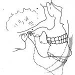
With cervico-facial localization of actinomycosis, the pathological process can occur with lesions of the skin, subcutaneous and intermuscular tissue. In this case, in addition to the skin of the face and neck, the tongue, lips, eye sockets, larynx and trachea are affected. Cervico-facial actinomycosis can lead to facial asymmetry.
With abdominal localization of actinomycosis, symptoms may resemble an acute attack of appendicitis or intestinal obstruction. Abdominal actinomycosis can involve the liver, kidneys, intestines, and spine.
Symptoms of actinomycosis in its taracal localization: weakness and cough. At first, the cough is dry, then it becomes moist with a mucopurulent sputum with a taste of copper.
Actinomycosis of the genitourinary system - pretty rare diseasedeveloping when the disease spreads from abdominal sick. With actinomycosis of the genitourinary system, pain occurs in the lower abdomen, the process of urination is disrupted, and cyanotic tumor-like formations are formed. An abscess can burst into the bladder or rectum.
With the spread of infection from other organs, actinomycosis of the joints and bones develops. Symptoms of bone actinomycosis are similar to those of osteomyelitis; the bones of the skull, ribs, and upper limbs may be affected. The actinomycosis of the joints is asymptomatic and does not cause a significant loss of their functions.
Actinomycosis of the central nervous system is characterized by an increase in body temperature, headache, dizziness, seizures, loss of consciousness, and impaired coordination. With actinomycosis of the central nervous system, the brain or spinal cord is affected: a cavity filled with pus is formed.
Diagnostics
 Diagnosis of actinomycosis is carried out by external signs: early stage actinomycosis has the form of a dense, slightly painful edema, which eventually acquires a bluish color, and at a later stage a fistula forms. Before treating actinomycosis, it is necessary to make sure that the diagnosis is correct (actinomycosis should be differentiated from other mycoses).
Laboratory confirmation of actinomycosis in 25% of cases is possible by analyzing the fistula discharge (if the internal organs are affected by actinomycosis, then the material is taken using a puncture biopsy of the affected organ): actinomycete druses are detected in it. In most cases (75%), actinomycete drusen are not detected; in this case, purulent discharge or biopsy material is inoculated.
Treatment
 The traditional treatment for actinomycosis is the daily intravenous administration of penicillin for 2-6 weeks. After the end of the injections, oral antibiotics (penicillin, amoxicillin) continue for another 6-12 months. Also, the patient is prescribed several courses of intramuscular or subcutaneous injection of actinolysate (a drug made from the cult of anaerobic actinomycetes). Physiotherapeutic methods such as iodine electrophoresis and ultraviolet irradiation of the skin are effective in the treatment of actinomycosis. Doses medicines are selected individually. A special diet for actinomycosis has not been developed.
With advanced actinomycosis, surgical treatment is performed: opening of abscesses and drainage of fistulas. With actinomycosis armpit or the groin area, the lesion is excised and cleaned, after which a suture is applied. Depending on the localization of actinomycosis, patients may need several operations. After surgical treatment In serious cases of actinomycosis, X-ray therapy is recommended.
Prevention
Specific prevention of actinomycosis has not been developed. General recommendations include timely treatment of infections of the oral cavity, gastrointestinal tract and respiratory system. It is necessary to observe personal hygiene and, if possible, avoid injury to the skin, as well as increase immunity.
ACTINOMYCOSIS, ACTINOBACILLOSIS AND RELATED DISEASES
Part /1 / 2 / 3 /
- General idea
- Actinomycosis
- Other diseases caused by fermenting actinomycetes
- Diseases caused by aerobic actinomycetes
- Nocardial infections
- Actinomycetoma
- Other diseases caused by aerobic actinomycetes
- Diseases caused by Rhodococcus spp.
- Diseases caused by Gordonia spp.
- Diseases caused by Tsukamurella spp.
- Diseases caused by Amycolatopsis and Pseudonocardia spp.
- Diseases caused by Oerskovia spp.
- Dermatophilosis
- Diseases caused by Actinobacillus spp.
- Actinomycetes as allergens
APPENDIX TO SECTION:
- Short-term treatment of actinomycosis: a case study and a literature review (Selvin S. Sudhakar and John J. Ross)
- literature review "Actinomycosis of the genital organs in women" (author Mirzabalaeva AK) journal "Problems of medical mycology" -2000-T.2 (2) .- P.11-16;
- Abdominal actinomycosis (literature review and description of two cases).
- The optimal duration of intravenous and oral administration of antibiotics in the treatment of thoracic actinomycosis.
- Actinomycosis of the pelvic organs. Is long-term antibiotic treatment necessary?
General idea
Actinomycosis, actinobacillosis, actinomycetoma and nocardiosis are diseases that are not related to each other in terms of etiology, epidemiology and therapy, but there are serious reasons for considering them together, since they have common history and nomenclature origin, as well as similar clinical and pathological manifestations. The taxonomic relationship between some of their causal agents is also similar.
The history of actinomycosis dates back to the early days of bacteriology. In 1877, the German veterinarian Otto Bollinger discovered that chronic tumor-like lesions of the jaws of cattle, thought of as a kind of sarcoma, contain small, opaque, yellowish, granular particles. Because their structure was like a group of crystals, he called them "Druses". Drusen were formed from thread-like, branching, mushroom-like structures, later characterized as gram-positive. Botanist Carl O Harz (1877) believed that this is a new type of mold and proposed a generic and specific designation Actinomyces bovis (radiant mushrooms, from the Greek aktis \u003d ray; mykes \u003d mushroom) due to the striking radial divergence of filaments in the granules. He also first introduced the term "actinomycosis" for this disease.
The first detailed description like pathological conditions in humans was published by the Berlin surgeon James Israel (Israel) in 1878. About a decade later, it was found that the most characteristic human pathogen, now called Actinomyces israelii or Actinomyces gerencseriae, and animal pathogen A. bovis are anaerobic, or at least facultatively anaerobic capnophil, bacteria that grow better in high CO 2 (Bujwid 1889, Mosselman and Lienaux 1890). It was only a few decades later that it was established that the causative agents of human and bovine actinomycosis are distinct species and that they are true, albeit filamentous, bacteria, not fungi, and that they were the first representatives of a large and heterogeneous group of bacteria, now belonging to orders Actinomycetales and Bifidobacteriales subclass Actinobacteridae in a newly defined class Actinobacteria (Stackebrandt, Rainey and Ward - Rainey 1997), but still often referred to simply as "actinomycetes".
Lignieres and Spitz in 1902 described a new disease in bovine animals in Argentina that clinically and pathologically resembles bovine actinomycosis. Organisms cultured from the respective lesions were tiny, short gram-negative bacterial rods that were markedly different from A. bovis... Due to the similarity between the clinical pictures of these two diseases, the pathogen was first named "actinobacillus", and then officially designated as Actinobacillus lignieresii (Brumpt 1910).
Before the anaerobic nature of the causative agents of actinomycosis in humans and animals was established, many attempts were made to grow microorganisms under aerobic conditions. In an extensive study of cases of actinomycosis in humans and cattle, Bostroem (1891) isolated filamentous microorganisms on aerobic gelatin or agar, which he regarded as pathogenic and to which he named " Actinomyces bovis"He also observed grain spines at the center of actinomycotic lesions and isolated culturally similar aerobic filamentous microorganisms from grass, grain, and other plant materials. In this regard, Bostroem concluded that grass or grain is an exogenous source of actinomycotic infection and that chewing on grass or grain could cause actinomycotic damage.This version persisted for a long time even after the studies of Naeslund (1925, 1931) proved that A. israelii is part of the congenital microflora of the human oral cavity, which does not occur in the environment, and, thus, the source of actinomycosis is always endogenous.
Towards the end of the 19th century, several researchers had identified pathogenic aerobic actinomycetes, similar to the causative agents of actinomycosis isolated by Bostroem. Nocard (1888) described an aerobic filamentous microorganism in farcin du boeuf, a cattle disease on the island of Guadeloupe. This pathogen was named Nocardia farcinicaTrevisan (1889). A similar branching bacterium was isolated from the affected lung in humans by Eppinger (1891), and this pathogen was subsequently designated as Nocardia asteroides Blanchard (1896). Another filamentous branching bacterium, first designated as " Streptothrix madurae", was isolated by Vincent (Vincent, 1894) from tumor-like lesions in India, called" Madura foot ". This organism was later called" Nocardia madurae"and is now known as Actinomadura madurae (Lechevalier and Lechevalier 1970).
Numerous additional genera and varieties of aerobic and anaerobic actinomycetes have been described since Bollinger's report. For the most part, these were safe inhabitants of the environment or body surfaces of humans and animals, and only a few could act as a pathogen in both humans and animals. This applies not only to some members of the traditional lineages. Actinomyces and Nocardiabut also to species of genera Bifidobacterium, Propionibacterium, Oerskovia, Gordonia, Rhodococcus, Tsukamurella, Actinomadura, Nocardiopsis, Streptomyces, Dermatophilus, Thermoactinomyces, Saccharopolyspora (Faenia), Saccharomonospora and Thermomonospora... Despite the growing spectrum of pathogenic actinomycetes, it would hardly be advisable to add, in addition to actinomycosis, nocardiosis, dermatophilosis, numerous further etiological designations such as propionibacteriosis, rhodococcosis, tsukamurellosis (tsukamurellosis), etc. On the other hand, for the sake of clarity, it would also not be entirely correct to use the term "actinomycosis" for any type of infection caused by actinomycetes, as has been common practice in the past. Likewise, the term nocardiosis does not encompass all types of nocardial infections or infections caused by other anaerobic actinomycetes. Thus, the classic designations of the disease "actinomycosis" and "nocardiosis" should be retained to denote a definite, clinically and etiologically disease with a characteristic clinical picture.
Diseases caused by fermenting actinomycetes
Fermenting carbohydrates anaerobic or capnophilic actinomycetes belonging to the families Actinomycetaceae, Propionibacteriaceae or Bifidobacteriaceae, act as etiological agents in various diseases in humans and animals. Among them, actinomycosis is the most characteristic manifestation of the disease. Other diseases that can be caused by fermenting actinomycetes: dental caries and periodontitis, lacrimal canaliculitis and other eye infections; infections associated with the use of intrauterine contraceptives and vaginal uterine rings, other inflammatory processes in humans, such as mastitis, peritonitis, pleurisy, septic abortion, abscesses and also a wide variety of purulent lesions in animals.
Actinomycosis
Actinomycosis is a subacute or rather chronic granulomatous disease that usually causes suppuration and abscess formation, and tends to form sinus tracts. The disease occurs in humans and animals. In addition to classic pathogens A. bovis and A. israelii, actinomycotic lesions can cause a diverse number of species of other enzymatic actinomycetes. Most of these agents belong to the family Actinomyces, but some are members of the genus Propionibacterium or Bifidobacterium... In addition, all typical actinomycotic lesions contain a variety of bacteria in addition to pathogenic actinomycetes. Thus, the term "actinomycosis" rather defines a polyetiological inflammatory syndrome than just a disease related to an individual pathogenic microorganism. To avoid introducing additional etiological terms and to remain bacteriologically correct, it was proposed to designate a group of closely related inflammatory processes by the term "actinomycosis" in plural (Schaal and Beaman 1984, Schaal 1996).
Actinomycosis in humans
Despite significant similarities in pathology, pathogenesis, and epidemiology, actinomycosis in humans and animals differ from each other in several important ways. Various species of actinomycetes are responsible for infections in humans and animals, and, in addition, bone involvement is rarely observed in humans, but very often in animals (Slack and Gerencser 1975).
Clinical manifestations of actinomycosis
Initial actinomycotic lesions usually develop in tissues adjacent to mucous membranes, which are the natural habitats of causative agents. The following areas are most often affected: cervico-facial, thoracic and abdominal. Rarely skin, bones, or central nervous system (CNS), which may also be involved in the pathological process (Slack and Gerencser 1975, Pulverer and Schaal 1984, Schaal and Beaman 1984, Schaal and Pulverer 1984, Schaal 1996). After the penetration of the pathogen into the tissues, the infection tends to progress slowly, regardless of the natural boundaries of the organ. Sometimes hematogenous spread is observed, in which the central nervous system (brain abscess) or natural cavities (empyema) may be involved. There is a characteristic tendency to both remission and exacerbation of symptoms, regardless of the conduct of antibiotic therapy. Due to the fact that actinomycosis in humans is endogenous infections, it is difficult or impossible to determine their incubation period. It is believed that before the first clinical signs it takes about 4 weeks, however numerous reports suggest that this period can be much longer or significantly shorter.
Cervico-facial actinomycosis
In the vast majority of cases, actinomycotic infection affects the face, neck, or both, the so-called cervico-facial region (data collected in Germany; see Table 1), but the numbers may vary in different geographic areas, especially in USA.
Table 1 Localization of actinomycosis in humans
| Localization | Of cases | % |
| Cervico-facial | 3249 | 97,6 |
| Thoracic, including chest wall | 43 | 1,3 |
| Abdominal, including pelvic organs | 22 | 0,7 |
| Limbs, skin | 22 | 0,7 |
| Brain | 4 | 0,1 |
| Blood (septicemia) | 2 | 0,06 |
| Total | 3329 | 100,0 |
Data collected at the Institute of Hygiene, University of Cologne, 1969-84, and at the Institute for Medical Microbiology and Immunology, University of Bonn, Germany, 1984-95.
Actinomycotic lesions are often preceded by a history of caries and tooth decay, tooth extraction, fracture of the jaw, periodontal abscess, mucosal damage by foreign bodies (bone fragments, fish bones, spines of grass or grain), or tonsil suppuration. It should be remembered that traumatic factors, local or general predisposing conditions, do not necessarily occur in all cases or may be overlooked in the history taking.
In cervicofacial actinomycosis, according to the analysis of 317 patients, the following tissues were most often involved in the process: adjacent to the lower jaw (53.6%), cheek (16.4%), chin (13.3%), mandibular ramus and angle (10.7%) , upper jaw (5.7%) and jaw joint (0.3%) (Herzog 1981). Other areas that are less commonly affected are neck, mastoid, sinuses, parotid, thyroid, tongue, lips, nasal septum, and ears (Slack and Gerencser 1975, Kingdom and Tami 1994). Direct bone and regional involvement lymph nodes This is very rare, but periostitis and post-traumatic osteomyelitis with fermenting actinomycetes are not uncommon (11.7% of cases reported by Herzog 1981).
Primary cervicofacial actinomycotic lesions are either acute, predominantly odontogenic, abscesses, or very sharp forms panniculitis, or as slow-forming, hard, reddish or deathly pale inflammatory infiltrates (Lentze 1969, Pulverer and Schaal 1978, Schaal 1979, 1981, 1996). Whereas, although chronic infiltrates are usually painless and acute forms of infection are painful, they can all lead to masticatory muscle trismus when the process develops near the temporomandibular joint.
To lead to a quick and complete healing, only a surgical incision and drainage in the vast majority of cases is not enough. Acute and especially chronic cases tend not to heal without specific antibiotic therapy. At best, there is a temporary regression of symptoms, after which relapses may develop after a few weeks or months. The longer both forms of actinomycosis persist, the sooner similar and very characteristic late signs of this disease develop in both cases. They include: regression and scarring of the central purulent focus, progression of hard, painless, deathly pale infiltrates in the periphery, the formation of multiple areas of softening and fistula formation. The latter appear spontaneously or form at the site of a surgical incision and, together with multiple abscesses, form a multi-chamber system of cavities in the affected tissue, which does not respond well to conventional therapy, including the administration of "standard" antibiotics, and shows a clear tendency to relapse after a temporary regression of inflammatory symptoms ... Without treatment or with inappropriate treatment, cervicofacial actinomycosis slowly progresses, even across the borders of the organ, and can become life threatening when introduced into the cranial cavity, mediastinum, or when invading large blood vessels (Herzog et al, 1984). Discharges from fistulas and pus from abscesses are usually yellowish and thicker in consistency than serous discharge and often contain particles originally called "drusen" or often referred to as "sulfur granules".
Thoracic actinomycosis
Thoracic lesions in actinomycosis are much less common than the cervico-facial form. The formation of the process is usually preceded by the aspiration of pathogenic material from the oral cavity, for example, plaque or calculus, the contents of the crypt of the amygdala or a foreign body contaminated with the microflora of the oral cavity, including pathogenic actinomycetes. Occasionally, this form of the disease develops due to local spread of the cervicofacial process, perforation of the diaphragm due to abdominal injury, or hematogenous spread from any distant focus of infection (Slack and Gerencser 1975).
First of all, thoracic actinomycosis may appear as a mediastinal tumor or bronchopneumonic infiltrate, necrotizing pneumonia, or lung abscess (Slack and Gerencser 1975, Schaal and Beaman 1984, Morris and Sewell 1994). Radiographs show discrete dense or multiple shadows in which cavities may form. At the onset of the disease, the main symptoms are chest pain, fever, cough with or without phlegm, weight loss, but hemoptysis is unusual. In the future, the infection can progress to pleural empyema, pericarditis, or chest wall involvement. If diagnosed late, or treatment is inadequate, the latter signs may include extensive subcutaneous chest wall abscesses, paravertebral or pelvic abscesses that form in the groin and are emptied by pus containing large numbers of actinomycotic druses.
Abdominal actinomycosis
Actinomycotic lesions of the abdominal and anorectal organs are quite rare (Table 1). Their development is associated with acute perforation of internal organs (appendicitis, diverticulitis, cryptitis, various ulcers), surgical or other traumatic injuries, including swallowed bone fragments or fish bones.
Another source of pelvic and abdominal actinomycotic infections has recently been identified. It turned out that in 10-20% of women with intrauterine contraceptives or vaginal uterine rings, the uterus and cervical canal are colonized by a mixed bacterial flora, which includes potentially pathogenic fermenting actinomycetes (Gupta, Hollander and Frost 1976, Gupta, Erozan and Frost 1978, Eibach and others. 1989, 1992, Schaal and Lee 1992, Chatwani and Amin-Hanjani 1994), as well as other predominantly anaerobic bacteria (Schaal and Lee 1992). They hardly occur in women who do not use these devices. This colonization may serve as an initial center for the development of aggressive actinomycosis with involvement of the pelvic organs and may even be the source of hematogenous metastatic hepatic or intracranial actinomycotic abscesses (Gupta, Erozan and Frost 1978).
The initial signs of abdominal actinomycosis are usually mild and vague. These include: fever, malaise, weakness, and pain that slowly but progressively increases. During the development of the process, it usually resembles slowly progressive tumors similar to malignant processes, such as cancers of the stomach, colon and rectum, anorectal region or cervix (Stein and Schaal 1984, Schaal 1985b, Ewig et al. 1993, Alvarado-Cerna and Bracho-Riquelme 1994, Skoutelis et al. 1995). Large subcutaneous abscesses, extensive lacrimal lumps, or fistulas may be observed, from which drusen is often the first characteristic sign of disease (Schaal and Beaman 1984). Without effective treatment abdominal actinomycosis can spread to any adjacent tissue or organ, including the liver, spleen, kidney, fallopian tubes, ovaries, uterus, testes, bladder, rectum, or abdominal wall (Slack and Gerencser 1975, Khalaff, Srigley and Klotz 1995, Mmller -Holzner et al. 1995).
Central nervous system actinomycosis
Actinomycosis of the brain and spinal cord is very rare, possibly due to the now more available effective antibiotic therapy, preventing hematogenous or direct spread of infection (Table 1). It is these mechanisms that are predominantly responsible for CNS involvement, especially when the primary lesion is located in the lungs or abdomen (Slack and Gerencser 1975, Jamjoom, Jamjoom and al-Hedaithy 1994, Voisin et al. 1998). The main manifestation of CNS actinomycosis is a cerebral abscess. Signs depend on localization, and are determined by the rate of development of an abscess, the degree of displacement or destruction of brain tissue. The main symptoms are - headache, increased intracranial pressure, focal symptoms, hemiparesis, aphasia, ataxia, and pathological reflexes (Slack and Gerencser 1975).
Actinomycosis of bones and skin
Unlike some animals, bone involvement is rare in human actinomycosis (Table 1). The process is usually caused by the direct spread of infection from the adjacent soft tissues. This leads to periostitis, which stimulates new osteogenesis, visible on radiographs. At the onset of the disease, limited areas of bone destruction can be observed, surrounded by denser bone tissue. In such cases, the mandible, ribs, and spine are most commonly involved. Although actinomycotic lesions of other bones have been described, they have not been confirmed by culture. Actinomycosis of the skin is extremely rare (Table 1). The source is mainly wounds contaminated with saliva or plaque, or human bites or injuries sustained in a fist fight. Hematogenous spread of the pathogen into the skin may also occur. The clinical picture of cutaneous or wound actinomycosis is very similar to the cervico-facial form.
Epidemiology of actinomycosis
The bacteria that are isolated from foci of actinomycotic lesions in humans essentially belong to the resident or transient congenital microflora of the mucous membranes. Thus, in addition to actinomycosis resulting from human bites or injuries in a fist fight, the disease always has an endogenous origin and is therefore not capable of epidemic outbreaks or transmission in the usual sense.
Although sporadic actinomycosis occurs worldwide, the incidence of actinomycosis appears to vary from continent to continent, from country to country, or even from region to region, possibly reflecting changing dental care standards and differences in the number and types of antibiotics used. ... Such factors may explain the lower absolute and relative incidence of cervicofacial actinomycosis in the United States compared with Europe, but the higher prevalence of thoracic and abdominal infections in the North American continent.
Based histological studies Hemmes (1963) calculated the incidence of actinomycotic infections in the Netherlands: 1 per 119,000 inhabitants per year. For the Cologne region in Germany until 1969, Lentze (1969) reported an incidence of 1 in 83,000. Later, this incidence was recalculated for 1970-85. and a range of 1 in 40,000 (acute and chronic cases combined) to 1 in 80,000 (chronic cases only) per year has been determined (Schaal 1979). This is significantly higher than the incidence of actinomycosis in other areas of Germany and other European countries. Such differences are difficult to explain, but they may more likely be related to local diagnostic characteristics than to true epidemiological differences.
It has long been known (Slack and Gerencser 1975, Pulverer and Schaal 1978, Schaal 1981, Schaal and Beaman 1984) that typical actinomycosis occurs 2.5-3.0 times more often in men than in women. In addition, epidemiological data show that the unequal distribution of the disease by sex is limited only to patients of puberty. Before puberty and at climacteric, actinomycosis is evenly distributed between the sexes (Pulverer and Schaal 1978, Schaal 1981). This suggests that the disease can occur in all age groups (Slack and Gerencser 1975, Pulverer and Schaal 1978, Schaal 1981). Among the patients observed, the youngest was 1.5 months old, and the oldest was 89 years old. However, the highest incidence of actinomycosis was observed in men aged 21 to 40 years and in women aged 11 to 30 years (Pulverer and Schaal 1978, Schaal 1981, 1992, Schaal and Beaman 1984).
Pathology and pathogenesis
initial stage acute illness - an inflammatory process that leads to the formation of an abscess or, if the course is chronic, tissue proliferation develops and multiple small abscesses form. More advanced processes are characterized by scar tissue in the center and with granulations on the periphery, which may include multiple purulent foci or cavities with multiple fistulous passages. Rarely, in cases. when bone is involved, osteoclastic and osteoblastic changes can occur.
In a purulent focus, druses of actinomycetes can be found. They are found in the contents of an abscess or in discharge from fistulas in about 25% of cases, which is of great diagnostic value. Druses are up to 1 mm in diameter and are visible to the naked eye. These are yellowish (with a reddish or brownish tinge) particles, resembling cauliflower at low magnifications. Under the microscope, after slight squeezing between the slide and the cover glass, one can see that they are composed of a different number of spherical lobes, which represent filamentous actinomycotic microcolonies formed in vivo and generally form a cauliflower-type structure. The surrounding tissues are usually infiltrated with polymorphonuclear leukocytes.
Fully crushed and Gram-stained granules at high magnification indicate that the material consists of clusters of gram-positive, intertwined branching mycelium filaments. The stained smears may also contain a varied number of other gram-positive and gram-negative rods and cocci, which represent the accompanying flora, as well as numerous leukocytes. Mainly in tissue material, and less often in purulent discharge, it can be observed that the tips of the peripheral filaments in the granule are covered with a club-like layer of hyaline material, which can help differentiate actinomycotic drusen from similar particles of other (microbial and non-microbial) origin. It should be emphasized that the term "sulfur granules", which is widely used to denote actinomycotic druses, refers only to the yellow color of the particles, and not at all to their high sulfur content.
The main natural habitat of all fermenting actinomycetes pathogenic to humans is the oral cavity of healthy adults, where they live in significant quantities. In the digestive and genital tracts, however, they seem to be present only sporadically or in low amounts. The same applies to the oral cavity of infants before teething and already edentulous adults. This may explain why cervicofacial actinomycosis is relatively less common at a very young or old age.
The relatively low incidence of the disease in comparison with the ubiquitous occurrence of the pathogen in adults is apparently associated with the need for tissue invasion rather than a simple defect in the mucous membrane. Such conditions are more likely to depend on local tissue damage at the site of implantation than on a defect in the function of the immune system as a whole. In this regard, a prerequisite for the establishment of fermenting actinomycetes in host tissues is the presence of a negative redox potential, on which both pathogenic actinomycetes and many accompanying bacteria depend. Such a local decrease in oxygen potential can be caused by impaired blood circulation due to general circulatory disorders or vascular disease, traumatic injuries with crushing of tissues or the introduction of foreign bodies, or with the necrotic ability of other microorganisms simultaneously present.
These so-called "accompanying microorganisms" act as a trigger for the actinomycotic process, producing local anaerobic conditions. In addition, they enhance the relatively low invasive ability of pathogenic fermenting actinomycetes by releasing aggressive enzymes such as hyaluronidases and toxins. Thus, actinomycosis is almost always a synergistic mixed infection, in which actinomycetes are a specific component, or a "leading" organism that determines the characteristics clinical course and characteristic symptoms of the disease. The composition of the accompanying microflora varies from case to case, but it is always present and often determines the initial clinical picture and some complications.
The varieties of fermenting actinomycetes that are capable of causing typical actinomycotic lesions in humans are presented in Table 2. Most often were identified A. israelii and A.gerencseriae, but the frequency of the latter is clearly underestimated, because they began to be separated from A. israelii since 1987.The third pathogen that can cause actinomycosis in humans is P. propionicum, but this species is rarely encountered. Previously classified as "Arachnia propionica" (Schaal 1986), he was recently transferred to the family Propionibacterium based on sequence similarity of 16S rRNA (Charfreitag, Collins and Stackebrandt 1988). It is not always easy to decide whether the other actinomycetes mentioned in Table 2 are significant pathogens or just members of a mixed bacterial flora with no particular significance.
Very little is known about the factors that may explain the pathogenicity of actinomycosis caused by fermenting actinomycetes. However, it has long been established (Slack and Gerencser 1975) that Actinomycesspp. can form a villous layer on its surface that can resemble hairs and which can promote adhesion of the pathogen to the host cells (Figdor and Davies 1997).
Tab. 2. Varieties of fermenting actinomycetes isolated from foci of actinomycotic lesions in humans.
| Kinds | Actinomycotic lesions (n \u200b\u200b\u003d 1376)% | Conditions associated with intrauterine contraceptives (n \u003d 114)% | Eyes, organs of lacrimation (n \u003d 98)% |
| Actinomyces israelii | 73.3 | 54.4 | 13.3 |
| A. gerencseriae * | 2.0 | 7.0 | 11.2 |
| A. naeslundii | 6.8 | 6.1 | 16.3 |
| A. viscosus | 4.9 | 16.7 | 19.4 |
| A. odontolyticus | 1.4 | 2.6 | 12.2 |
| A. meyeri | 0.6 | 3.5 | 0.0 |
| A. georgiae | 0.2 | 0.0 | 0.0 |
| A. neuii | 0.2 | 0.0 | 0.0 |
| Propionibacterium propionicum | 3.3 | 4.4 | 16.3 |
| Bifidobacterium dentium | 0.4 | 2.6 | 2.2 |
| Corynebacterium matruchotii | 0.9 | 0.0 | 2.2 |
| Rothia dentocariosa | 0.4 | 0.6 | 5.1 |
| Not identified | 5.7 | 2.0 | 2.0 |
| Total | 100.0 | 100.0 | 100.0 |
Data collected at the Institute of Hygiene, University of Cologne, 1969-84, and at the Institute for Medical Microbiology and Immunology, University of Bonn, 1984-95.
* Note: the differences between A. israelii and A. gerencseriae until 1987, usually not produced.
The accompanying microflora in the foci of actinomycosis can consist of both aerobic and anaerobic microbes. In more than 50% of the cases studied by Schaal, the accompanying microorganisms consisted exclusively of anaerobes (Table 3). In other cases, both obligate (strict) anaerobes and facultative anaerobes or aerobes were found. On average, 2-4 species of accompanying bacteria were present in the focus, but in some cases up to 10 were identified.
Table 3. Aerobic microorganisms associated with fermenting actinomycetes obtained from human lesions
| Kinds | ||
| No aerobic growth | 47,2 | 32,1 |
| Coagulase-negative staphylococci | 27,9 | 14,8 |
| Staphylococcus aureus | 12,7 | 6,2 |
| alpha hemolytic streptococcus | 11,2 | 11,1 |
| beta hemolytic streptococcus | 4,9 | 11,1 |
| Streptococcus pneumoniae | 0,0 | 0,0 |
| Enterococci | 0,0 | 16,1 |
| Cutaneous corynobacteria | 0,0 | 3,7 |
| Haemophilusspp. | 0,1 | 1,2 |
| Enterobacteriaceae | 2,5 | 13,6 |
| Gardnerella vaginalis | 0,0 | 6,2 |
| Non-fermenting | 0,2 | 0,0 |
| Yeast | 0,1 | 0,0 |
| Total | 100,0 | 100,0 |
Among the aerobic contaminants (Table 3), the most common were coagulase-negative staphylococci, Staphylococcus aureus, alpha-hemolytic and beta-hemolytic streptococci. Anaerobic and capnophilic (microaerophilic) accompanying microflora is much more diverse and numerous. Synergistic interactions likely exist between A. israelii and A. gerencseriae from Actinobacillus (Haemophilus) actinomycetemcomitans... The latter microorganism, the name of which refers to its characteristic similarity with actinomycetes, is often the cause especially chronic course diseases and ineffective treatment. This pathogen can maintain an inflammatory process with similar symptoms even after chemotherapy has completely eliminated actinomycetes. Other common actinomycete companions are black pigmented Bacteroidaceae (Prevotellaspp., Porphyromonas spp.), unpigmented Prevotella and Bacteroides spp., Fusobacteria, the so-called microaerophilic streptococci, which belong mainly to the variety Streptococcus anginosus(milleri), propionibacteria and Eikenella corrodens (Table 4).
Table 4. Anaerobic microorganisms associated with fermenting actinomycetes obtained from human lesions
| Kinds | Cervico-facial actinomycosis (n \u003d 3197)% | Conditions associated with intrauterine contraceptives (n \u003d 81)% |
| Actinobacillus actinomycetemcomitans | 22,9 | 2,5 |
| Microaerophilic streptococci | 29,3 | 22,2 |
| Peptostreptococcusspp. | 18,2 | 29,6 |
| Black pigmented Bacteroidaceae | 37,7 | 58,0 |
| Unpigmented Bacteroides / Prevotellaspp. | 14,0 | 69,1 |
| Fusobacterium spp. | 32,5 | 22,2 |
| Leptotrichia buccalis | 20,4 | 2,5 |
| Eikenella corrodens | 16,5 | 46,9 |
| Capnocytophaga spp. | 0,4 | 6,2 |
| Campylobacter / Selenomonas spp. | 0,1 | 3,7 |
| Propionibacterium spp. | 30,5 | 24,7 |
| Bifidobacteriumspp. | 0,1 | 1,2 |
| Lactobacillus spp. | 0,5 | 42,0 |
| TOTAL | 100,0 | 100,0 |
According to Schaal and Lee (1992).
Very little is known regarding the humoral and cellular immune responses of patients suffering from actinomycosis. Antibodies against fermentative actinomycetes can be detected in human serum different methods, including methods of immunofluorescence and enzyme immunoassay. Most of these antibodies react more actively with antigens A. naeslundii and A. viscosusthan with A. israelii, A. gerencseriae, or P. propionicum... In addition, antibodies are primarily associated with the presence of periodontal disease and, rarely, with antecedent or existing invasive actinomycosis. Thus, it was established that the antibody response in actinomycosis is insignificant or sporadic. In addition, antibodies probably have no protective effect against actinomycetes, and their presence is in no way a sign of self-healing from this disease.
On the other hand, it was established quite a long time ago (Lentze 1938) that the immune system of patients with actinomycosis can be stimulated by the introduction of cells killed by formalin or extracts of cells of pathogenic actinomycetes (actinolysate). This leads to the formation of an antibody response that can be measured. More importantly, however, after the introduction of actinomycotic antigens, a so-called local reaction occurs, that is, a temporary increase in inflammation. The immune response formed after repeated injections helps to overcome the disease. These observations provide a basis for the vaccine treatment of actinomycosis, which was used before antimicrobial therapy (Lentze 1938, 1969).
Diagnostics
The diagnosis of human actinomycosis is mainly based on the isolation and identification of causative agents because clinical symptoms are often misleading and histopathology and serology are low specific and low sensitivity. The presence of druses, which sometimes impart pus appearance semolina should initiate the search for actinomycetes. However, given that only 25% of samples of actinomycotic pus contain these granules, their absence does not exclude the diagnosis of actinomycosis.
Collection and transportation of pathogenic material.
Pathological material suitable for bacteriological analysis of actinomycosis is pus, discharge from fistulas, bronchial secretions, granulation and biopsies. During collection, precautions should be taken against contamination by congenital mucosal microflora. Whenever possible, pus or tissue should be obtained by percutaneous puncture. To diagnose thoracic actinomycosis, bronchial secretions must be obtained transtracheally. Sputum examination is unreliable because it usually contains oral actinomycetes, including pathogenic species. Transthoracic percutaneous biopsy or percutaneous puncture of suspicious abdominal abscesses are often the only means of obtaining satisfactory specimens for diagnosis. Transport of samples to the bacteriological laboratory should be fast enough. If long-term transport is unavoidable, special transport media such as Stuart's should be used, although fermenting actinomycetes are less susceptible to oxidative damage than severe anaerobes.
Microscopic examination
When drusen are present, this makes it possible to quickly and relatively reliably make a preliminary diagnosis after examination at low magnification (d 100) of an actinomycotic granule placed under a cover glass and with a 1% solution of methylene blue introduced into the drop. Actinomycotic drusen appear as cauliflower-like particles with an uncolored center and a blue periphery, in which leukocytes and short filaments, sometimes with "clubs", radiate from the center of the granule. Gram-stained swabs obtained by squeezing granules between two slides show filamentous, branching, gram-positive structures that represent pathogenic actinomycetes, as well as a variety of other gram-negative and gram-positive bacteria that indicate the presence of accompanying microorganisms. The presence of these bacteria is necessary to distinguish actinomycotic drusen from granules formed by various aerobic actinomycetes ( Nocardia, Actinomadura, Streptomyces), which never contain accompanying microflora. Direct and indirect immunofluorescence for the detection of specific antibodies can also be used to identify the species of actinomycetes present in the pellet without culture isolation.
Cultural diagnostics
To obtain reliable results, it is advisable to use transparent media so that the plates can be carefully inspected for characteristic filamentous colonies and to grow the culture for at least 14 days. Cultures can be tested every 2-3 days without changing anaerobic conditions if Fortner's (1928) method is used to obtain a low oxygen potential. If anaerobic flasks or cups are used, it is advisable to inoculate on two or three media at the same time in order to examine them to determine the growth of actinomycetes after 3, 7 and 14 days. Removing the plates from the anaerobic environment usually stops further growth of microorganisms that need long-term incubation without changing anaerobic conditions.
The preliminary results of the culture study are obtained after 2-3 days, when characteristic arachnid microcolonies can be seen under the microscope A. israelii, A. gerencseriae or P. propionicum... Confirmation of preliminary microscopic or early cultural diagnoses by unambiguous identification of a pathogenic species of actinomycetes can take 14 days or more. This is necessary in order to reliably reveal the differences between fermenting actinomycetes and morphologically similar contaminants obtained from the patient's mucous membranes, as well as similar aerobic actinomycetes of childbirth. Nocardia, Actinomadura and Streptomyces... A detailed bacteriological analysis of the accompanying microflora can also be useful for the selection of appropriate antibiotic therapy.
Molecular methods, such as genetic studies or polymerase chain reactions (PCR), are currently under development and in the future may allow for a faster diagnosis of actinomycosis.
Serological diagnostics.
Actinomycotic infection does not necessarily stimulate a humoral immune response, which can be detected by available laboratory methods. However, none of the methods used, with a wide variety of antigens used, provided satisfactory results due to problems with sensitivity and specificity (Holmberg, Nord and Wadstrmm 1975, Holmberg 1981, Persson and Holmberg 1985).
Treatment
Surgical dissection of actinomycotic foci and drainage of purulent contents is always the basis for the treatment of actinomycosis. However, it is known that even radical surgery often only ends with a temporary reduction in symptoms and may be accompanied by one or more relapses. In order to overcome these problems, in the past they tried to use substances such as iodides, thymol, copper sulfate, hydrogen peroxide, silver nitrate, arsenic preparations, which, however, did not improve long-term results. Only subcutaneous injection of killed actinomycete cells (heterovaccine Lentze 1938) has shown a definite efficacy.
Progress in the treatment of actinomycosis came when sulfonamides and penicillin became available. Penicillin was active against pathogenic actinomycetes in vitro and in vivo... Given that many patients had little or no benefit from penicillin therapy, treatment with high doses of penicillin was often recommended for at least 3 months and up to 12–18 months (Harvey, Cantrell, and Fisher 1957).
Poor response to penicillin treatment is often due to the presence of concomitant bacteria that are penicillin-resistant. In addition, drugs poorly penetrate through the dense tissue of actinomycotic cells and into drusen. Finally, A. actinomycetemcomitans - are usually resistant to penicillin, although they do not produce beta-lactamases. Thus, penicillin in the treatment of actinomycosis is only effective when not present. A. actinomycetemcomitans and when the accompanying microflora does not contain any producers of beta-lactamases.
Aminopenicillins are somewhat more active against pathogenic actinomycetes than penicillin, and, in addition, they inhibit growth A. actinomycetemcomitans... However, given that they are not resistant to beta-lactamases, microorganisms that produce beta-lactamases can interfere with their therapeutic efficacy. This is rarely the case with cervicofacial actinomycosis, but with thoracic and especially abdominal infections, beta-lactamase producers are usually present. Therefore, the currently used treatment regimens include drugs that are effective against actinomycetes and the potential of producers of beta-lactamases of the type S. aureus, gram-negative anaerobes, and, in cases of abdominal actinomycosis, Enterobacteriaceae.
The current recommendations for antibiotic therapy for actinomycosis are as follows: therapy of choice for cervicofacial actinomycosis: amoxicillin plus clavulanic acid, or possibly ampicillin plus sulbactam. The initial dose is 2 g 3 times a day of amoxicillin plus 0.2 g 3 times a day of clavulanic acid per day for 1 week, and 1 g 3 times a day of amoxicillin plus 0.1 g of clavulanic acid 3 times a day per day for another week. In rare cases chronic infections the cervico-facial area may require up to 4 weeks of treatment. The specified treatment regimen can be used for thoracic actinomycosis, but in these cases it is recommended to give a high dose for a longer period - within 3-4 weeks. In long-term chronic cases of pulmonary actinomycosis, in order to increase tissue concentration, an increased dose of ampicillin may be necessary. Depending on the accompanying flora, aminoglycosides may also be needed, especially when there is a persistent presence Enterobacteriaceae type Klebsiellaspp. or Enterobacter spp. The latter are generally usually present in abdominal actinomycosis. The therapy of choice for treating these infections is the combination of amoxicillin and clavulanic acid with metronidazole (or clindamycin) for severe anaerobes plus tobramycin or gentamicin. Imipenem may be a suitable alternative, but it is rarely used to treat actinomycotic infections (Edelmann et al. 1987, Yew et al. 1999).
It is important to note that neither metronidazole nor clindamycin can be used to treat actinomycotic infection alone without added antimicrobial agents, especially aminopenicillins, because clindamycin is almost ineffective against A. actinomycetemcomitans (Niederau et al. 1982, Schaal 1983, Schaal et al. 1984) and metronidazole is inactive against pathogenic actinomycetes (Schaal and Pape 1980, Niederau et al. 1982). For the treatment of patients who are allergic to penicillins, tetracyclines or cephalosporins can be used instead of aminopenicillins, however, the clinical efficacy of these drugs is much less than that of aminopenicillins or a combination of aminopenicillins with beta-lactamase inhibitors.
Forecast
Before the advent of modern antibiotics in practice, the prognosis of actinomycosis was dubious - closer to unfavorable. Even now, patients receiving inadequate therapy can suffer from actinomycosis for many years and even die from this disease or its complications. This is especially true for thoracic and abdominal infections, which are often diagnosed only in the last stage. If the diagnosis is made early and antibiotic therapy is adequate, then the prognosis of cervicofacial and cutaneous actinomycosis is generally good. Thoracic, abdominal and systemic manifestations, however, remain a serious problem and require active treatment.
Other diseases caused by fermenting actinomycetes
Fermenting actinomycetes can also cause some other diseases, but they differ significantly from typical actinomycotic lesions in clinical manifestations, prognosis and treatment; therefore, they should not be referred to as "actinomycosis". However, some of them are no less important than actinomycosis, from both medical and economic points of view.
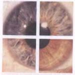
Canaliculitis and other eye infections
Most frequent illnessnot associated with trauma and caused by fermenting actinomycetes - lacrimal canaliculitis with conjunctivitis and without conjunctivitis. This disorder is usually characterized by yellowish to brownish adhesions within the tubule and pus in the inner corner of the eye. The most important causative actinomycetes are P. propionicum, A. viscosus and A.israelii (Table 2). Less often isolated A. naeslundii, A. gerencseriae and Actinomyces odontolyticus (Schaal and Lee 1992). Associated bacteria are often, but not always, present. Excluding availability Streptococcus pneumoniae or Haemophilus influenzae in the eyes and A. actinomycetemcomitans with the cervico-facial form of actinomycosis, the accompanying flora in both lesions is very similar.
In addition to lacrimal canaliculitis, ocular infections caused by fermenting actinomycetes can also present as conjunctivitis, keratitis, dacryocystitis, inflammation of the eyelid glands, and even a periobital abscess, granuloma, or intraocular infection (Schaal 1986, Schaal and Lee 1992). A reliable diagnosis of lacrimal canaliculitis and other actinomycotic lesions of the eyes is carried out in accordance with the bacteriological procedures mentioned above. Removal of lacrimal adhesions, which are usually found in canaliculitis, and topical antibiotic use almost always results in a quick cure in cases where there is a non-invasive process. Invasive infections (abscesses, granulomas, intraocular infections) require systemic therapy with appropriate antibacterial drugs.
Conditions associated with intrauterine contraceptives (IUDs).
As discussed earlier in the section on abdominal actinomycosis, the uterus and cervical canal of women in the presence of intrauterine contraceptives or vaginal uterine rings are often colonized by a complex bacterial flora, which consists of enzymatic actinomycetes and various other aerobic and anaerobic bacteria (Eibach et al. 1989, Schaal et al. 1989, Schaal et al. Lee 1992). These microorganisms are especially abundant directly on the IUD filament within the cervical canal, and very much resemble the characteristic polymicrobial flora of actinomycotic lesions. The predominant actinomycetes under these circumstances are A. Israelii.Relatively often found A. viscosus... Other species were also sometimes isolated (see Table 2). The accompanying flora in these cases is very similar, but not identical to cervicofacial actinomycosis (see Tables 3 and 4). Of the aerobic bacteria in the IUD, enterococci are more or less often found, Enterobacteriaceae and Gardnerella vaginalis(Table 3). Although anaerobes and capnophils (microaerophiles) are usually present (Table 4), a much lower excretion rate should be noted. A. actinomycetemcomitans and even a lower frequency of detecting fusobacteria in the IUD than cervicofacial actinomycosis, while the non-pigmented varieties Bacteroidesand Prevotella, E. corrodens and lactobacilli are more often isolated from the IUD. The presence of fermenting actinomycetes and characteristic concomitant bacteria on the IUD and in the cervical canal is not necessarily associated with symptoms of an aggressive actinomycotic infection, which requires specific treatment. However, approximately 28% of patients with actinomycetes in the cervical canal or on the IUD had symptoms of infection of the lower genital tract, and the other 26% had an infection of the upper genital tract (Eibach et al. 1989, 1992). Symptoms such as fever, pain, or vaginal discharge, usually disappear within 4-8 weeks after removal of the IUD, at least with infections of the lower genital tract.
When typical actinomycetes are found on the IUD or in the cervical canal, the use of the IUD should be discontinued. After the microflora returns to normal levels, the IUD can be used again without increasing the risk of developing genital actinomycosis.
Other suppurative infections
Fermenting actinomycetes can also cause other inflammatory processes. These include pharyngitis, otitis media, urethritis, funisitis (inflammation of the umbilical cord) (Wright et al. 1994), cutaneous and subcutaneous suppurative lesions, abscesses with or without associated mixed anaerobic flora, empyema, and septicemia (Schaal 1986).
These infections can cause not only "classic" Actinomycesspp., like A. naeslundii, A. viscosus, A. odontolyticusand Actinomyces meyeribut also some others Actinomyces spp. and Arcanobacterium haemolyticum,such as the: Actinomyces pyogenes, Actinomyces neuiisubsp ... neuii, Actinomyces neuiisubsp ... anitratus (Funke et al. 1994), Actinomyces bernardiae (Funke et al. 1995), Actinomyces radingae, Actinomyces turicensis (Wмst and others. 1995), Actinomyces europaeus (Funke et al. 1997) and Acinomyces graevenitzii (Ramos et al. 1997). It has also been described as a new actinomycete-like species Acinobaculum schaalii(Lawson et al. 1997), which was isolated from patients.
Diseases caused by aerobic actinomycetes
Aerobic actinomycetes with an oxidative type of carbohydrate metabolism make up a large and very heterogeneous group of filamentous bacteria that have recently been subdivided into Micrococcineae, Corynebacterineae, Micromonosporineae, Pseudonocardineae, Streptomycineae, Streptosporangineae, Frankineae and Glycomycineae order Actinomycetales within a newly defined class Actinobacteria (Stackebrandt, Rainey and Ward - Rainey 1997). They are widely represented in nature, especially in soil, and many play a significant role in the circulation of organic remains. Only a few of these microorganisms are of medical importance as infectious agents or as sources of strong allergens. They belong to families Cellulomonadaceae, Dermatophilaceae, Nocardiaceae, Gordoniaceae, Tsukamurellaceae, Pseudonocardiaceae, Streptomycetaceae, Nocardiopsaceae and Thermomonosporaceae... Depending on the species of actinomycete involved, its site and mechanism of introduction, as well as the immune status of the host, aerobic actinomycetes can cause a variety of diseases in humans and animals. In addition, as has recently been established, these microorganisms can be the cause of nosocomial infections, such as catheter-associated sepsis or postoperative wound infections. The most common pathogens responsible for these diseases belong to the genera Nocardia and Actinomadura, but other actinomycetes of the type are also sometimes isolated from patients Amycolatopsis, Gordonia, Nocardiopsis, Pseudonocardia, Rhodococcus, Saccharothrix, Streptomyces and Tsukamurella (Schaal and Lee 1992, McNeil and Brown 1994).
Actinomycosis
Actinomycosis is a chronic disease of cattle, pigs and animals of other species, as well as humans, characterized by the formation of specific granulomas in various tissues and organs (skin, bones, parenchymal organs).
Actinomycetes (Greek mykos - mushroom; actis - ray) unicellular microorganisms - radiant fungi.
Order Actinomycetales
Family Actinomycetaceae
Genus Actinomyces
The disease is most often caused by Actinomyces bovis. It is a coccoid or branching filamentous form.
In the affected tissues, it looks like rods and threads or forms characteristic clusters in the form of a bush or rosette (drusen), consists of a central ball of Gr + threads. Drusen can be seen with the naked eye in pus, where they are present in the form of small grains of yellow-ash or brown color... Thread width 0.2-1.2; length 100-600 microns.
Cultivation
Isolation of the primary culture of actinomycosis is carried out under anaerobic conditions at a temperature of 37 ° C, inoculated on Sabouraud agar or glucose-blood agar. The culture is developing slowly. On the 15-20th day after sowing, small yellowish colonies appear in the thickness of the agar. The colonies firmly grow together with the environment, their surface is, as it were, sprinkled with lime powder - this is an aerial mycelium, at the ends of which spores develop, giving the colonies a yellowish or red color.
Can be grown on Kitta-Tarozzi medium, MPA (with the addition of whey), MPF, MPB, in milk and on potatoes.
Biochemical activity is poorly expressed. Ferments with the formation of CG - glucose, galactose, glycine, liquefies the fermented fat.
Antigenic structure
The pathogen has two serological variants: 1 and 2, which differ in surface antigens. They can be identified in the RIF.
Sustainability
Actinomycetes are resistant to drying out, especially their spores. At a temperature of 70-80 ° C, actinomycetes die after 5 minutes, the sun's rays kill them after 3 hours, 5% chloramine solution - after 3 hours, 5% lysol solution - after 30 minutes, 3% formaldehyde solution - after 20 minutes.
Pathogenicity and pathogenesis
Pathogenicity - insufficiently studied. Pathogenic actinomycetes are believed to contain endotoxin, an exotoxin of a necrotoxin type that promotes tissue necrotization.
Spread. The causative agent of actinomycosis is widespread in nature. It is found in soil, water, rotting fruits, grains of cereals, in animal organisms in the oral cavity, the hollows of the teeth, in the tonsils, in the upper respiratory tract, urinary tract.
Infection occurs through the penetration of the pathogen in violation of the integrity of the skin, through the mucous membrane of the oral cavity, pharynx or intestines. The pathogen spreads through the blood through the body, forming metastases in the internal organs, bone tissue or skin.
Actinomycosis affects cattle, but can affect pigs, horses, goats, dogs, rabbits.
Pathogenesis. Once in damaged tissues, actinomycetes settle at the site of introduction or migrate through the intercellular spaces into various tissues. Through damaged lymphatic vessels, the pathogen reaches the lymph nodes, and once in the blood, reaches various parts of the body. At the site of the introduction of actinomycetes, colonies are formed in the form of druses. On the periphery of the druse, the mycelium forms a dense plexus, and in the center it is more rare.
In the actinomycotic focus, proliferative phenomena develop, accompanied by the formation of granulation tissue. In the center of the infiltrate, purulent softening of tissues occurs, which leads to a breakthrough of pus outward.
Laboratory diagnostics
Diagnosis is by clinical signs and by the presence of drusen at the site of the swelling. For this purpose, microscopy of stained and unstained preparations from pus, histological sections of pieces of affected tissues is performed.
Isolation of a pure culture and biological assay are rare.
The duration of a full laboratory study is 15-20 days, microscopic - 1 day.
Immunity and funds specific prevention
After the transferred disease does not form, the disease may recur. In recovered animals, precipitins, agglutinins, KS antibodies are formed in the blood, which are not indicators of resistance. In the course of the disease, delayed-type hypersensitivity develops.
There are no specific prophylaxis and therapy tools. Until now, the main treatment is the surgical method.
For treatment, antibiotics can be used in combination with sulfa drugs.
Iodine therapy gives good results, especially in the initial stage before the formation of an actinomycotic abscess.
Actinomycosis is an infectious disease caused by actinomycetes (radiant fungi). It proceeds in an acute and chronic form, manifests itself as dense granulomas, fistulas and abscesses, affects the skin and internal organs. For diagnostics, inoculation on nutrient media is used, it allows you to detect the characteristic mycelium in the separated and the growth of specific colonies. For treatment, immunostimulants and antibiotics are used, ultraviolet irradiation of the skin and electrophoresis are prescribed. In severe cases, surgical intervention is required - treatment of fistulas, opening of abscesses, drainage of the affected cavities.
Features of actinomycosis
The causative agents of actinomycosis are the radiant fungi Actinomyces albus, Actinomyces bovis, Actinomyces israelu, Actinomyces violaceus. In the presence of a nutrient medium, they actively reproduce and form colonies of various shapes with projections similar to rays. This type of pathogenic microorganisms is found not only in humans, but also in animals. Most often - in the form of yellowish lumps (druses) with a diameter of 1-2 mm. When viewed through a microscope, clusters of mycelium filaments are visible in the center of the lumps, and bulges in the form of flasks at the edges. There are druses without radial protrusions. Radiant mushrooms die when exposed to benzylpenicillin, chloramphenicol, streptomycin, tetracycline, erythromycin. The incubation period can last from several days to several years. Therefore, for a long time, the state of health with actinomycosis does not worsen, and the disease does not manifest itself in any way.

There are more than ten clinical forms of actinomycosis:
- Cervico-facial (maxillofacial).
- Dermal.
- Osteoarticular.
- Thoracic.
- Abdominal.
- Genitourinary.
- Nervous (actinomycosis of the central nervous system).
- Mycetoma (Madurian foot or foot actinomycosis).
- Other, more rare forms.
Actinomycosis is widespread everywhere, people and farm animals get sick with it. The causative agent of the disease is present in the environment, in the human microflora - in the mouth, on the tonsils, the gastrointestinal mucosa. There are internal and external ways infection. How different forms of actinomycosis look can be seen in the photos below.

Symptoms of actinomycosis
From the moment the radiant fungi enter the body until the first symptoms appear, it can take several weeks or even years. At the initial stage, purple or cyanotic infiltrates of a spherical shape (seals resembling atheromas) are formed. They cause aesthetic discomfort, but do not worsen well-being. After some time, the seals soften and then open. Fistulas form inside the infiltrates, bloody pus is released from them. Sometimes grains are found inside the fistula yellow color - these are the accumulations of pathogenic fungi. Over time, necrosis develops, ulcers form at the site of the fistulas. Cough is also a characteristic symptom. At first it is dry, then it turns into moist with the release of phlegm, the smell of which is similar to the smell of earth. When going to chronic form lumps and fistulas appear on the chest, lower back and hips. If symptoms of actinomycosis appear, you should immediately consult a doctor and receive qualified treatment.
Causes of actinomycosis
The name of the causative agents of the disease indicates that they form colonies in the form of an accumulation of filaments with flask-shaped processes. When stained with hematoxylin-eosin, the clusters turn blue, and the rays turn pink. Thanks to this, under the microscope, the colonies acquire a very unusual appearance. Pathogenic fungi (actinomycetes) are present in normal human microflora, but in a calm state they do not pose a danger. They can be found in the oral cavity, on dental plaque during caries, on the tonsils, bronchi, in the stomach, rectum and anus. In nature, radiant mushrooms are present in soil, water, dry grass. Therefore, infection can be both exogenous (multiplication of fungi on the surface of the skin) and endogenous - the development of the disease from within the body. The most effective remedy fight against radiant mushrooms - antibacterial drugs... In many cases, the source of infection cannot be identified. Sometimes it is contact with a carrier of actinomycosis, sometimes it is an infection from the environment.

There are a number of main ways of infection with actinomycosis:
- Contact (household).
- Airborne.
- Aerogenic (by inhalation of contaminated dust).
- Ingestion with food, water.
In the absence of favorable conditions for actinomycetes, they remain dormant for some time (saprophytic existence). With a pathogenic effect, they actively multiply, cause local inflammation, hematogenous or lymphogenous spread of the infection occurs throughout the body.
In men, actinomycosis is diagnosed twice as often as in women, the risk group includes men and women aged 21 to 40 years. The effectiveness and results of treatment depend on the immune system, the incidence of diseases increases in the cold season.
Actinomycosis in children
According to statistics, actinomycosis in children affects the lungs in 15% of cases, the intestines in 20%, and the face and neck in 50%. The affected area becomes cyanotic, dense to the touch. Fistulas with light yellow pus appear in the lesions. In most cases, this is maxillofacial or bone actinomycosis. It is divided into cutaneous, subcutaneous and musculocutaneous, primary and secondary. The provoking factor in the primary - sick teeth, in the secondary - soft tissue damage. A typical clinical picture of actinomycosis in children is actinomycotic granuloma.
As for the bone tissue, in children it is resistant to the necrotic process. However, with the active course of the disease, a large amount of pus accumulates, which leads to resorption of bones, the formation of cavities and fistulas in them. Bone actinomycosis has two forms. For the first, pronounced plastic changes are characteristic, for the second, necrotic processes in the bone tissue that are invisible at first glance (bone abscess). At the initial stage, the disease has no characteristic signs, so it is very difficult to identify it.
To reduce the risk of the disease, you need, first of all, to monitor the condition of the child's teeth. With timely diagnosis and treatment, skin and bones are restored. It takes a long time to heal a child. complex therapy with breaks for 1-2 months.
Diagnosis of actinomycosis
Only a doctor can diagnose the disease. Injuries, chronic infections, and surgeries are important. At the initial stage, actinomycosis is difficult to identify, therefore, the diagnosis can be confirmed only with a characteristic skin lesion. For this, laboratory and instrumental studies are assigned:
- The culture of actinomycetes is distinguished in the purulent contents of the fistulas.
- Crops for Saburo's Wednesday are being studied.
- Microscopic analysis of the grown colonies is carried out.

The preliminary result can be obtained in 3 days, the final result in 12 days.
In addition, isolation of the actinomycete culture may be required. Macroscopically detect granulomas, purulent transformations and tissue decay. Microscopically, cell decay and necrosis, fibrosis and fibrous structures around the lesions are detected.
There are 2 stages of actinomycosis - initial (destructive) and secondary (destructive-productive). In the first case, there is the formation of granulation tissue, a tendency to suppuration and cell disintegration, in the second - the attachment of plasma, lymphoid, xanthoma, epithelioid cells, collagen fibers, drusen.
When making a diagnosis, the doctor may prescribe:
- RIF (reaction of immunofluorescence to determine the species of actinomycetes).
- RSK with actinolysate (complement binding reaction).
- X-ray (with suspicion of damage to internal organs).
- Ultrasound (with an abdominal form of the disease).
- Clinical blood test, urinalysis, biochemical blood test (auxiliary methods).
Treatment of actinomycosis
 Treatment of actinomycosis is a set of measures aimed at relieving symptoms and eliminating the causes. The maximum effect is provided by a combination of antibiotics and immune drugs. The treatment regimen depends on the form and degree of the disease.
- In the cervico-facial (maxillofacial) form - phenoxymethylpenicillin (2 g per day for 6 weeks), tetracycline (0.75 g 4 times a day for 4 weeks or 3 g per day for the first 10 days, then 0.5 g 4 times a day for another 3 weeks), erythromycin (0.3 g 4 times a day for 6 weeks).
- For abdominal and pulmonary actinomycosis - intravenous benzylpenicillin (10,000,000 units per day or more for 1-1.5 months), then phenoxymethylpenicillin (2-5 g per day for 2-5 months).
- With the development of secondary staphylococcal infection - dicloxacillin or antibiotics of the tetracycline group, anaerobic - metronidazole.
- In case of violation of the immune system - actinolysate subcutaneously or intramuscularly (3 ml 2 times a week for 3 months, at least 20 injections per course).
- With empyema and abscess - surgical intervention (opening, drainage).
- In case of damage to the lung tissue - lobectomy.
The most effective drugs in the treatment of actinomycosis are antibiotics of the tetracycline group, phenoxymethylpenicillin and erythromycin. There are no actinomycetes resistant to them to date.
Folk remedies
It is important to understand that traditional medicine is an auxiliary measure in drug therapy, but not a separate way to get rid of the disease. The basis of treatment is antibiotics, they increase the effectiveness and consolidate the result - recipes of traditional medicine, but they can be used only after consulting a doctor.
- Onion. Peel the onion, grind into a gruel, squeeze. Lubricate damaged skin areas, use only freshly squeezed juice.
Garlic. Pour 6 cloves of finely chopped garlic with 250 ml of alcohol or vodka, leave for 2-3 days in a cool dark place, then store in the refrigerator, closed. - Lubricate the affected areas or apply compresses diluted with distilled water in a 1: 2 ratio.
- Eleutherococcus. The finished tincture is sold at the pharmacy. Take 40 drops 2 times a day, this will increase immunity and speed up the healing process.
- Eucalyptus. Mix 2 tbsp. tablespoons of birch buds, horsetail leaves and eucalyptus, pour 500 ml of boiling water. Add lemon balm and St. John's wort if desired, let it brew, strain. Drink 60 ml each time after meals.
Complications of actinomycosis
The most mild form the disease is considered to be maxillofacial actinomycosis, but even its treatment does not exclude the development of relapses. If untreated, complications that can be dangerous to health and life can occur. In the event that the fungus affects the internal organs, untimely therapy can lead to serious conditions and death. In general, the prognosis for recovery is favorable, in order to avoid complications, you need to be under the supervision of a doctor, follow his recommendations, and take preventive measures.
Prevention of actinomycosis
Prevention of actinomycosis does not require much effort, it is enough to lead a healthy lifestyle, give up bad habits, monitor your health and follow simple rules. In order not to get sick or to speed up recovery:
- Practice good hygiene.
- Treat teeth, gastrointestinal tract in a timely manner.
- Destroy all possible foci of infection as early as possible, carry out sanitation.
- Maintain immunity, avoid hypothermia and too frequent colds.
- Get preventive medical examinations.
With bronchial asthma, chronic enterocolitis, liver cirrhosis, Crohn's disease and other concomitant chronic diseases see your doctor regularly. Remember: if actinomycosis of the skin and other organs is not diagnosed in time, if you do not receive medical assistance at the initial stage, the disease can be fatal. Irreparable harm to health will cause self-medication, as well as the use of funds
Actinomycosis (another name is radiant-fungal disease) is a chronic pathology, the occurrence of which is provoked by different types of actinomycetes. In actinomycosis, the defeat of various organs and tissues consists in the formation of compacted infiltrates, which after a while fester with the appearance of fistulas (pathological passages), as well as a specific lesion of the skin around them.
Table of contents:Etiology. Pathogen characteristics
Most often, actinomycosis is caused by these types of pathogen:
- Actinomyces Israeli;
- Actinomyces bovis;
- Actinomyces albus;
- Actinomyces violaceus.
 This mushroom was called radiant because it, growing on one or another nutrient medium, forms peculiar colonies, often characterized by radiant edges. In the studied pathological material separate lumps of yellowish color are distinguished, having a diameter of 1-2 mm - they are also called druses... Microscopic examination in the middle of the lumps shows accumulations of mycelium filaments (in fact, the "body" of the fungus), which on the periphery of the drusen turn into bulges, similar to flasks (sometimes they are absent). The microbiological picture when stained with a microbiological dye is peculiar and memorable: the center of the drusen is blue in color, and the flasks are pink.
Actinomycetes are characterized by sensitivity to such as:
- benzylpenicillin (it is better known as simply penicillin) - at a dose of 20 U / ml;
- streptomycin - at 20 μg / ml;
- tetracycline - at 20 μg / ml;
- chloramphenicol - at 10 μg / ml;
- erythromycin - at 1.25 μg / ml.
Actinomycetes cause disease not only in humans, but also in farm animals. However, cases of human infection from an animal, as well as from another person, have not been recorded. Interestingly, actinomycetes were more than once accidentally found in other people, when an examination was carried out to clarify another diagnosis. They were found:
- in the mouth;
- plaque on the teeth;
- on the palatine tonsils;
- on the mucous membrane of the digestive tract.
Epidemiology
The prevalence of the disease is extensive - actinomycosis is diagnosed in patients in all countries. Pathogens are widespread in nature. They were found in soil, on living plants, hay, straw, and other natural structures.
Pathogenesis
With plants, actinomycetes enter the body and settle on mucous membranes in the form of saprophytes - a type of condition when a microorganism lives in the host's body, but does no harm, living its own life.
- gets inflamed;
- suppurates;
- multiple abscesses appear - limited small cavities filled with pus;
- the wall of abscesses does not withstand the overflow of purulent contents and breaks through, fistulous passages are formed.
Secondary infection joins, as the body is weakened by the fight against the infectious agent. In most cases it is. They also contribute to the suppuration of infiltration granules created by actinomycetes. Pathological process it is aggravated by the fact that the antigens of radiant fungi provoke a specific sensitization of the body, as a result of which it becomes more sensitive to any antigens - in particular, microorganisms that attack it at the moment. Allergic restructuring of the body is manifested:
- delayed-type hypersensitization;
- hypersensitization of the tuberculin type;
- the formation of antibodies (complement-binding, agglutinins, precipitins, and others).
Incubation period and clinical signs
The incubation period can vary over a very wide range - from 2-3 weeks to several years.
There are the following clinical types of actinomycosis:

The disease refers to primary chronic infections, therefore it is characterized by:
- long course;
- incessant progression.
A visual representation of what tissue changes provokes actinomycosis gives a visual picture when the skin is involved:

Symptoms of maxillofacial actinomycosis
This is the most common form of actinomycosis.. By the severity of the process, the following forms can be distinguished:
- cutaneous, or superficial;
- subcutaneous;
- muscular, or deep.
With the cutaneous form of actinomycosis, which is quite rare, the course in comparison with other forms is quite favorable . Infiltrates are ball-like or hemispherical, located shallow under the skin. Changes can capture:
- cheeks;
- one or both lips;
- language throughout its length;
- tonsils;
- eye socket area;
- larynx.
With the muscular variety, pathological changes concern mainly the masticatory muscles (localized under the connective tissue fascia that covers them). They can form an infiltrate of increased density (consistency - like cartilage) in the area of \u200b\u200bthe mandibular angle. The face takes on a characteristic appearance:
- it is asymmetrical;
- there is trismus (muscle spasm, which does not allow the lower jaw to move).
Symptoms of CNS actinomycosis in the initial stages are as follows:
- an increase in body temperature - first to subfebrile numbers, then higher;
- , which becomes more pronounced over time;
- seizures.
When further development the disease shows pronounced signs of a violation of the central nervous system:
- and not related to food intake;
- painful;
- frequent;
- lack of coordination (such patients may even fall).
Symptoms of the actinomycosis of the genitourinary system
The urinary and urinary organs with actinomycosis are rarely affected, and the defeat of the genital organs is generally extremely rare.
Primary actinomycosis of the genital organs arises as a secondary lesion in abdominal actinomycosis due to the active spread of the infiltrate on them and means that a primary focus should be sought in the abdominal cavity. Formed in the bladder, the infiltration can pass to the tissues around the prostate in men and the uterus in women, but the prostate and uterus themselves may not be affected, despite the proximity of these organs to the pathological process.
Symptoms:
- aching;
- purulent discharge from the urinary tract;
- in the later stages - the formation of fistulas of the soft tissues of the abdomen in the pelvic area.
What is mycetoma (maduromatosis, madura foot). Symptoms
Mycetoma is a kind of actinomycosis that affects the foot. As a nosology has long been known, it was especially often found in patients living in tropical latitudes.
The onset of the disease is manifested by the appearance of knots on the foot (mainly on the sole) with characteristic signs:
- dense in consistency;
- having clear boundaries;
- the size of a pea and more;
- covered at first with unchanged skin, which then, above the seal itself, acquire a characteristic red-violet or brown color.
After a while, new nodules appear near the primary nodes, the foot swells, grows, looks deformed - its shape becomes ugly, like the foot of a mythical lizard with numerous warts. The nodes "ripen" into abscesses, the wall of which breaks after a while, they open. And although the nodes are superficial - the formed fistulous passages go deep, which indicates a deep lesion by infiltration of the tissues of the foot. A purulent (sometimes mixed with blood) liquid with drusen flows from the fistulas, often with a characteristic disgusting odor.
 There is a slow but steady progress of the process, and over time, the entire sole becomes covered with knots disfiguring it. They are not subjectively disturbing, since they are practically painless. Sometimes the sole is deformed so that the toes are turned upward. Having filled the entire surface of the sole, knots begin to appear on the dorsum of the foot. The deformation progresses to such an extent that the foot does not look like an anatomical structure, but like an ugly mass covered with pigment spots, in which many fistulous passages and cavities are revealed that form in place of nodes after tissue expulsion.
The process does not stop and can spread deeper into muscle mass, tendons and bones. In some cases, atrophic changes in the muscles of the lower leg are noted. The appearance of the limb with the Madura foot is characteristic and frightening - a thin shin and a swollen deformed foot.
Typically, mycetoma affects only one foot. The disease itself can last for a long time, for years or even decades (on average, 10 to 20 years).
Diagnostics
If the process has gone far, up to the formation of fistulas and characteristic changes in the skin around them, the diagnosis of actinomycosis is not difficult. The initial stages of the development of the disease without additional research methods are much more difficult to determine.
The most reliable diagnostic result is provided by the identification of a radiant fungus microbiologically in:
- purulent contents from fistulas;
- tissue particles taken for analysis by biopsy;
- druses (sometimes only mycelium filaments can be found in them).
For analysis for the purpose of bacteriological confirmation of actinomycosis, biological material is used - these are:
- purulent discharge from the fistulous passages;
- sputum (bronchial secretion);
- (in particular, from infiltrates).
To avoid misdiagnosis during the collection of biological material, one must be careful not to contaminate samples of congenital actinomycotic saprophytic microflora from the mucous membranes. For this purpose, biological material (pus or tissue) in all possible cases is obtained:
- with percutaneous puncture;
- for the diagnosis of the thoracic form of actinomycosis - by means of transtracheal puncture.
Such punctures bypassing congenital foci of actinomycosis are often the only way to obtain satisfactory samples of biological material for diagnostic research. But it should be remembered that when using a biomaterial taken thanks to a transtracheal puncture, the diagnosis may also be unreliable, because the contents of the oral cavity in which actinomycetes live (both saprophytic and pathogenic) can enter the bronchi.
Actinomycetes are characterized by the ability to ferment, therefore they are easily oxidized (although less than anaerobes). This means that it is necessary to transport biological material after collection to the bacteriological laboratory promptly, until it has undergone changes that can distort the results of the study and affect the diagnosis. If, nevertheless, a long-term transportation is to be done, then it is necessary to use special media developed for transportation - according to the type of Stewart's medium.
The presence of druses allows a preliminary diagnosis of actinomycosis to be made. They can be determined by viewing under a microscope an actinomycotic granule placed on a cover glass with the addition of methylene blue dye. Antimycotic drusen are visualized as cauliflower particles, in which:
- the center is not painted;
- the periphery is colored blue.
In these particles, leukocytes and filaments of mycelium (in some cases with characteristic flasks painted in pink) are visible, which diverge from the center of the granule to its periphery. The benefit of microbiological research is that, in addition to actinomycetes, concomitant microflora is determined in the smear, so that concomitant infectious diseases can be correctly assessed.
You can isolate the culture of the radiant mushroom by sowing the material on the so-called Saburov's medium. When sowing crops, the first results are obtained in 2-3 days. In these terms, the characteristic spider-like microcolonies Actinomyces israelii or Actinomyces gerencseriae can be visualized under a microscope.
When determining different types of actinomycetes in a granule, the detection of specific antibodies, which is carried out using immunofluorescence, will help:
- straight;
- indirect.
This method allows you to identify different types of pathogen, even without sowing crops.
In some cases, for the purpose of diagnosis, an intradermal form with actinolysate is used (it is positive in 80% of all patients). In this case, only positive and sharply positive results should be taken into account - weakly positive results are often observed in patients of dental clinics (for example, with alveolar pyorrhea, manifested by the release of pus from the alveoli). On the other hand, a negative test does not mean the absence of actinomycosis. It is determined by:
- patients suffering from severe forms of this disease; negative result the test is explained by a significant and sharply onset inhibition of cellular immunity;
- (in this case, a negative result will always be observed).
In many cases, actinomycosis, developing rapidly, requires detailing in a short time. For the sake of faster diagnostics, molecular methods are now being developed such as:
- genetic research;
- polymerase chain reaction ().
In some cases, the diagnosis is complicated due to the fact that:
- clinical symptoms are more typical for other diseases than for actinomycosis;
- (tissue examination) and serological (antibody determination) studies are low specific and low sensitivity . In other words, they can be positive not only with actinomycosis, but also with other diseases - on the other hand, with actinomycosis they give rather weak results, and this casts doubt on the fact that the disease is provoked precisely by the radiant fungus.
 Drusen detection is not 100% accurate when diagnosing actinomycosis: drusen contained only 25% of samples isolated from fistulas, their absence did not mean the absence of actinomycosis, which in these cases was confirmed using other diagnostic methods.
Instrumental diagnostic methods, with the help of which it is possible to determine the presence of abscesses, act as auxiliary ones, because the detection of abscesses does not mean that they are of actinomycotic origin. On the other hand, instrumental methods can be an important aid in diagnosis if, due to anatomical specifics, it is difficult to take pus or biopsy tissues. So, with actinomycosis, the central nervous system is performed with contrast, while the abscesses are visualized in the form of foci with the following characteristics:
- round or irregular;
- with reduced density;
- surrounded by shadows in the form of wide rings.
Differential diagnosis
Actinomycosis of the lungs must be differentiated from:
- lung tumors;
- abscesses of the chest cavity organs;
- other mycoses (primarily aspergillosis, nocardiosis, histoplasmosis - previously rare, now more common diseases);
- lung damage.
Abdominal actinomycosis must be distinguished from surgical diseases:
- spicy;
- local;
- spilled peritonitis.
Actinomycosis of other organs and systems (joints, bones, kidneys, genitals, and so on) should be distinguished from their nonspecific purulent diseases.
Treatment
For the treatment of actinomycosis, a combination is used:
- etiotropic therapy - that is, aimed at the cause of the disease (antibacterial agents);
- immunotherapy.
- long-term, for 1-3 months. Apply:
- penicillins;
- preparations of the tetracycline series.
These drugs have a good effect on the radiant fungus - insensitive to them strains of actinomycetes have not been isolated.
For the purpose, actinolysate is used, which:
- stimulates the phagocytic process ("eating" foreign cells by phagocytic cells);
- enhances the production of antibodies to a wide range microorganisms; it is valuable when multi-infection has joined.
 Surgical treatment is used to eliminate abscesses. Abscesses and the focus of empyema (diffuse purulent tissue damage without walls, like an abscess) are opened and drained. If actinomycosis has led to massive damage to the lung tissue and its purulent fusion, a lobectomy is performed (removal of the affected lobe of the lung).
Since relapses are possible, convalescents (cured people who no longer show signs of the disease) should be under dispensary supervision for a long time - from 6 to 12 months .
Prevention
There is no specific prophylaxis of actinomycosis, vaccines have not been developed. Specific measures in the outbreak, if detected, are not carried out.
Non-specific prophylaxis includes a number of fairly simple measures that protect not only from the causative agents of actinomycosis:
- thorough oral hygiene;
- timely visits to the dentist not only with a therapeutic, but also a preventive purpose, detection of the smallest dental problems and their elimination;
- timely treatment of inflammatory changes in the mucous membrane of the oral cavity and tonsils, prevention of tonsillitis.
Forecast
Without etiotropic therapy, the prognosis for health and life is unfavorable.