The outer cover of the visual organ is the conjunctiva of the eyeball, which can be subject to mechanical damage and other negative influences. The mucous or conjunctival membrane is located from the extreme region of the eyelids to the cornea. Such an ocular structure performs various functions, but mainly it serves as a protective layer and stabilizes the eyes, is responsible for the synthesis of mucin and the work of the lacrimal glands. If the conjunctiva becomes inflamed, changes color, folds or other abnormalities appear on it, then it is recommended to contact an ophthalmologist as soon as possible and not delay treatment.
Anatomy and function
The conjunctiva or the mucous membrane of the eye is the most vulnerable structure of the visual organ, since it is daily exposed to various external factors. With its help, the upper and lower vaults are formed, which are blind pockets. Through the latter, the eyeball can move freely. The conjunctiva has the following functional features:
- Thanks to the multilayer epithelial tissue on the surface, it is possible to protect the eyeball from injury and other lesions. The cells provide protection against foreign body entry.
- The production of exudate by the lacrimal glands, through which small particles are removed from the conjunctiva.
- Protection against pathogenic microorganisms due to the production of lysozyme and immunoglobulins by glandular cells. With their help it is possible to reduce the likelihood of inflammatory reactions in the area of \u200b\u200bthe eyeball.
Characteristics and structure
 The structure of the mucous membrane of the organ of vision consists of the conjunctiva and the eyelid, which are interconnected and complement each other.
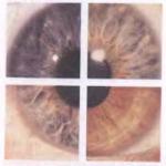
The surface of the mucous membrane of the eyeball is subdivided into the conjunctiva of the eye and eyelid. When the latter are closed, the membranes are connected, as a result of which the lower or upper fornix of the conjunctiva is formed. In the structure of the ocular structure there is a rudimentary formation, which in ophthalmology is also known as the third eyelid. The duplication forms a kind of semilunar fold. It is localized in the area of \u200b\u200bthe medial angle of the optic organ.
In some nationalities, especially the Mongoloid type, the rudimentary formation is emphasized more sharply than in others, which gives the eyes more expressiveness.
According to the histological structure, there is a bulbar, palpebral conjunctiva and a mucous membrane of the fornix. The following layers of cells of the visual organ are distinguished:
- Epithelial, including squamous epithelium with several layers, which contains various glands.
- Subepithelial. Consists of loose connective tissue, lymphocytes and a small number of glandular cells.
A lot of blood enters the conjunctiva through the arteries of the eyelids. In inflammatory reactions, transition colors from white to deep red can be observed. A similar phenomenon is associated with the expansion of a large number of vascular plexuses. Also, various diseases of the conjunctiva can lead to impaired sensory innervation. The patient may experience unpleasant symptoms such as frequent blinking.
Why does it develop and what symptoms are accompanied by the pathology?
 Due to its location on the surface of the eye, inflammatory processes often occur, one of which is conjunctivitis.
 The conjunctiva of the eyelids and sclera are often subject to various diseases, a fold can form on it, color change and other pathologies occur. This is due to the fact that it is located at the very top of the mucous membrane of the eyeball. To a greater extent, the diseases are inflammatory in nature and are known in ophthalmology as conjunctivitis. The disease is of several types, while the tarsal conjunctiva and other parts of the eye structure are affected. The following reasons can affect the disease:
- contact with the mucous membrane of the eyeball of bacteria, viruses and other infections;
- mechanical damage;
- an allergic reaction to eye drops, pollen or animal hair;
- unsuitable contact lenses that provoke allergies;
- negative impact on the conjunctiva of household cosmetics, disinfectants, polluted air.
When the conjunctiva or cornea becomes inflamed, the patient is worried about pain in the eyes and other unpleasant symptoms, which can be stronger or weaker, depending on the form and type of ailment. It is possible to recognize the disease by the following signs:
- pain syndrome when blinking;
- dilated conjunctival vessels, as a result of which the mucous membrane of the eyeball turns red;
- itchy and burning sensations;
- purulent and other pathological discharge from the eyes, which should not normally be present;
- neoplasms on the conjunctiva or in the lower eyelid;
- drying out of the mucous membrane against the background of dystrophic changes.
Ophthalmologists diagnose inflammation of the conjunctival membrane of unclear etiology if the cause of the disease has not been established.
Diagnostic measures
 In case of the formation of undesirable manifestations on the organs of vision, it is necessary to consult a doctor who will conduct an examination and determine the ailment.
 If the conjunctiva does not completely cover the eyeball, its anatomy is impaired or there are other disorders that adversely affect visual function, then you should immediately contact an ophthalmologist. Also, medical advice is required in case of an inflammatory reaction, redness of the mucous membrane of the eye and in the presence of other unpleasant symptoms. In most cases, a visual examination of the eyeball or the use of ophthalmic techniques is sufficient for a specialist. If you establish an accurate diagnosis and find out why the bulbar conjunctiva or other ocular structures are inflamed, then the following diagnostic manipulations are prescribed:
- ultrasound diagnostics of the eyeball;
- biomicroscopy, which involves examining the visual organs with a slit lamp;
- bacteriological laboratory studies of pathological secretions, through which it is possible to determine the pathogenic microflora.
The pterygium is formed from the tissue of the conjunctiva, which has undergone degenerative changes, and grows from the limbus towards the middle of the cornea. The pterygium can be of different sizes - from a couple of millimeters to large formations that close the cornea and significantly reduce the patient's quality of life.
What is a pterygium?
The pterygium, or pterygoid hymen, is an abnormal formation located on the inner corner of the eye, having a triangular shape.
The development of pathology can be rapid, characterized by rapid growth, or slow.
Prevalence
Epidemiology is directly related to a person's place of residence. For example, in the United States, in those geographic areas that are above 40-degree latitude, the incidence of pathology does not exceed 2% of 100% of the population.
In settlements located at a latitude of 28-36 degrees, the incidence rises to 10%.
According to experts, this is due to an increase in the amount of solar radiation received by humans. 
In females, the pathology develops less frequently than in men, which is due to the more frequent presence of men under the scorching rays of the sun in connection with the type of work. The first signs of pterygium are usually noted at a young and mature age (25-40 years). Until the age of 20, the disease is rarely recorded.
Causes of the disease
The reasons for the development of the disease are: the high frequency and duration of the effect of ultraviolet radiation on the eye area, which is inherent in residents of regions with a hot climate, work in open areas, neglect of methods and means of eye protection. The hereditary disposition to the appearance of signs of pterygium has also been proven.
Ptergium symptoms
In the early stages of the disease, there may be no symptoms at all. Later, signs of eye irritation develop, conjunctival redness, a feeling of the presence of sand, "fog" in the eyes, eyelid swelling, a slight decrease in visual function. 
Diagnostic methods
Examination by an ophthalmologist includes a visual acuity test and a visual examination using a special lamp. If there are phenomena of myopia, astigmatism, keratotopography is prescribed. Dynamic tracking of ongoing processes allows you to calculate the rate of development of the disease.
Consequences and complications
Among the unpleasant symptoms that can join as the pterygium progresses, there are:
- incomplete vision of objects, distortion of their outlines;
- significant drop in vision;
- pain in the eyes, severe irritation, inflammation of the conjunctiva due to rubbing, scratching;
- the appearance of adhesions, scars on the cornea, eyelids, etc.;
- fusion of pterygium tissues with other parts of the organ of vision, a decrease in the mobility of extraocular muscles, as a result of which the eyeball may lose mobility;
- double vision ().
The phenomena of diplopia most often develop due to partial paralysis of the external muscle. If the patient has undergone surgery for the pterygium, such unpleasant consequences can be observed as a result of the detachment of the muscle tendon from the area of \u200b\u200bits attachment.
A rare complication of pterygium is corneal degeneration with pronounced thinning, which is observed against the background of regular contact of the cornea with the bulging part of the formation.
The most dangerous, but rarest consequence of the disease may be its transformation into a malignant tumor.
Ptergium treatment
To reduce the rate of the course of the disease, drops of the "artificial tear" type, moisturizing gels and ointments are used. Patients are advised to wear glasses with UV filters at all times when outdoors. To eliminate the symptoms of pterygium, eye ointments and drops with glucocorticosteroids are used.
Surgical treatment
A radical way to eliminate the formation in the area of \u200b\u200bthe inner corner of the eye is a surgical operation. It is carried out to restore the aesthetic appeal of the face, as well as for therapeutic purposes (to normalize visual acuity, eliminate discomfort, irritation and other symptoms).
Surgical removal of the pterygium can be performed according to various methods, but all of them are aimed at excision of abnormally overgrown tissues.
It was noted that removal of the pterygium without subsequent drug treatment leads to its reappearance in half or more cases.
To prevent this from happening, immediately after the operation, treatment with immunosuppressants (cytostatics) is performed, courses of therapy with β-radiation are carried out, the affected area is treated with cryocoagulants, etc.
If postoperative therapy was carried out in full, the probability of recurrence of the pterygium is no more than 10%.
If the pterygium is of considerable size, it may be necessary to transplant (insert or suture) a conjunctival autograft or special artificial membranes to hide the resulting cosmetic defect.
The operation is not difficult and is often performed under local anesthetic. In parallel with anti-relapse treatment, antibiotic therapy is prescribed, drops to prevent inflammation.
In some cases, the operation leads to the development of complications. These can be: eye infection, graft rejection, tissue inflammation in the suture area, visual dysfunctions (for example, double vision), scarring of the cornea.
The most rare, but still occurring complications - perforation of the eyeball, the penetration of blood into the vitreous. Against the background of treatment with cytostatics and radiation therapy, the cornea may become thinner, sometimes scleral ectasia occurs.
The conjunctival membrane is the integumentary layer that is located around the eyeball. The mucous membrane originates at the marginal surface, and then passes to the eyeball itself and reaches. If the patient twists the eyelid, the conjunctiva becomes accessible for examination.
The structure of the conjunctiva of the eye
The entire surface of the mucous membrane of the eye can be divided into two sections:
- Conjunctiva of the eyeball;
- Conjunctiva of the century.
When the eyelids are closed, the conjunctiva joins to form two sacs (lower and upper). If the eyelids are open, the mucous membrane forms two corresponding vaults. There is also a rudimentary formation that bears the name of the third century. It is located in the region of the medial corner of the eye and is better expressed in some nationalities, in particular, the Mongoloid type. This fold was well pronounced in our ancestors, but over time it lost its purpose.
From the point of view of histology, the conjunctiva consists of two layers of cells:
1. The epithelial layer includes stratified squamous epithelium, which contains a large number of glandular cells.
2. The subepithelial layer includes loose connective tissue, lymphocytes and a small number of glandular cells.

The mucous membrane of the eye is very well supplied with blood. The blood flow comes from the arteries of the eyelids, as well as from the pool of the ciliary arteries. If an inflammatory process develops in the area of \u200b\u200bthe eye surface, then the mucous membrane acquires a red tint. This is due to the expansion of the abundant number of blood vessels. In addition, during the inflammatory process, pain occurs, which is associated with irritation of the branches of the trigeminal nerve. In addition, the development of so-called reflected pains, which are caused by the involvement of inflammatory reactions of other branches of the trigeminal nerve, is possible. In particular, pain that occurs in diseases of the ENT organs can radiate to the eye.
Physiological role of the conjunctiva of the eye
The main functions of the conjunctiva are associated with the structure of this membrane of the eye:
1. The protective role is associated with the presence of stratified epithelium on the surface. These cells protect the eyeball itself from small foreign objects.
2. Produce fluid, which also helps to remove small particles from the mucosal surface.
3. Lysozyme produced by glandular cells, as well as immunoglobulins, provide protection against pathogenic flora and reduce the risk of developing an inflammatory reaction.
Video about the structure of the conjunctiva of the eye
Symptoms of conjunctival lesions
The direct manifestations of conjunctival pathologies depend on the pathological process itself. Among them are:
- Pain in the eye area, aggravated by blinking movements;
- conjunctiva due to vasodilation;
- Changes in the nature of the discharge (the appearance of pus, etc.);
- and burning;
- An increase in the amount of fluid;
- Neoplasm on the surface of the conjunctiva;
- Dryness of the mucous membrane associated with dystrophy.
Diagnostic methods for lesions of the conjunctiva of the eye
A number of studies are used to diagnose pathologies of the mucous membrane:
- (carried out using a slit lamp);
- Bacteriological examination of the discharge for the presence of infectious agents.

It should be noted once again that the conjunctiva belongs to the important organs of the optical system and protects the eyeball from external damage. In addition, due to the presence of lysozyme and immunoglobulins, the conjunctiva is able to resist pathogenic microflora.
Diseases of the conjunctiva of the eye
Among the pathologies that can affect the conjunctival membrane are:
- , which consists in the formation of a wen on the surface of the mucous membrane;
- is an inflammatory reaction that is associated with the invasion of pathogens or an allergic attack.
- Tumor neoplasms of a benign or malignant nature (fibroma, nevus, etc.).
- Keratoconjunctivitis dry, which is a sign of degenerative processes.
The main role of the conjunctiva is protection from external factors, providing comfort, which is achieved through the work of numerous glands that produce mucin, as well as additional lacrimal glands. The production of mucin and tear fluid forms a stable tear film that protects and moisturizes the eye. Therefore, with diseases of the conjunctiva, for example, conjunctivitis, there is pronounced discomfort and the form of a burning sensation, a foreign body or sand in the eyes.
Conjunctival structure
The conjunctiva is a thin transparent mucous membrane that covers the back surface of the eyelids, where it is very tightly connected to the cartilage, then forms the conjunctival vaults: upper and lower.The vaults are areas of a relatively free conjunctiva that look like pockets and provide freedom of movement of the eyeball, and the upper fornix is \u200b\u200btwice as large as the lower. The conjunctiva of the arches passes to the eyeball, located above the dense tenon membrane, reaching the limbus region. In this case, the epithelium of the conjunctiva - its surface layer directly passes into the epithelium of the cornea.

The blood supply to the conjunctiva of the eyelids is provided by the same vessels as the eyelids themselves. In the conjunctiva of the eyeball, the superficial and deep layers of blood vessels are isolated. Superficial is formed by the perforating arteries of the eyelids and the anterior ciliary arteries. The deep layer of the conjunctival vessels is formed by the anterior ciliary arteries, forming a dense network around the cornea.
The venous vascular system corresponds to the arterial one. In addition, the conjunctiva is rich in accumulations of lymphoid tissue and lymphatic vessels. The sensitivity of the conjunctiva is provided by the lacrimal, subblock and infraorbital nerves.
Symptoms of defeat
The conjunctiva, like a mucous membrane, reacts to any external irritation with inflammation. An irritant can be fever, allergens, chemicals, and most commonly, a bacterial or viral infection. The main manifestations of inflammation of the conjunctiva are: lacrimation, redness, itching, burning or dryness, pain when blinking and moving the eyeball with an increase in the lymphoid tissue of the eyelid conjunctiva. The sensation of a foreign body may appear when the cornea is involved in the process. Discharge from the eyes during inflammation of the conjunctiva can be different: from watery-mucous to purulent with crusts, depending on the damaging irritant agent. In acute viral lesions, hemorrhages may appear under the conjunctiva, it becomes edematous.
With insufficient function of the lacrimal glands and certain cells, the conjunctiva can dry out, which leads to various degenerative conditions. The conjunctiva of the eyeball, the vault, and then the eyelids can grow together, limiting the movement of the eyeball.
Normally, the conjunctiva does not spread to the cornea, but in some people, especially in windy environments and / or dusty work, the conjunctiva slowly grows on the corneal area and upon reaching a certain size. This growth, called pterygium, can impair vision.
In the conjunctiva, pigment inclusions in the form of brownish-dark spots may be normal, but they must be observed by an ophthalmologist.
Diagnostic and treatment methods
For a detailed examination of the conjunctiva, an ophthalmologist needs a slit-lamp examination. At the same time, the conjunctiva of the eyelids, eyeball and fornices, the degree of dilatation of its vessels, the presence of hemorrhages, edema, the nature of the resulting secretions, the involvement of other eye structures in the inflammatory or degenerative process are evaluated.Treatment for conjunctival diseases depends on the cause. From washing and antibacterial and anti-inflammatory treatment for chemical burns, infections, to surgical treatment for pterygium and simblepharon.
In the conjunctiva, two sections are distinguished: the conjunctiva of the eyelids and the eyeball. In the places of transition, it forms two vaults, and when the eyes are closed, two closed cavities are conjunctival sacs (the spaces between the upper and lower eyelids and the eyeball). Into these anatomical formations, medicinal substances are injected in the form of eye drops or ointments. In the inner corner of the eye, the conjunctiva forms a lunar fold and a lacrimal meatus (a rudiment of the third century, inherited from our ancestors).
The conjunctiva has two layers: epithelial and subepithelial. The epithelial layer consists of a stratified epithelium with a large number of glandular cells (lacrimal glands of Krause, Wolfirng, mucin producing glands (Manza), the secret of which moisturizes and disinfects the mucous membrane of the eye. The subepithelial layer is represented by loose tissue with the inclusion of glands and an accumulation of lymphoid tissue.
Innervation and blood supply
The conjunctiva receives blood supply from the vessels of the eyelids, as well as from the anterior ciliary vessels, which form two layers: superficial and deep. With inflammatory diseases or irritation, a reflex dilation of the vessels of the mucous membrane occurs - the eyes "turn red" (conjunctival hyperemia).
The mucous membrane of the eye has a sensitive innervation (branches of the trigeminal nerve).
Functions
The main function of the conjunctiva is protective. With the help of a tear and the substances it contains (mucin, lysozyme, immunoglobulins), small foreign bodies and bacteria are removed from the surface of the eyeball, and the cornea is moistened.
Diseases
Diseases of the conjunctiva can be divided into three groups, for reasons causing them:
1.Infectious: viral or bacterial.
2.Allergic conjunctivitis: pollen, medicines, cosmetics, animals, etc.
3.Dystrophic diseases of the conjunctiva: (wen), keratoconjunctivitis dry, etc.
4. Neoplasms of the conjunctiva (malignant and benign): nevi, cysts, fibromas.