Surgical pathology of the digestive organs in dogs is actual problem veterinary surgery. Pathologies associated with the presence of obstruction gastrointestinal tract accounts for 20 to 30% of all surgical pathology of the gastrointestinal tract in dogs. The complexity of diagnostics and surgical techniques during operations on the abdominal organs requires a more thorough study of this problem by veterinary specialists. The most difficult, from the point of view of diagnosis and surgical treatment are foreign bodies in the esophagus. The purpose of our work is to determine the main diagnostic criteria, compare methods of surgical treatment of foreign bodies in the esophagus, and also outline the basic principles for the prevention of postoperative complications.
According to our observations, based on treatment statistics, at the Department of Veterinary Surgery of the Federal State Educational Institution of Higher Professional Education MGAVMiB named after K.I. Scriabin from 2003 to 2009. 49 dogs with obstruction of the gastrointestinal tract (GIT), 62% of animals had foreign objects, 14% had intussusception, 18% had neoplasms and 6% had adhesive disease. In dogs with foreign objects in the gastrointestinal tract, the location of obstruction is distributed in the following proportions: 11% is due to obstruction of the esophagus, 27% is localized in the stomach, 56% is located in sections small intestine, and 6% for localization in the large intestine. Despite the fact that dogs are predators and anatomical structure The mouth, pharynx and esophagus are determined by the ability to swallow large pieces of food; obstruction of the esophagus in 90% of cases occurs in the area of the diaphragm, since the esophageal opening of the diaphragm does not have the ability to expand greatly. Most of the foreign objects we removed from the esophagus were bone fragments, but there were cases of rubber balls, sponges, rags, etc. being removed.
Diagnosis of foreign bodies in the esophagus involves taking a history and X-ray examination. According to the anamnesis, regurgitation is observed immediately (1-3 minutes) after ingestion of roughage. In some cases, the animal can consume liquid in small quantities, then they speak of partial obstruction of the esophagus. With partial obstruction of the esophagus, the ability to take fluid in doses may cease on day 2-3 due to swelling of the esophageal wall. To confirm the diagnosis, radiography is performed in a standing lateral projection (Fig. 1), so that it would be possible to determine the level free liquid in the abdominal cavity. If the foreign body is not radiopaque, then radiography is performed immediately after oral administration of a radiopaque substance (barium sulfate with kefir).
Rice. 1 Lateral x-ray of the chest wall of a dog in a standing position.
A foreign body is visible in front of the diaphragm; there is no free fluid.
When the diagnosis of a foreign body in the esophagus is confirmed, surgical treatment is started immediately. This is due to the possibility of perforation of the esophageal wall by a foreign body. In this case, the contents of the esophagus flow into the chest cavity, which will undoubtedly lead to purulent pleurisy, and this is a deadly complication.
Removal of a foreign object from the esophagus can be carried out by intrathoracic esophagotomy, intraabdominal gastrotomy and using a gastroscope with operational functions. The gastroscopy method is quite simple to use: after inserting a gastroscope into the esophagus, fragments of the foreign body are fragmented and removed in parts. However, the high cost of a gastroscope and sets of manipulators does not yet allow its widespread use in veterinary practice.
The choice of surgical techniques depends on several factors: if the foreign body is in the esophagus for more than 4 days, if not x-ray free fluid is visible in the abdominal cavity and there is an increase in general temperature, that is, when perforation of the esophagus occurs, intrathoracic esophagotomy is performed. If there is no perforation of the esophagus and less than 3 days have passed, intra-abdominal gastrotomy.
When planning the operation, we recommend taking into account the chosen method of operation. Preparation for surgery consists of medication preparation, we recommend not performing induction anesthesia with xylazine to avoid the gag reflex. The surgical field is prepared in the preumbilical region of the anterior chest wall and in the area of the 4-10 right intercostal space.
The technique of intrathoracic esophagotomy consists of surgical access to the chest cavity on the right in the area of the 7th intercostal space. The trachea is intubated and a ventilator is connected. An incision is made in the skin, subcutaneous tissue, intercostal muscles and pleura. With the help of a rib expander, the lungs are spread apart, the lobe of the lung is retracted to the side, thereby providing access to the esophagus. After assessing the size and position of the foreign object, a perpendicular incision is made into the esophagus while removing the esophageal contents using a surgical suction device. Extract foreign object, after which an intestinal two-story suture is placed on the wall of the esophagus. Chest wall sutured in layers, vacuum drainage is installed. The release of fluid from the drainage is monitored daily for 5 days; the drainage is removed on the 5th day after surgery. Postoperative therapy according to generally accepted methods.
The intra-abdominal gastrotomy technique involves surgical access to the abdominal cavity in the pre-umbilical region. The stomach is removed into the surgical wound, after which a gastrotomy is performed, with a long incision of 4-6 cm closer to the cardiac part. At the same time, assistants administer gastric tube into the esophagus, 5-7 ml is poured into the esophageal cavity. Vaseline oil. The surgeon inserts his hand into the stomach, opens the cardiac sphincter with his fingers and inserts his fingers into the esophagus, palpating the foreign body. While the assistant brings the probe to the foreign body from the other side and fixes it to prevent displacement cranially. After such fixation, the surgeon with the other hand, using an Alice or Kocher clamp, under the control of his hand, removes the foreign body through the cardiac part and an incision in the stomach wall. After this, the surgeon palpates the integrity of the esophageal wall. If a wall rupture is observed, then the second stage of the operation will be intrathoracic suturing of the esophageal rupture (see above). If no rupture of the esophagus is detected, the operation is completed: suturing the surgical wound of the stomach wall, washing the abdominal cavity and suturing the anterior abdominal wall. On the 4th day after surgery, a control x-ray or ultrasound is performed to detect free fluid in the abdominal and chest cavities. Postoperative therapy according to the generally accepted method for gastrotomy.
According to our observations, when prompt removal foreign objects a large number of complications (23% of the total number of operated animals) accounted for purulent complications, the reason for which is non-compliance with asepsis and antisepsis, inadequate postoperative antibiotic therapy, failure of sutures on the esophagus or stomach.
In conclusion, we can conclude that although diagnosing foreign bodies in the esophageal cavity does not pose a problem for most doctors, surgical treatment requires high professionalism, especially if the method of intrathoracic removal of the foreign body is chosen. In addition, it is necessary to carefully monitor the postoperative course of the disease and prevent the development of purulent processes in the chest or abdominal cavity.
SUMMARY
The surgical pathology of digestive organs at dogs is an actual problem of veterinary surgery. The pathologies connected with the presence of impassability of a gastroenteric path makes from 20 to 30% of all surgical abdominal pathology at dogs.Complexity of diagnostics and operative receptions at operations on bodies of a belly cavity demands more careful studying of the given problem by veterinary experts . The most difficult, from the point of view of diagnostics and operative treatment foreign matters in a gullet are. The purpose of our work is to define the basic diagnostic criteria to compare techniques of operative treatment of alien bodies in a gullet and as to state main principles of preventive maintenance of postoperative complications.
Literature
1. Anatomy of a dog Slesarenko N.A. Lan, St. Petersburg 2004
2. Surgery of the stomach and spleen in dogs, Timofeev S.V., Pozyabin S.V. and others. M.:Zoomedlit, 2009
3. Veterinary radiography Khan K., Hurd Ch. M.: Aquarium, 2006.
4. X-ray diagnosis of surgical diseases of the abdominal cavity in dogs Pozyabin S.V., Timofeev S.V. Veterinary medicine M.: 2006.- No. 4.-P.36-37
The curiosity of our four-legged explorers knows no bounds. They are ready to try not only new treats, but also everything that comes their way. Is it any wonder that at one point they swallow something, be it a stick, paper or a piece of a rubber toy. Fortunately, in most cases, these things pass through the gastrointestinal tract without problems, surprising the owners at the exit with the quirkiness of the pet’s culinary preferences. However, sometimes the animal’s luck changes, and the foreign body becomes firmly stuck in the stomach or intestines.
Without a timely response, such a situation can cost the health and even the life of your four-legged pet, which is why it is so important to recognize the danger in time and seek help.
How to tell if your dog has eaten a foreign body
Even if you did not notice how an inedible object disappeared into the dog’s mouth, you should be alerted by signs indicating a possible obstruction:
- Vomit. Involuntary eruption of eaten food or water occurs immediately after eating or drinking. However, if it is not the stomach, but the intestines that are clogged, from the moment of eating it can take from a few minutes to a couple of hours. The main thing that should alert the owner is the regularity of vomiting. That is, everything that the dog tries to swallow comes back after a short time.
- Diarrhea. Liquid feces often contain large amounts of mucus or traces of blood. If a dog has swallowed a sharp object that has injured the walls of the stomach or intestines, the stool may be black - a sign of heavy internal bleeding.
- Abdominal pain. ABOUT pain says the animal's pose - a hunched back and a tense, toned stomach. The dog does not allow itself to be touched and whines when the peritoneum is touched.
- Lack of appetite. The dog is not only the usual food, but also a treat. Most often, the animal does not even approach the bowl, or, becoming interested for a second, sniffs and turns away.
- Tension during defecation. The dog sits down several times, straining, groans and grunts, sometimes squeals during the act of defecation. As a rule, when the gastrointestinal tract is blocked by a foreign body, only small portions of feces come out of the animal. This, by the way, is another of the main signs of obstruction.
- Weakness. The loss of fluid and electrolytes important for life (potassium, sodium) leads to dehydration of the body and, as a result, weakness and depression. You can check how dehydrated your pet’s body is using a simple test: grab your dog’s skin with two fingers and pull it as far as possible. If the skin does not smooth out within a few seconds, fluid loss has reached a critical level.
- Change in behavior. Lack of interest in life, depression and reluctance to communicate indicate feeling unwell dogs. In addition, manifestations of aggression are possible when trying to feel the belly or examine the pet's mouth.
- Cough. If a foreign body is stuck in the throat or respiratory tract, the dog may try, trying to get rid of the object. In this case, increased salivation and convulsive attempts to swallow may be observed.
 The insidiousness of this condition is that the symptoms of obstruction do not appear immediately. The dog may feel fine for several days or even weeks after swallowing the object, but the above signs may appear periodically or not at all. However, then the animal’s condition deteriorates sharply.
Medical diagnosis
At the first sign of a foreign body in the gastrointestinal tract, we recommend that you immediately consult a doctor. Remember that it is very difficult to diagnose such a problem, as they say, “by eye” - only clinical researches can confirm or refute the diagnosis.
- Palpation of the abdominal cavity. If the foreign body is quite large and dense, for example, a rubber ball, it is quite possible that it can be felt through the walls of the stomach. However, even if nothing is found during palpation, this is not a reason to exhale with relief. A huge number of objects, for example, a rag, a bag or thread, cannot be felt by hand.
- X-ray. During the examination, stones, metal and rubber objects are clearly visible. Or, if the foreign body is not detected, the doctor may notice changes internal organs, characteristic of the presence of a foreign body.
- Radiographic examination. To track the progress of an object through the stomach and intestines, a contrast agent (most often barium) is used, which is given orally to the dog.
- Endoscopy. Today it is considered the most the best method foreign body diagnosis.
- Laboratory research. To rule out other causes of your pet’s illness, the doctor may prescribe a blood test for a biochemical test.
What to do?
The main problem in this situation is the critical amount of time allocated to the choice of therapy and the treatment itself. The foreign body compresses vital vessels, leading to tissue necrosis and the development of peritonitis. This is why it is so important for owners to listen to the veterinarian’s recommendations and follow his instructions, because we are talking about the life of the pet.
If the object is stuck shallowly and, you can try to remove it with your hand, tweezers or medical forceps. To avoid injury, a special clamp is inserted into the animal’s mouth to prevent jaw compression.
If ingestion of a foreign body is noticed immediately, the best way out will induce vomiting in the dog using a 1.5% hydrogen peroxide solution. Peroxide, entering the gastrointestinal tract, expands, irritating the walls of the stomach. Generally, if vomiting is induced within 2 hours of ingestion, the item will come out without causing much harm.
Another effective way induce vomiting - pour a tablespoon of salt onto the root of the dog’s tongue (the dose is given for large dog). Irritation of the receptors leads to an involuntary gag reflex. Just remember to offer your dog water later - salt and subsequent vomiting causes extreme thirst.
To envelop a foreign body and facilitate its passage through the gastrointestinal tract, Vaseline oil is used, which is poured into the dog’s mouth. Due to the fact that this substance is not absorbed by the walls of the stomach, it helps to contract the intestinal muscles and smoother passage of the object through the digestive tract.


If the foreign body does not come out on its own, your doctor may recommend surgery. During the operation, the veterinarian opens the intestinal wall and removes the object. If necrotic areas are detected, resection (excision) of part of the stomach or intestine is performed.
After the operation is completed, the animal should be kept under to prevent the opening of internal bleeding or the development of peritonitis.
What not to do
Sometimes, wanting to help a pet, owners, unwittingly, significantly worsen its condition by performing unnecessary or dangerous actions. What should you never do?
- Pull an object out of your throat or anus yourself. By trying to remove a protruding object, you can further injure the walls of the stomach or larynx. Removing hard or sharp objects, as well as bodies with jagged surfaces, is especially dangerous. It is no less dangerous to pull out various threads or ropes. As they pass through the gastrointestinal tract, they can become entangled or, when caught on something, lead to ruptures in the walls of the stomach or intestines.
- Give antiemetic drugs. Medicinal substances, blocking the urge to vomit, do not improve the situation in any way, but only deprive the animal of the chance to get rid of the foreign body on its own and lubricate clinical picture diseases.
- Do an enema. Firstly, the enema irritates the intestinal walls, and secondly, if a foreign body leads to blockage, water, not finding a way out, can lead to rupture of internal organs and peritonitis.
- Give food or water. Any products entering the gastrointestinal tract cause new attacks of vomiting, which leads to rapid dehydration of the animal.
The following items pose a particular danger to our pets:
- Batteries. The acid contained in batteries can enter the dog's stomach, causing chemical burns and...
- Magnets. Small magnetic balls swallowed by an animal are unevenly distributed in the gastrointestinal tract and, through the walls of the stomach or intestines, literally stick to each other, pinching living tissues together. Necrosis and foci of inflammation very quickly form at the junction site.
- Cotton swabs. By absorbing water and increasing in size, tampons, firstly, accelerate dehydration, and secondly, they tightly clog the lumen, practically not moving due to the fleecy cotton structure.
- Threads and elastic bands. A long thread, despite its thinness, can cause great trouble. The rings of the gastrointestinal tract are literally strung on it and assembled into an accordion, also causing necrosis and rupture of sections of the intestine. An elastic band, when contracted, can cut tissue like a fishing line.
- Fillers for cat litter. Any liquid that gets on the filler granules causes them to stick together into a lump. Once in the dog’s stomach, the filler increases in size several times and causes obstruction.
How to keep your dog safe
To avoid the horrors described above, simply do not let your dog eat inedible or dangerous objects:
- If your pet is prone to this, keep it on a leash while walking or wear a muzzle that covers its mouth.
- Do not give it to him with sharp edges; it is better to exclude boiled bones from the diet altogether.
- Offer for leisure large size that are impossible to swallow. The safest toys are those made from solid rubber, from which it is impossible to bite off a piece.
- Let your pet chew dried treats only in your presence and take away small pieces in a timely manner.
- At home, remove all small and unsafe objects from sight. Hide all kinds of magnetic construction sets and puzzles out of harm's way.
And, most importantly, spend as much time as possible with your dog, teaching him not to pick up anything on the street or in the apartment, and if he takes something in his mouth, to spit it out on command. This way you are guaranteed to save your own nerves, as well as the life and health of your beloved four-legged friend.
In most cases, foreign bodies in dogs are tennis balls, small toys, buttons, paper or foil, plastic bags, rags. In this situation, there is a high risk of developing complete or partial blockage of the stomach, volvulus of the digestive tube, intestinal obstruction. If objects are sharp, then internal bleeding and perforation of the walls of internal organs may develop. If foreign objects get into respiratory system the pet may die from asphyxia.
Symptoms in a dog: the animal makes frequent movements of the jaws, there is profuse salivation, retching or full-blown vomiting, or food flows out without active movements from the abdominal press, the dog refuses food, it has severe diarrhea, if there is a complete blockage, then it does not consume water at all, if the intestines are damaged by acute objects, diarrhea mixed with blood is observed, defecation is difficult, difficulty breathing, cyanosis of the mucous membranes develops, pain in the abdomen, apathy and lethargy.
It is strictly prohibited to give laxatives and antiemetics. You should not give a sick pet cleansing enemas, which can lead to the movement of a sharp foreign object through the intestinal tube and perforation of internal organs.
The owner must provide home complete peace. It is not recommended to independently remove swallowed objects from the throat, as well as those protruding from the rectum. It is strictly forbidden to feed and water the animal.
In a specialized institution They will conduct a full examination, prescribe an ultrasound and x-ray examination. In most cases, X-ray diagnostics are used with the help of preliminary drinking of barium salts (more often this is done with kefir). The contrast method allows you to determine the presence and location of foreign objects that are invisible on a regular x-ray.
After identifying the object, the veterinarian begins to remove the foreign body from the dog. The operation can be performed using several methods. The simplest and most effective is use of a gastroscope equipped with operational functions. With its help, a veterinary surgeon defragments the foreign body and removes it. The disadvantage of this method is its high cost.
 Removing chicken bones using an endoscope
 If the image shows no accumulation of fluid in the abdominal cavity, there is no perforation of the esophagus, and no more than 3 days have passed since the object was swallowed, intra-abdominal gastrotomy. Access to the esophagus is through the stomach. During the operation, a gastric tube is inserted. After removal, sutures are placed on the stomach, fluid is removed from the abdominal cavity, and then sutures are placed on the peritoneum. If perforation of the esophagus is detected, its walls are first sutured.
If the foreign body is in the digestive tube for more than 4 days, in case of perforation of the esophagus, a life-saving procedure is performed. intrathoracic esophagotomy. Operative access to the esophagus is carried out with right side in the area of the 7th rib. After removing the foreign object, vacuum drainage is installed for a period of at least 5 days.
A foreign body is removed from the intestine by laparotomy. In some cases, a veterinary surgeon resorts to resection of a section of the intestinal tube if necrosis has occurred. In small pets, the intestine is sutured with a one-story suture; for surgical intervention in large animals, a two-story suture is used. Postoperative care carried out according to generally accepted surgical technique with diet and antibiotic therapy.
If a foreign body is found in the throat, the veterinarian can remove it using long surgical tweezers or forceps.
Read more in our article about helping an animal and options for removing a foreign object by a veterinarian.
One of the most common emergencies in the life of a four-legged pet owner is the ingestion of an inedible object. In most cases, foreign bodies in dogs are tennis balls, small toys, buttons, paper or foil, plastic bags, rags.
The danger of this situation is that there is a high risk of the animal developing complete or partial obstruction (blockage) of the stomach, volvulus of the digestive tube, and intestinal obstruction. If the object is sharp, then internal bleeding and perforation of the walls of internal organs may develop. If foreign objects enter the respiratory system, the pet may die from asphyxia. Knowing the symptoms of a foreign body in a dog will help the owner recognize the danger.
Veterinary specialists, based on many years of practice, believe that the following signs can be used to suspect that a pet has swallowed an inedible object:

The owner should know that if a foreign body is in the dog’s stomach, then clinical manifestations obstructions may occur some time after ingestion.
What to do if swallowed
The owner, suspecting that his four-legged friend has swallowed an inedible object, must first of all know that it is strictly forbidden to give any laxatives or antiemetics. You should not give a sick pet cleansing enemas, which can lead to the movement of a sharp foreign object through the intestinal tube and perforation of internal organs.
Veterinary specialists, when asked by the owner what to do if the dog has swallowed a foreign body, first of all recommend giving the animal complete rest. It is not recommended to independently remove swallowed objects from the throat, as well as those protruding from the rectum. Foreign bodies may be sharp or jagged, which will result in injury to the mucous membrane of the internal organs.
Diagnosis of an animal
In a specialized institution, the sick pet will undergo a full clinical examination. If the veterinarian suspects that the animal has swallowed an inedible object, an ultrasound and x-ray examination will be prescribed.
If there is a possibility that the pet has swallowed radiopaque substances (metal objects, sharp bones), it is easy to detect them on a regular x-ray. The procedure is carried out, as a rule, in a lateral projection in order to determine the level of fluid in the peritoneum.
 Foreign body is located in the stomach
 In most cases, veterinary practice uses x-ray diagnostics using preliminary drinking of barium salts (more often this is done with kefir). This contrast method allows you to determine the presence and location of foreign objects that are not visible on a regular x-ray.
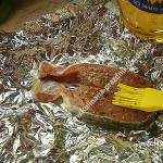
 Foreign body (toy rubber ball) located in the esophagus
 Differential diagnosis is carried out in relation to poisoning, acute viral infection, intussusception not associated with the penetration of a foreign body, etc.
Foreign body removal and surgery
Having detected a foreign object and determined its location, the veterinarian immediately begins to remove the foreign body from the dog. The urgency of surgical intervention is dictated by the high risk of perforation of the walls of the esophagus, stomach or intestines with the subsequent development of bleeding and peritonitis.
If an object unnatural for the body is detected in the respiratory tract, urgent surgery is dictated to save the pet from asphyxia.
If in the stomach, intestines, esophagus
In veterinary practice, surgery to remove a foreign body from a dog is performed using several methods. The simplest and most effective is the use of a gastroscope equipped with operational functions. With its help, a veterinary surgeon defragments the foreign body and removes it. The disadvantage of this method is its high cost. High-tech equipment is available only in megacities.
If the X-ray does not reveal fluid accumulation in the abdominal cavity, there is no perforation of the esophagus, and no more than 3 days have passed since the object was swallowed, the veterinarian performs an intra-abdominal gastrotomy.
Access to the esophagus is through the stomach. During the operation, a gastric tube is inserted into the esophagus. After removing the foreign body from the dog's esophagus, the veterinary surgeon will stitch the stomach, remove fluid from the abdomen, and then stitch the peritoneum. If perforation of the esophagus is detected, its walls are first sutured.
In the event that the owner does not apply immediately, the foreign body is in the digestive tube for more than 4 days; in case of perforation of the esophagus, as a rule, an intrathoracic esophagotomy is performed to save the animal’s life. Operative access to the esophagus is carried out on the right side in the area of the 7th rib. After removing the foreign object, vacuum drainage is installed for a period of at least 5 days.
 The dog underwent surgery to remove part of his intestines. The dog swallowed a sharp bone, resulting in intestinal perforation and peritonitis.
 If a foreign body is found in the dog's intestines, it is removed by laparotomy. In some cases, a veterinary surgeon resorts to resection of a section of the intestinal tube if necrosis has occurred. In small pets, the intestine is sutured with a one-story suture; for surgical intervention in large animals, a two-story suture is used.
Postoperative care for your four-legged friend is carried out according to generally accepted surgical techniques with mandatory adherence to diet and antibacterial therapy.
To see how bones are removed from a dog's stomach, watch this video:
If in the throat, larynx, trachea
If a foreign body is found in a dog's throat, a veterinarian can remove it using long surgical tweezers or a forceps. For this procedure, the animal's jaws are fixed using a special jaw, which provides access to the larynx. This procedure is possible when a foreign object is located shallowly. After extraction, the mouth is irrigated antiseptic solution. For this purpose, use a solution of furatsilin, potassium permanganate.
The owner should understand that untimely assistance in a situation where a foreign body is in the dog’s trachea can lead to such serious complications as pleurisy, pneumothorax. Typically, a veterinarian will perform endoscopic removal of the foreign object. The operation requires general anesthesia.
In some cases, the surgeon resorts to tracheotomy. Surgical intervention using a tracheotube (a special instrument that is inserted into the dissected trachea), most effective when the foreign object is located in the lower parts of the bronchial tube.
 Removing a foreign object (rubber ball) using a forceps
 If it is impossible to remove the swallowed object using an endoscope and tracheotomy, the veterinary surgeon performs an operation by quick access through the chest.
Prevention
The following advice from veterinary specialists and experienced dog breeders will help the owner prevent such trouble as swallowing or inhaling an inedible object:
- When walking, an animal prone to picking up inedible objects should be kept on a leash.
- It is necessary to exclude bones from the diet, which are often the cause of perforation of the stomach and intestinal mucosa.
- Toys for exercising with your pet should be chosen only in a safe size made of solid rubber.
- The room where the dog is kept must be clean. The owner needs to regularly ensure that small objects (toys, sewing accessories, parts of construction sets and puzzles) are not within the reach of a curious pet.
Restless four-legged friends often become victims of their curiosity. Swallowing an inedible object is fraught with serious complications - from the development of aspiration bronchopneumonia to internal bleeding and the development of peritonitis.
Diagnosis is based on clinical examination, palpation and radiographic examination, including the use of a contrast agent. Treatment in most cases is surgical. Veterinary surgeons have various methods of accessing a foreign object, depending on its location.
Useful video
For symptoms, diagnosis and options for removing foreign bodies in dogs, watch this video:
We eat stones, sand, toys.
Let's start with the oral cavity, i.e. graze. Most often, the dog suffers from sharp bones or unplaned sticks. If your pet greedily crunches bones, and then suddenly begins to rub his face with his paws, saliva comes out with blood, it means that he has a piece of treat in his mouth. The same picture will arise if, during training, the dog bites off a piece of a stick or gets a splinter in its mouth.
What should the owner do?
You need to carefully examine the oral cavity; if you notice a foreign body, try to remove it.
Does not work? Call a veterinarian at home or urgently go to the nearest animal clinic.
Did you remove the splinter yourself? Congratulations. Now you need to thoroughly lubricate the wound with iodine to prevent infection. If the wound is too large, the help of doctors will be needed to apply stitches.
The pharynx and esophagus are affected.
During the game a pet can accidentally swallow anything, from a pebble to a needle. Also, along with food, dogs often swallow bones of chickens or fish, which safely get stuck in the esophagus or pharynx.
If the animal has eaten needles or bones, i.e. sharp objects, symptoms may be: vomiting, restlessness, excessive salivation. You may see blood. Some pets start eating soil. All this should alert you; you cannot ignore the symptoms.
Did your dog swallow a smooth or round object? Coughing, drooling and vomiting are then possible. It is likely that the first day will pass quietly, and you will not even know what your pet ate with lunch today. On the second day, all the food that the pet may try to eat will come out through the mouth. Usually, if an object is stuck in the upper part of the esophagus, you can even feel it with your hands.
What to do? You can try to remove the foreign object from the throat yourself. Take a spoon, press the root of the tongue, open the animal’s mouth wide and try to grab the object with your fingers. You can use tweezers instead of fingers, but be careful not to hurt your pet. If all your attempts end only with your bad words and your pet's plaintive whining, contact your veterinarian. A specialist will definitely help you.
Unhealthy food.
During puppyhood, dogs try to eat a funny yellow ball or get a mouth full of stones. All this “good” settles in the stomach or intestines and causes many unpleasant moments for both the pet and its owner. If small items can come out naturally, the more significant ones will be stuck for a long time. In addition, they can injure the gastric mucosa, even pierce it, and this will lead to surgery.
How can you tell if your pet has eaten stones or balls? He will refuse his usual food, drink frequently, and will start vomiting. If an object is firmly stuck in the intestines and causes obstruction, then the pet may not have regular bowel movements. Try patting your pet's belly. Is he moaning pitifully? Most likely, he ate extraneous things.
In this case, there can be only one help - contacting a veterinarian. And the sooner you take your pet to the clinic, the better. An x-ray will help identify a foreign object, and the doctor will prescribe treatment. Giving Castor oil or other laxatives alone are not recommended. You can only make things worse.
They ate it and... almost died.
The rectum may contain the same things as the stomach or mouth. If the dog whines, constantly looks at its tail, tries to go to the toilet, but cannot, then there is a foreign object in the rectum.
Are you ready to do anything for your pet? Then put on a rubber glove and try to explore the rectum with your fingers to remove the foreign object. But it's better to take me pet V veterinary clinic. Remember that the sooner you seek help from a specialist, the greater the chance that your animal will be safe and sound.
Job digestive system(more precisely, coordinated and “error-free” work) is extremely important for the health of our dogs. The slightest violations are fraught with serious consequences, including severe digestive disorders, exhaustion and, in some cases, death. Even a seemingly “harmless” esophagitis can cause a lot of trouble for your pet.
So called inflammation of the esophageal mucosa. The prevalence in dogs is unknown, but it is likely to be quite widespread. The problem is inadequate diagnosis associated with poor equipment in many veterinary clinics.
Please pay Special attention on the pet, which begins a few minutes after any anesthesia. It is very likely that he has reflux esophagitis. No geographical or age-sex predisposing factors have yet been identified (most likely, they simply do not exist). Dogs of all breeds, sexes and ages are affected. Due to certain reasons (teeth wear, periodontal disease), people get sick somewhat more often
Although… Certain breeds(for example, brachycephalic varieties and, in particular,) are subject to increased risk hernia development hiatus diaphragm, pathological dysfunction of the lower esophageal sphincter. Practicing veterinarians note that there are more cases of esophagitis in such animals.
Bitches are also at risk (according to the global veterinary community), but there are no real studies to confirm this. In addition, so far no one has been able to explain what could cause this.
Causes and main predisposing factors
Most often, mechanical or chemical injury to the mucous membrane is to blame. That is, in cases where a dog eats food contaminated with household chemicals or eats greedily, the prospects for the esophagus are not very bright... It happens that inflammation develops against the background of persistent or frequent vomiting appearing against the background of poisoning or action.
Read also: Diarrhea in dogs: causes, symptoms and treatment

Often, inflammation of the esophagus occurs in those dogs whose owners forcefully try to feed them deworming tablets, without even trying to first crush them to a more “sane” state. Also interesting are cases of the disease that appeared after ingestion of various foreign bodies. As a rule, “ill-mannered” dogs who love to visit all the local garbage cans suffer from this. By the way, esophagitis in cats is often caused by hairballs, which are a “headache” for many representatives of long-haired breeds.
The disease is often associated with anesthesia, or more precisely, with improper preparation for it. If your veterinarian says your pet shouldn't eat anything before surgery, he shouldn't eat anything! Vegetative feature nervous system is that during anesthesia it becomes too “autonomous”, and therefore if there is semi-digested contents in the stomach, it torrent will go into the esophagus. The mucous membrane of this organ is not designed to resist hydrochloric acid, which dissolves tissue and causes inflammation. And this, by the way, the best option, because aspiration pneumonia is much worse and often leads to death!
Certain medications(for example, doxycycline, clindamycin, bisphosphonates) do not have a very favorable effect on the mucous membranes, so their administration should coincide with the feeding time of the animal. Finally, esophagitis often affects dogs treated with radiotherapy. However, with successful treatment of cancer, inflammation of the esophagus is a trifle.
Read also: Why is my dog itching? Looking for dangerous symptoms
Clinical picture
Basic clinical sign- vomiting, but this is an extremely unreliable symptom. But! If a poisoned animal vomits “on schedule” and it is clear that something is clearly wrong with the pet, then with esophagitis the dog can vomit “out of the blue,” even in the middle of the apartment. This happens suddenly, spontaneously. Are there any other symptoms of esophagitis? Yes, there are a lot of them.
Important! Dysphagia (pain when swallowing), gagging, hypersalivation (constant drooling), constant neck twitching, lip licking, weight loss, lack of appetite and coughing can all be observed in a sick animal.
In rare cases, signs of classic symptoms develop, but the pathological mechanism of this has not yet been studied. In addition, sick animals often experience normal shortness of breath.

Watching your dog while eating - very important diagnostic method , which is not always given due importance. By the way, how exactly can an accurate diagnosis be made? An esophagoscopy is required. The damaged mucous membrane is distinguished by the following visual characteristics: signs of pathology:
- She is very red (hyperemic).
- Erosion, extensive and numerous ulcers may be observed; in severe cases, abundant leaks of exudate are found on the walls of the esophagus. At chronic course disease, the organ undergoes fibrosis - due to proliferation connective tissue the esophagus narrows.
- The surface of the mucosa becomes “grainy”, its structure changes greatly compared to normal.
Interestingly, in some cases there are no obvious signs of inflammation. This often occurs in humans, but in dogs such development of pathology is poorly described, since there are no reliable statistical data and clinical research results. In such a situation, a diagnosis of iatrogenic esophagitis in dogs may be made. It should not be confused with idiopathic (in this case the cause is unknown, but signs of inflammation are more than obvious).