The pleura is a serous membrane lining the inner surface of the chest wall and outer surface lungs, forming two isolated bags (Fig.).
Borders of the pleura and lungs in front (1) and behind (2): the dotted line is the border of the pleura, the solid line is the border of the lungs.
The pleura lining the walls of the chest cavity is called parietal, or parietal. It distinguishes between the costal pleura (covering the ribs and intercostal spaces, the diaphragmatic pleura, lining the upper surface of the diaphragm, and the mediastinal pleura, limiting. The pulmonary, or visceral, pleura covers the outer and interlobar surfaces of the lungs. It is tightly fused with the lungs, and its deep layers form septa separating the pulmonary lobules.Between the visceral and parietal layers of the pleura there is a closed isolated space - a slit-like pleural cavity.
Closed pleural injuries occur when struck by blunt objects. There are bruises and ruptures of the pleura as a result of a concussion, bruise or fracture of the ribs.
Pleural injuries are observed in all penetrating chest wounds. In this case, traumatic (see) and hemothorax (see) occurs with possible infectious complications subsequently - pleurisy and pyopneumothorax (see).
Inflammatory diseases pleura - see.
Among benign tumors pleura are observed, lipomas, angiomas, etc. Specific symptoms with these tumors no. Primary malignant tumors of the pleura are often multiple in nature and are accompanied by a sharp thickening of the pleura with the development of secondary pleurisy. With them, pain appears relatively early with deep breathing and coughing with irradiation into the chest, and later - shortness of breath and fever. Serous effusion in the pleural cavity then becomes hemorrhagic. bad. Metastases occur in the pleura malignant tumors from other organs.
Pleura (from the Greek pleura - side, wall) is a serous membrane covering the lungs and the inner surface of the chest, forms two symmetrical isolated sacs located in both halves of the chest. The pleura develops from the inner (splanchnopleura) and outer (somatopleura) layers of splanchnotomes of the mesoderm.
Anatomy, histology. The visceral pleura (pleura visceralis, s. pleura pulmonalis) covers the entire surface of the lungs, plunges into their grooves and leaves only a small area uncovered in the area of the hilum of the lung. The parietal pleura (pleura parietalis) is divided into costal (pleura costalis), diaphragmatic (pleura diaphragmatica) and mediastinal (pleura inediastinalis). Pulmonary ligaments (ligg. pulmonalia) represent a duplicate of the serous membrane, located in the frontal plane and connecting the visceral and mediastinal pleura. Between the visceral and parietal pleura there is a slit-like microscopic cavity, reaching when the lungs collapse large sizes. The sections of the pleura in which one parietal sheet passes into another, forming gaps unfilled with lung tissue, are called pleural sinuses (recessus pleuralis). There are costophrenic, costomediastinal and phrenic-mediastinal sinuses.
Like other serous membranes, the pleura has a layered structure. The visceral pleura includes 6 layers: 1) mesothelium; 2) limiting membrane; 3) superficial fibrous collagen layer; 4) superficial elastic network; 5) deep elastic network; 6) deep lattice collagen-elastic layer (Fig. 1). All fibrous layers of the pleura are penetrated by a plexus of reticular fibers. In some places, in the deep lattice collagen-elastic layer there are strands of smooth muscle fibers. The parietal pleura is much thicker than the visceral pleura and is distinguished by structural features of the fibrous structure. Among the cellular forms of the pleura there are fibroblasts, histiocytes, fat and mast cells, and lymphocytes.
Rice. 1. Diagram of the fibrous structure of the pleura (according to Wittels): 1 - mesothelium; 2 - boundary membrane; 3 - superficial fibrous collagen layer; 4 - superficial elastic network; 5 - deep elastic network; 6 - deep lattice collagen-elastic layer.
In the entire visceral pleura and in the predominant area of the parietal pleura, blood and lymphatic vessels lie only in the deepest layer. They are separated from the pleural cavity by a fibrous serous-hemolymphatic barrier, which includes most layers of the pleura. In certain places of the parietal pleura (intercostal spaces, the area of the transverse thoracic muscle, the lateral parts of the tendon center of the diaphragm) there is a serous-lymphatic barrier of a “reduced” type. Thanks to this, the lymphatic vessels are as close as possible to the pleural cavity. In these places specially differentiated resorption apparatuses are located cavity fluid- suction hatches (see Peritoneum). In the visceral pleura of adults, blood capillaries located superficially (closer to the pleural cavity) predominate quantitatively. In the parietal pleura in the areas where the suction hatches are concentrated, lymphatic capillaries are quantitatively predominant, extending to the surface in these places.
In the pleural cavity there is a continuous change of cavity fluid: its formation and absorption. During the day, a volume of fluid passes through the pleural cavity, approximately equal to 27% of the volume of blood plasma. Under physiological conditions, the formation of cavity fluid is carried out mainly by the visceral pleura, while this fluid is absorbed mainly by the costal pleura. The remaining areas of the parietal pleura normally do not take a noticeable part in these processes. Thanks to morphological and functional features various parts pleura, among which the different permeability of its vessels is of particular importance; fluid moves from the visceral to the costal pleura, that is, directed circulation of fluid takes place in the pleural cavity. Under pathological conditions, these relationships change radically, since any area of the visceral or parietal pleura becomes capable of both the formation and absorption of cavity fluid.
The blood vessels of the pleura arise mainly from the intercostal and internal mammary arteries. The visceral pleura is also supplied with vessels from the phrenic artery system.
The outflow of lymph from the parietal pleura occurs parallel to the intercostal vessels in The lymph nodes, located at the heads of the ribs. From the mediastinal and diaphragmatic pleura, lymph follows the sternal and anterior mediastinal path to the venous angle or thoracic duct, and along the posterior mediastinal path - to the peri-aortic lymph nodes.
The pleura is innervated by the vagus and phrenic nerves, bundles of fibers extending from the V-VII cervical and I-II thoracic spinal nodes. The greatest number of receptor endings and small nerve ganglia are concentrated in the mediastinal pleura: in the region of the root of the lung, pulmonary ligament and cardiac depression.
Table of contents of the topic "Pleura. Pleural cavity. Mediastinum.":In the chest cavity there are three completely separate serous sacs - one for each lung and one, middle, for the heart. The serous membrane of the lung is called the pleura. It consists of two sheets: visceral pleura, pleura visceralis, And pleura parietal, parietal, pleura parietalis.
Visceral pleura, or pulmonary, pleura pulmonalis, covers the lung itself and fuses so tightly with the substance of the lung that it cannot be removed without damaging the integrity of the tissue; it enters the grooves of the lung and thus separates the lobes of the lung from each other. Villous protrusions of the pleura are found on the sharp edges of the lungs. Covering the lung on all sides, the pulmonary pleura at the root of the lung directly continues into the parietal pleura. Along the lower edge of the lung root there are serous layers of the anterior and back surfaces the roots are united into one fold, lig. pulmonale, which descends vertically down the inner surface of the lung and attaches to the diaphragm.
Parietal pleura, pleura parietalis, represents the outer layer of the serous sac of the lungs. With its outer surface, the parietal pleura fuses with the walls of the chest cavity, and with its inner surface it faces directly to the visceral pleura. The inner surface of the pleura is covered with mesothelium and, when moistened with a small amount of serous fluid, appears shiny, thereby reducing friction between the two pleural layers, visceral and parietal, during respiratory movements.
Pleura plays a vital role in the processes of transudation (excretion) and resorption (absorption), the normal relationships between which are sharply disrupted during disease processes of the thoracic cavity organs.

With macroscopic homogeneity and a similar histological structure, the parietal and visceral pleura are performed different function, which is obviously due to their different embryological origins. The visceral pleura, in which blood vessels predominate over lymphatic vessels, primarily performs the function of excretion. Parietal pleura, which has in its costal region specific suction devices from serous cavities and a predominance lymphatic vessels above the blood vessels, carries out the function of resorption. The slit-like space between the parietal and visceral layers adjacent to each other is called pleural cavity, cavitas pleuralis. In a healthy person, the pleural cavity is macroscopically invisible.
At rest, it contains 1-2 ml of liquid, which, with a capillary layer, separates the contacting surfaces of the pleural layers. Thanks to this fluid, adhesion occurs between two surfaces that are under the influence of opposing forces: inspiratory stretch of the chest and elastic traction of the lung tissue. The presence of these two opposing forces: on the one hand, the elastic tension of the lung tissue, on the other, the stretching of the chest wall, creates negative pressure in the pleural cavity, which is thus not the pressure of some gas, but arises due to the action of the mentioned forces. When the chest is opened, the pleural cavity artificially increases, since the lungs collapse due to the balancing of atmospheric pressure both on the outer surface and from the inside, from the bronchi.

Parietal pleura is one continuous sac surrounding the lung, but for purposes of description it is divided into sections: pleura costalis, diaphragmatica and mediastinalis. In addition, the upper part of each pleural sac is called the dome of the pleura, cupula pleurae. The dome of the pleura covers the apex of the corresponding lung and stands out from the chest in the neck area 3 - 4 cm above the anterior end of the first rib. On the lateral side, the dome of the pleura is limited by mm. sca-leni anterior et medius, medially and anteriorly lie a. and v. subclaviae, medially and posteriorly - trachea and esophagus. Pleura costalis- the most extensive section of the parietal pleura, covering the ribs and intercostal spaces from the inside. Under the costal pleura, between it and chest wall, there is a thin fibrous membrane, fascia endothoracica, which is especially pronounced in the area of the pleural dome.
Pleura diaphragmatica covers the upper surface of the diaphragm, with the exception of the middle part, where the pericardium is directly adjacent to the diaphragm. Pleura mediastinalis located in the anteroposterior direction, extends from the posterior surface of the sternum and the lateral surface of the spinal column to the root of the lung and limits the mediastinal organs laterally. At the back of the spine and in front of the sternum, the mediastinal pleura passes directly into the costal pleura, below at the base of the pericardium into the diaphragmatic pleura, and at the root of the lung into the visceral layer.
It is the serous membrane of the lung. It is divided into visceral (pulmonary) and parietal (parietal). Each lung is covered with pulmonary pleura, which along the surface of the root passes into the parietal pleura, lining the walls of the chest cavity adjacent to the lung and delimiting the lung from the mediastinum. The visceral (pulmonary) pleura tightly fuses with the tissue of the organ, covering it on all sides, and extends into the cracks between the lobes of the lung. Down from the root of the lung, the visceral pleura, descending from the anterior and posterior surfaces of the lung root, forms a vertically located pulmonary ligament, which lies in the frontal plane between the medial surface of the lung and the mediastinal pleura and descends almost to the diaphragm.
The parietal (parietal) pleura is a continuous sheet. It fuses with the inner surface of the chest wall and forms a closed sac in each half of the chest cavity, containing the right or left lung, covered with visceral pleura. Based on the position of the parts of the parietal pleura, it is divided into the costal, mediastinal and diaphragmatic pleura. The costal pleura covers the inner surface of the ribs and intercostal spaces. It lies on the intrathoracic fascia. In front near the sternum and behind at the spinal column, the costal pleura passes into the mediastinal pleura. The mediastinal pleura is adjacent to the mediastinal organs on the lateral side, located in the anteroposterior direction, extending from the inner surface of the sternum to the lateral surface of the spinal column.
The mediastinal pleura on the right and left is fused with the pericardium. On the right it also borders with the superior vena cava and azygos vein, as well as with the esophagus, on the left - with thoracic aorta. In the region of the root of the lung, the mediastinal pleura covers it and passes into the visceral pleura. At the top, at the level of the superior aperture of the chest, the costal and mediastinal pleura pass into each other and form a dome of the pleura; it is limited on the lateral side by the scalene muscles. Behind the dome of the pleura are the head of the 1st rib and the long neck, covered with the prevertebral plate of the cervical fascia, to which the dome of the pleura is fixed. Anteriorly and medially to the dome of the pleura are adjacent subclavian artery and vein. Above the dome of the pleura is the brachial plexus. Below, the costal and mediastinal pleura passes into the diaphragmatic pleura; it covers the muscular and tendon parts of the diaphragm, with the exception of its central sections, where the pericardium is fused with the diaphragm. Between the parietal and visceral pleura there is a slit-like closed space - the pleural cavity. The cavity contains a small amount of serous fluid, which moistens the adjacent smooth layers of pleura covered with mesothelial cells and eliminates friction between them. When breathing, increasing and decreasing the volume of the lungs, the moistened visceral pleura slides freely along the inner surface of the parietal pleura.
In the places where the costal pleura transitions into the diaphragmatic and mediastinal pleura, depressions of greater or lesser size are formed - pleural sinuses. These sinuses are reserve spaces of the right and left pleural cavities, as well as receptacles in which pleural (serous) fluid can accumulate if the processes of its formation or absorption are disrupted, as well as blood, pus in case of damage or diseases of the lungs and pleura. Between the costal and diaphragmatic pleura there is a clearly visible deep costophrenic sinus reaching largest sizes at the level of the midaxillary line (here its depth is about 3 cm). At the junction of the mediastinal pleura and the phrenic pleura there is a not very deep, sagittally oriented diaphragiomediastinal sinus. A less pronounced sinus (depression) is present at the place where the costal pleura (in its anterior section) transitions into the mediastinal pleura. Here the costomediastinal sinus is formed.
The dome of the pleura on the right and left reaches the neck of the 1st rib, which corresponds to the level of the spinous process 7 cervical vertebra(behind). In front, the dome of the pleura rises 3-4 cm above 1 rib (1-2 cm above the collarbone). The anterior border of the right and left costal pleura extends differently. On the right, the anterior border from the dome of the pleura descends behind the right sternoclavicular joint, then goes behind the manubrium to the middle of its connection with the body and from here descends behind the body of the sternum, located to the left of the midline, to the 6th rib, where it goes to the right and passes into the lower border of the pleura .
The lower border of the pleura on the right corresponds to the line of transition of the costal pleura to the diaphragmatic pleura. From the level of the junction of the cartilage of the 6th rib with the sternum, the lower border of the pleura is directed laterally and downward, along the midclavicular line the 7th rib crosses, along the anterior axillary line - the 8th rib, along the midaxillary line - the 9th rib, along the anterior axillary line - the 10th rib, along scapular line - 11th rib and approaches the spinal column at the level of the neck of the 12th rib, where the lower border passes into the posterior border of the pleura.
On the left, the anterior border of the parietal pleura from the dome goes the same way on the right, behind the sternoclavicular joint (left). Then it is directed behind the manubrium and the body of the sternum down to the level of the cartilage of the 4th rib, located closer to the left edge of the sternum. Here, deviating laterally and downwards, it crosses the left edge of the sternum and descends near it to the cartilage of the 6th rib (runs almost parallel to the left edge of the sternum), where it passes into the lower border of the pleura. The lower border of the costal pleura on the left is located slightly lower than on right side. At the back, as on the right, at the level of the 12th rib it becomes the posterior border. The posterior border of the pleura (corresponds to the posterior line of transition of the costal pleura to the mediastinal) descends from the dome of the pleura down along the spinal column to the head of the 12th rib, where it passes into the lower border. The anterior borders of the costal pleura on the right and left are located unequally. Over the course of ribs 2 to 4, they run along the side of the sternum parallel to each other, and diverge at the top and bottom, forming two triangular spaces free from the pleura - the upper and lower interpleural fields. The upper interpleural field, facing downward with its apex, is located behind the manubrium of the sternum. In the area of the upper space in children lies thymus, and in adults - the remains of this jelly and fatty tissue. The lower interpleural field, located with its apex upward, is located behind the lower half of the body of the sternum and the adjacent anterior sections of the fourth and fifth left intercostal spaces. Here the pericardial sac is in direct contact with the chest wall. Boundaries of the lung and pleural sac (both right and left) basically correspond to each other. However, even at maximum inhale lung does not fill the pleural sac completely, since it is larger than the organ located in it. The boundaries of the pleural dome correspond to the boundaries of the apex of the lung. The posterior border of the lungs and pleura, as well as their anterior border on the right, coincide. The anterior border of the parietal pleura on the left, as well as the lower border of the parietal pleura on the right and left, differ significantly from these borders in the right and left lungs.
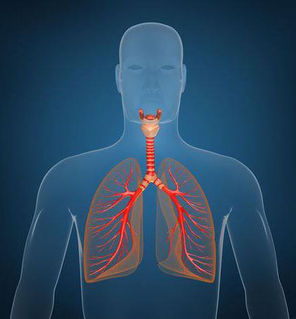
Lungs ( pulmones; Greek pneumones) are paired respiratory organs, which are hollow sacs of a cellular structure, divided into thousands of separate sacs (alveoli) with moist walls, equipped with a dense network of blood capillaries (Fig. No. 230). The branch of medicine that studies the structure, function and diseases of the lungs is called pulmonology.
The lungs are located in a hermetically closed thoracic cavity and are separated from each other by the mediastinum, which includes the heart, large vessels (aorta, superior vena cava), esophagus and other organs (Fig. No. 231). By lung shape resembles an irregular cone with the base facing the diaphragm and the apex protruding 2-3 cm above the collarbone in the neck area. On each lung there are 3 surfaces: diaphragmatic, costal and medial, and two edges: anterior and inferior. The costal and diaphragmatic surfaces are separated from each other by a sharp lower edge and are adjacent, respectively, to the ribs, intercostal muscles and the dome of the diaphragm. The medial surface, facing the mediastinum, is separated from the costal surface by the anterior edge of the lung. On the mediastinal (mediastinal) surface of both lungs there are the pulmonary gates, through which the main bronchi, vessels and nerves that make up the root of the lung pass.
Each lung is divided into lobes through grooves (Fig. No. 230). IN right lung there are 3 lobes: upper, middle and lower, in the left there are 2 lobes: upper and lower. The lobes are divided into segments, of which there are approximately 10 in each lung. The segments consist of lobules, and the lobules consist of acini (Fig. No. 232, 233). Acini (bunches) are structural and functional units of the lung, which perform the main function of the lungs - gas exchange. Each pulmonary lobe includes 16-18 acini. The acini starts from the terminal bronchiole, which is dichotomously divided into respiratory bronchioles of the 1st-2-3rd order and passes into the alveolar ducts and alveolar sacs with the alveoli of the lungs located on their walls. The number of pulmonary acini in one lung reaches 150,000. Each acini includes a large number of alveoli.
Alveoli- these are protrusions in the form of vesicles with a diameter of up to 0.25 mm, the inner surface of which is lined with single-layer squamous epithelium located on a network of elastic fibers and braided on the outside with blood capillaries. The inside of the alveoli is covered with a thin film of phospholipid - surfactant (Fig. No. 236), which performs many important functions:
1) reduces the surface tension of the alveoli;
2) increases the compliance of the lungs;
3) ensures the stability of the pulmonary alveoli, preventing their collapse, adhesion and the appearance of atelectasis;
4) prevents the transudation (exit) of fluid to the surface of the alveoli from the plasma of the capillaries of the lungs.
The thickness of the alveolar wall at the points of contact (adjacent) of the nuclear-free areas of lung epithelial cells and capillary endothelium is about 0.5 microns. On the free surface of epithelial cells there are very short cytoplasmic projections facing the cavity of the alveoli, which increases the total area of contact of air with the surface of the epithelium. The number of alveoli in both lungs in an adult reaches from 600 to 700 million, and the total respiratory surface of all alveoli is about 100 sq.m.
In addition to the respiratory function, the lungs regulate water metabolism, participate in thermoregulation processes, and are a blood depot (from 0.5 to 1.2 liters of blood).
IN clinical practice it is necessary to determine the boundaries of the lungs: anterior, lower and posterior (Fig. No. 234, 235). The apices of the lungs protrude 2-3 cm above the collarbone. The anterior border (projection of the anterior edge) descends from the apices of both lungs along the sternum, running almost parallel at a distance of 1-1.5 cm to the level of the cartilage of the 4th rib. Here, the border of the left lung deviates to the left by 4-5 cm, forming a cardiac notch. At the level of the cartilage of the sixth ribs, the anterior borders of the lungs pass into the lower ones. The lower border of the lungs corresponds along the midclavicular line to the VI rib, along the midaxillary line to the VIII rib, along the scapular line to the X rib, and along the paravertebral line to the XI rib. The lower border of the left lung is located 1-2 cm below the given border of the right lung. With maximum inspiration, the lower edge of the lung descends 5-7 cm. The posterior border of the lungs runs along the paravertebral line (along the heads of the ribs).
On the outside, each lung is covered with a serous membrane - pleura, consisting of two sheets: wall(parietal) and pulmonary(visceral). Between the layers of the pleura there is a capillary gap filled with serous fluid - pleural cavity. This fluid reduces friction between the layers of the pleura during breathing movements. In places where one part of the parietal pleura transitions to another, spare spaces are formed - pleural sinuses, which fill the lungs at the moment of maximum inhalation. In case of pathology, inflammatory exudate can accumulate in them. Especially big costophrenic sinus located in the lower part of the pleural cavity. The right and left pleural cavities do not communicate with each other. Normally, there is no air in the pleural cavity, and the pressure in it is always negative, i.e. below atmospheric. During a quiet inhalation it is 6-8 cm of water. Art. below atmospheric, during a quiet exhalation - by 4-5 cm of water. Art. Due to the negative pressure in the pleural cavities, the lungs are in an expanded state, taking on the configuration of the wall of the chest cavity.
Negative intrathoracic pressure value:
1) helps to stretch the pulmonary alveoli and increase the respiratory surface of the lungs, especially during inhalation;
2) ensures venous return of blood to the heart and improves blood circulation in the pulmonary circle, especially during the inhalation phase;
3) promotes lymph circulation;
4) helps the food bolus move through the esophagus.
Pneumonia is called pneumonia, inflammation of the pleura - pleurisy. The accumulation of fluid in the pleural cavity is called hydrothorax, blood - hemothorax, purulent exudate - pyothorax.
As you know, a person cannot live without air for more than three minutes. At this point, the reserves of oxygen dissolved in the blood are depleted, and brain starvation occurs, which is manifested by fainting, and in severe cases, coma and even death. Of course, people trained in a certain way were able to extend the airless period to five, seven and even ten minutes, but to an ordinary person This is unlikely to be possible. Exchange processes, occurring in the body, require a constant supply of oxygen molecules, and copes well with this task respiratory system.
Stages of breathing
Oxygen exchange between the body and the external environment occurs in four stages:
- Air will enter the lungs from the external environment and fill all available space.
- Diffusion of gases, including oxygen, occurs through the wall of the alveoli (the structural unit of the lungs) into the blood.
- Hemoglobin, which is found in red blood cells, binds most oxygen and carries it throughout the body. Small part dissolves in the blood unchanged.
- Oxygen leaves hemoglobin compounds and passes through the vessel wall into the cells of tissues and organs.
Note that in this process the respiratory system participates only in initial stage, the rest depends on the nature of the blood flow, its properties and the level of tissue metabolism. In addition, the lungs participate in heat exchange, excretion toxic substances, voice formation.
Anatomy
The entire respiratory system is divided into two sections, depending on the relative position of the organs.
The upper respiratory tract consists of the nasal and nasopharynx, oropharynx, pharynx and pharynx. And for the most part they are cavities formed by the walls of the skull bones or the muscular-connective tissue frame.

The lower respiratory tract includes the larynx. The alveoli are not included in this classification, since they are an integral part of the lung parenchyma and the terminal section of the bronchi at the same time.
Briefly about each component unit respiratory tract.
Nasal cavity
This is an osteochondral formation that is located on the facial part of the skull. It consists of two non-communicating cavities (right and left) and a partition between them, which forms a tortuous passage. Inside it is covered with a mucous membrane, which has a large amount of blood vessels. This feature helps warm the passing air during inhalation. And the presence of small cilia allows you to filter out large dust particles, pollen and other dirt. Moreover, it is nasal cavity helps a person distinguish odors.
The nasopharynx, oropharynx, pharynx and pharynx serve to pass warmed air into the larynx. The structure is closely related to the anatomy of the skull and almost completely replicates its musculoskeletal frame.
Larynx
The human voice forms directly in the larynx. This is where the vocal cords are located, which vibrate as air flows through them. These are similar to strings, but due to their structure (length, thickness), their capabilities are not limited to one tone. The sound of the voice is amplified due to the close proximity of the intracranial sinuses or cavities, which create a certain resonance. But the voice is not yet speech. Articulate sounds are formed only with the coordinated work of all the components of the upper respiratory tract and nervous system.

The trachea, or windpipe, is a tube that consists of cartilage on one side and ligaments on the other. Its length is ten to fifteen centimeters. At level five thoracic vertebra it is divided into two main bronchi: left and right. The structure of the organs of the lower respiratory tract is mainly represented by cartilage, which, when connected, form tubes that conduct air deep into the lung parenchyma.
Isolation of the respiratory system
The pleura is the outer thin shell lung, represented by serous connective tissue. Outwardly, it can be mistaken for a shiny protective coating, and this is not too far from the truth. It covers the internal organs on all sides and is also located on the inner surface of the chest. Anatomically, two parts of the pleura are distinguished: one actually covers the lungs, and the second lines chest cavity from the inside.

Visceral leaf
That part of the shell that is on top internal organs, called the visceral or pulmonary pleura. It is tightly soldered to the parenchyma (the actual substance) of the lungs, and it can only be separated surgically. It is thanks to such close contact and repetition of all the contours of the organ that the grooves dividing the lung into lobes can be distinguished. These areas are called the interlobar pleura. Having passed along the entire surface of the lungs, the connective tissue surrounds the root of the lung to protect the vessels, nerves and main bronchus entering it, and then passes to the chest wall.
Parietal leaf
Starting from the transition point, the leaf connective tissue is called the parietal or parietal pleura. This is due to the fact that its attachment will no longer be to the lung parenchyma, but to the ribs, intercostal muscles, their fascia and the diaphragm. Important feature it is believed that the serous membrane remains intact throughout its entire length, despite differences in topographic names. For their own convenience, anatomists distinguish between the costal, diaphragmatic and mediastinal sections, and the part of the pleura above the apex of the lung is called the dome.
Cavity
Between the two layers of the pleura there is a small gap (no more than seven tenths of a millimeter), this is the lungs. It is filled with secretion, which is produced directly by the serous membrane. Fine healthy man produces only a few milliliters of this substance daily. Pleural fluid is necessary to soften the frictional force that occurs between the sheets of connective tissue during breathing.

Pathological conditions
Mostly, pleural diseases are inflammatory in nature. As a rule, this is more of a complication than an independent disease; as a rule, it is considered by doctors in conjunction with other clinical symptoms. Tuberculosis is the most common reason why the pleura becomes inflamed. This infection widespread among the population. IN classic version Primary infection occurs through the lungs. The structure determines the transition of inflammation and pathogens from the parenchyma to the serous membrane.
In addition to tuberculosis, the culprits of inflammation of the pleura can be tumors, allergic reactions, pneumonia caused by streptococci, staphylococci and pyogenic flora, injuries.
Pleurisy in nature can be dry (fibrinous) and effusion (exudative).
Dry inflammation
In this case, the vascular network inside the connective tissue sheets swells, and a small amount of fluid leaks out of it. It coagulates in the pleural cavity and forms dense masses that are deposited on the surface of the lungs. In severe cases, there are so many of these plaques that a hard shell forms around the lung, which prevents the person from breathing. This complication can be corrected without surgical intervention impossible.
Exudative inflammation
If pleural fluid is produced in a significant amount, then it is said to be divided into serous, hemorrhagic and purulent. It all depends on the nature of the liquid that is located between the connective tissue sheets.

If the liquid is clear or slightly cloudy, yellow color- then this is a serous effusion. It contains a lot of protein and a small amount of other cells. It can be in such a volume that it fills the entire chest cavity, compressing the organs of the respiratory system and preventing them from working.
If the doctor saw during a diagnostic puncture that chest there is red liquid, this indicates that there is damage to the vessel. The reasons may be different: from penetrating injury and closed fracture ribs with displacement of fragments until the lung tissue melts into a tuberculous cavity.
Presence in exudate large quantity leukocytes makes it cloudy, with a yellow-green tint. This is pus, which means the patient has bacterial infection with serious complications. Purulent pleurisy otherwise called empyema. Sometimes accumulations of inflammatory fluid cause complications in the heart muscle, causing pericarditis.
As we can see, the respiratory system consists of more than just the lungs. It includes the nose and mouth, pharynx and larynx with ligaments, trachea, bronchi, lungs and, of course, pleura. This is a whole complex of organs that works harmoniously, delivering oxygen and other gases to the body. atmospheric air. In order to maintain this mechanism in order, it is necessary to regularly undergo fluorography, avoid acute respiratory infections and constantly improve your immunity. Then negative impact environment will have less impact on respiratory function.