The vast majority of organisms that live on Earth consist of cells that are in many ways similar in their chemical composition, structure and life. In every cell, metabolism and energy conversion take place. Cell division underlies the processes of growth and reproduction of organisms. Thus, the cell is a unit of structure, development and reproduction of organisms.
The cell can exist only as an integral system, indivisible into parts. Cell integrity is provided by biological membranes. A cell is an element of a system of a higher rank - an organism. Parts and organelles of a cell, consisting of complex molecules, are integral systems of a lower rank.
A cell is an open system connected with the environment through the exchange of matter and energy. This functional system in which each molecule performs a specific function. The cell has stability, the ability to self-regulate and self-reproduce.
The cell is a self-governing system. The controlling genetic system of a cell is represented by complex macromolecules - nucleic acids (DNA and RNA).
In 1838-1839. German biologists M. Schleiden and T. Schwann summarized the knowledge about the cell and formulated the main position cell theory, the essence of which lies in the fact that all organisms, both plant and animal, consist of cells.
In 1859, R. Virchow described the process of cell division and formulated one of the most important provisions of the cell theory: "Every cell comes from another cell." New cells are formed as a result of the division of the mother cell, and not from non-cellular substance, as previously thought.
The discovery by the Russian scientist K. Baer in 1826 of mammalian eggs led to the conclusion that the cell underlies the development of multicellular organisms.
Modern cell theory includes the following provisions:
1) a cell is a unit of structure and development of all organisms;
2) the cells of organisms from different kingdoms of wildlife are similar in structure, chemical composition, metabolism, and the main manifestations of vital activity;
3) new cells are formed as a result of division of the mother cell;
4) in a multicellular organism, cells form tissues;
5) Organs are made up of tissues.
With the introduction to biology of modern biological, physical and chemical methods research has made it possible to study the structure and functioning of the various components of the cell. One of the methods for studying cells is microscopy. A modern light microscope magnifies objects 3000 times and allows you to see the largest organelles of a cell, observe the movement of the cytoplasm, and cell division.
Invented in the 40s. 20th century An electron microscope gives magnification of tens and hundreds of thousands of times. In an electron microscope, instead of light, a stream of electrons is used, and instead of lenses, electromagnetic fields. Therefore, the electron microscope gives a clear image at much higher magnifications. With the help of such a microscope, it was possible to study the structure of cell organelles.
The structure and composition of cell organelles is studied using the method centrifugation. Crushed tissues with destroyed cell membranes are placed in test tubes and rotated in a centrifuge at high speed. The method is based on the fact that different cell organelles have different mass and density. More dense organelles are deposited in a test tube at low centrifugation speeds, less dense - at high ones. These layers are studied separately.
widely used cell and tissue culture method, which consists in the fact that from one or more cells on a special nutrient medium, you can get a group of the same type of animal or plant cells and even grow a whole plant. Using this method, you can get an answer to the question of how various tissues and organs of the body are formed from one cell.
The main provisions of the cell theory were first formulated by M. Schleiden and T. Schwann. A cell is a unit of structure, life, reproduction and development of all living organisms. To study cells, methods of microscopy, centrifugation, cell and tissue culture, etc. are used.
Cells of fungi, plants and animals have much in common not only in chemical composition, but also in structure. When a cell is examined under a microscope, various structures are visible in it - organelles. Each organelle performs specific functions. There are three main parts in a cell: the plasma membrane, the nucleus and the cytoplasm (Figure 1).
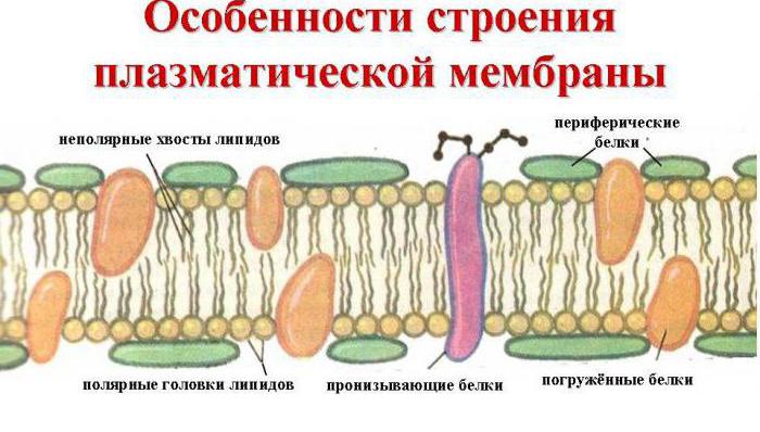
plasma membrane separates the cell and its contents from the environment. In figure 2 you can see: the membrane is formed by two layers of lipids, and protein molecules penetrate the thickness of the membrane.
The main function of the plasma membrane transport. It ensures the supply of nutrients to the cell and the removal of metabolic products from it.
An important property of the membrane is selective permeability, or semi-permeability, allows the cell to interact with the environment: only certain substances enter and leave it. Small molecules of water and some other substances enter the cell by diffusion, partly through the pores in the membrane.
In the cytoplasm, the cell sap of vacuoles plant cell, dissolved sugars, organic acids, salts. Moreover, their concentration in the cell is much higher than in environment. The greater the concentration of these substances in the cell, the more it absorbs water. It is known that water is constantly consumed by the cell, due to which the concentration of cell sap increases and water enters the cell again.
The entry of larger molecules (glucose, amino acids) into the cell is provided by the transport proteins of the membrane, which, by combining with the molecules of the transported substances, carry them through the membrane. Enzymes that break down ATP are involved in this process.
Figure 1. Generalized scheme of the structure of a eukaryotic cell.
(click on image to enlarge image)
Figure 2. The structure of the plasma membrane.
1 - piercing squirrels, 2 - submerged squirrels, 3 - external squirrels 
Figure 3. Scheme of pinocytosis and phagocytosis.
Even larger molecules of proteins and polysaccharides enter the cell by phagocytosis (from the Greek. phagos- devouring and kitos- vessel, cell), and drops of liquid - by pinocytosis (from the Greek. pinot- drink and kitos) (Fig. 3).
Animal cells, unlike plant cells, are surrounded by a soft and flexible "fur coat", formed mainly by polysaccharide molecules, which, by attaching to some membrane proteins and lipids, surround the cell from the outside. The composition of polysaccharides is specific for different tissues, due to which the cells "recognize" each other and connect with each other.
Plant cells do not have such a "fur coat". They have a pore-filled membrane above the plasma membrane. cell wall composed predominantly of cellulose. Threads of the cytoplasm stretch from cell to cell through the pores, connecting the cells to each other. This is how the connection between cells is carried out and the integrity of the body is achieved.
The cell membrane in plants plays the role of a strong skeleton and protects the cell from damage.
Most bacteria and all fungi have a cell membrane, only its chemical composition is different. In fungi, it consists of a chitin-like substance.
The cells of fungi, plants and animals have a similar structure. There are three main parts in a cell: nucleus, cytoplasm and plasma membrane. The plasma membrane is made up of lipids and proteins. It ensures the entry of substances into the cell and their release from the cell. In the cells of plants, fungi, and most bacteria, there is a cell membrane above the plasma membrane. It performs a protective function and plays the role of a skeleton. In plants, the cell wall consists of cellulose, while in fungi, it is made up of a chitin-like substance. Animal cells are covered with polysaccharides that provide contacts between cells of the same tissue.
Do you know that the bulk of the cell is cytoplasm. It consists of water, amino acids, proteins, carbohydrates, ATP, ions of non-organic substances. The cytoplasm contains the nucleus and organelles of the cell. In it, substances move from one part of the cell to another. The cytoplasm ensures the interaction of all organelles. This is where chemical reactions take place.
The entire cytoplasm is permeated with thin protein microtubules, forming cell cytoskeleton due to which it retains its permanent shape. The cell cytoskeleton is flexible, since microtubules are able to change their position, move from one end and shorten from the other. Various substances enter the cell. What happens to them in the cage?
In lysosomes - small rounded membrane vesicles (see Fig. 1), molecules of complex organic substances are broken down into simpler molecules with the help of hydrolytic enzymes. For example, proteins are broken down into amino acids, polysaccharides into monosaccharides, fats into glycerol and fatty acids. For this function, lysosomes are often referred to as the "digestive stations" of the cell.
If the membrane of lysosomes is destroyed, then the enzymes contained in them can digest the cell itself. Therefore, sometimes lysosomes are called "tools for killing the cell."
Enzymatic oxidation of small molecules of amino acids, monosaccharides, fatty acids and alcohols formed in lysosomes to carbon dioxide and water begins in the cytoplasm and ends in other organelles - mitochondria. Mitochondria are rod-shaped, filamentous or spherical organelles, delimited from the cytoplasm by two membranes (Fig. 4). The outer membrane is smooth, while the inner membrane forms folds - cristae which increase its surface. Enzymes involved in the reactions of oxidation of organic substances to carbon dioxide and water. In this case, energy is released, which is stored by the cell in ATP molecules. Therefore, mitochondria are called the "powerhouses" of the cell.
In the cell, organic substances are not only oxidized, but also synthesized. The synthesis of lipids and carbohydrates is carried out on the endoplasmic reticulum - EPS (Fig. 5), and proteins - on ribosomes. What is an EPS? This is a system of tubules and cisterns, the walls of which are formed by a membrane. They permeate the entire cytoplasm. Through the ER channels, substances move to different parts of the cell.
There is a smooth and rough EPS. Carbohydrates and lipids are synthesized on the surface of smooth EPS with the participation of enzymes. The roughness of EPS is given by small rounded bodies located on it - ribosomes(see Fig. 1), which are involved in the synthesis of proteins.
Synthesis of organic substances occurs in plastids found only in plant cells.
Rice. 4. Scheme of the structure of mitochondria.
1.- outer membrane; 2.- inner membrane; 3.- folds of the inner membrane - cristae. 
Rice. 5. Scheme of the structure of rough EPS.
Rice. 6. Scheme of the structure of the chloroplast.
1.- outer membrane; 2.- inner membrane; 3.- internal contents of the chloroplast; 4. - folds of the inner membrane, collected in "stacks" and forming grana. 
In colorless plastids - leucoplasts(from Greek. leukos- white and plastos- created) starch accumulates. Potato tubers are very rich in leukoplasts. Yellow, orange, red color is given to fruits and flowers chromoplasts(from Greek. chrome- color and plastos). They synthesize the pigments involved in photosynthesis, - carotenoids. In plant life, the importance chloroplasts(from Greek. chloros- greenish and plastos) - green plastids. In figure 6, you can see that chloroplasts are covered with two membranes: outer and inner. The inner membrane forms folds; between the folds are bubbles stacked in piles - grains. The grains contain chlorophyll molecules that are involved in photosynthesis. Each chloroplast contains about 50 grains arranged in a checkerboard pattern. This arrangement ensures maximum illumination of each grain.
In the cytoplasm, proteins, lipids, carbohydrates can accumulate in the form of grains, crystals, droplets. These inclusion- spare nutrients, which are consumed by the cell as needed.
In plant cells, part of the reserve nutrients, as well as decay products, accumulate in the cell sap of vacuoles (see Fig. 1). They can account for up to 90% of the volume of a plant cell. Animal cells have temporary vacuoles that occupy no more than 5% of their volume.
Rice. 7. Scheme of the structure of the Golgi complex.
In Figure 7 you see a system of cavities surrounded by a membrane. This golgi complex, which performs various functions in the cell: participates in the accumulation and transportation of substances, their removal from the cell, the formation of lysosomes, cell wall. For example, cellulose molecules enter the cavity of the Golgi complex, which, with the help of bubbles, move to the cell surface and are included in the cell membrane.
Most cells reproduce by dividing. This process involves cell center. It consists of two centrioles surrounded by dense cytoplasm (see Fig. 1). At the beginning of division, centrioles diverge towards the poles of the cell. Protein filaments diverge from them, which are connected to chromosomes and ensure their uniform distribution between two daughter cells.
All organelles of the cell are closely interconnected. For example, protein molecules are synthesized in ribosomes, they are transported through ER channels to different parts cells, and proteins are destroyed in lysosomes. The newly synthesized molecules are used to build cell structures or accumulate in the cytoplasm and vacuoles as reserve nutrients.
The cell is filled with cytoplasm. The cytoplasm contains the nucleus and various organelles: lysosomes, mitochondria, plastids, vacuoles, ER, cell center, Golgi complex. They differ in their structure and functions. All organelles of the cytoplasm interact with each other, ensuring the normal functioning of the cell.
Table 1. STRUCTURE OF THE CELL
| ORGANELLES | STRUCTURE AND PROPERTIES | FUNCTIONS |
| Shell | Consists of cellulose. Surrounds plant cells. Has pores | It gives the cell strength, maintains a certain shape, protects. Is the skeleton of plants |
| outdoor cell membrane | Double membrane cell structure. It consists of a bilipid layer and mosaically interspersed proteins, carbohydrates are located outside. Semi-permeable | Limits the living content of the cells of all organisms. Provides selective permeability, protects, regulates the water-salt balance, exchange with the external environment. |
| Endoplasmic reticulum (ER) | single membrane structure. The system of tubules, tubules, cisterns. Penetrates the entire cytoplasm of the cell. Smooth ER and granular ER with ribosomes | Divides the cell into separate compartments where chemical processes take place. Provides communication and transport of substances in the cell. Protein synthesis takes place on the granular endoplasmic reticulum. On the smooth - lipid synthesis |
| golgi apparatus | single membrane structure. The system of bubbles, tanks, in which the products of synthesis and decay are located | Provides packaging and removal of substances from the cell, forms primary lysosomes |
| Lysosomes | Single-membrane spherical cell structures. Contains hydrolytic enzymes | Provides the breakdown of macromolecular substances, intracellular digestion |
| Ribosomes | Non-membrane mushroom-shaped structures. Composed of small and large subunits | Contained in the nucleus, cytoplasm and on the granular endoplasmic reticulum. Participates in protein biosynthesis. |
| Mitochondria | Two-membrane oblong organelles. The outer membrane is smooth, the inner one forms cristae. filled with matrix. There are mitochondrial DNA, RNA, ribosomes. Semi-autonomous structure | They are the energy stations of the cells. They provide the respiratory process - oxygen oxidation of organic substances. ATP synthesis in progress |
| Plastids Chloroplasts | characteristic of plant cells. Two-membrane, semi-autonomous oblong organelles. Inside they are filled with stroma, in which the grana are located. Grana are formed from membrane structures - thylakoids. Has DNA, RNA, ribosomes | Photosynthesis takes place. On the membranes of the thylakoids, reactions of the light phase take place, in the stroma - of the dark phase. Synthesis of carbohydrates |
| Chromoplasts | Two-membrane spherical organelles. Contains pigments: red, orange, yellow. Formed from chloroplasts | Give color to flowers and fruits. Formed in autumn from chloroplasts, give the leaves a yellow color |
| Leucoplasts | Two-membrane unstained spherical plastids. In the light they can transform into chloroplasts | Stores nutrients in the form of starch grains |
| Cell Center | non-membrane structures. Composed of two centrioles and a centrosphere | Forms a spindle of cell division, participate in division. Cells double after division |
| Vacuole | characteristic of the plant cell. Membrane cavity filled cell sap | Regulates osmotic pressure cells. Accumulates nutrients and waste products of the cell |
| Core | The main component of the cell. Surrounded by a bilayer porous nuclear membrane. filled with karyoplasm. Contains DNA in the form of chromosomes (chromatin) | Regulates all processes in the cell. Provides transmission of hereditary information. The number of chromosomes is constant for each species. Supports DNA replication and RNA synthesis |
| nucleolus | Dark formation in the nucleus, not separated from the karyoplasm | Site of ribosome formation |
| Movement organelles. Cilia. Flagella | Outgrowths of the cytoplasm surrounded by a membrane | Provide cell movement, removal of dust particles (ciliated epithelium) |
The most important role in the vital activity and cell division of fungi, plants and animals belongs to the nucleus and the chromosomes located in it. Most of the cells of these organisms have a single nucleus, but there are also multinucleated cells, such as muscle cells. The nucleus is located in the cytoplasm and has a round or oval shape. It is covered with a shell consisting of two membranes. The nuclear membrane has pores through which the exchange of substances between the nucleus and the cytoplasm takes place. The nucleus is filled with nuclear juice, which contains the nucleoli and chromosomes.
Nucleoli are "workshops for the production" of ribosomes, which are formed from ribosomal RNA formed in the nucleus and proteins synthesized in the cytoplasm.
The main function of the nucleus - the storage and transmission of hereditary information - is associated with chromosomes. Each type of organism has its own set of chromosomes: a certain number, shape and size.
All body cells except sex cells are called somatic(from Greek. catfish- body). The cells of an organism of the same species contain the same set of chromosomes. For example, in humans, each cell of the body contains 46 chromosomes, in the fruit fly Drosophila - 8 chromosomes.
Somatic cells usually have a double set of chromosomes. It is called diploid and denoted 2 n. So, a person has 23 pairs of chromosomes, that is, 2 n= 46. Sex cells contain half as many chromosomes. Is it single or haploid, kit. Person 1 n = 23.
All chromosomes in somatic cells, unlike chromosomes in germ cells, are paired. The chromosomes that make up one pair are identical to each other. Paired chromosomes are called homologous. Chromosomes that belong to different pairs and differ in shape and size are called non-homologous(Fig. 8).
In some species, the number of chromosomes may be the same. For example, in red clover and peas 2 n= 14. However, their chromosomes differ in shape, size, nucleotide composition of DNA molecules.
Rice. 8. A set of chromosomes in Drosophila cells.
Rice. 9. The structure of the chromosome.
To understand the role of chromosomes in the transmission of hereditary information, it is necessary to get acquainted with their structure and chemical composition.
The chromosomes of a non-dividing cell look like long thin threads. Each chromosome before cell division consists of two identical threads - chromatids, which are connected between the constriction fins - (Fig. 9).
Chromosomes are made up of DNA and proteins. Since the nucleotide composition of DNA differs between different types, the composition of chromosomes is unique for each species.
Every cell except bacteria has a nucleus containing nucleoli and chromosomes. Each species is characterized by a specific set of chromosomes: number, shape and size. In the somatic cells of most organisms, the set of chromosomes is diploid, in the sex cells it is haploid. Paired chromosomes are called homologous. Chromosomes are made up of DNA and proteins. DNA molecules provide storage and transmission of hereditary information from cell to cell and from organism to organism.
Having worked through these topics, you should be able to:
- Tell in what cases it is necessary to use a light microscope (structure), a transmission electron microscope.
- Describe the structure of the cell membrane and explain the relationship between the structure of the membrane and its ability to exchange substances between the cell and the environment.
- Define the processes: diffusion, facilitated diffusion, active transport, endocytosis, exocytosis and osmosis. Point out the differences between these processes.
- Name the functions of structures and indicate in which cells (plant, animal or prokaryotic) they are located: nucleus, nuclear membrane, nucleoplasm, chromosomes, plasma membrane, ribosome, mitochondrion, cell wall, chloroplast, vacuole, lysosome, smooth (agranular) and rough (granular) endoplasmic reticulum, cell center, Golgi apparatus, cilium, flagellum, mesosome, pili or fimbriae.
- Name at least three signs by which a plant cell can be distinguished from an animal cell.
- List the major differences between prokaryotic and eukaryotic cells.
Ivanova T.V., Kalinova G.S., Myagkova A.N. "General Biology". Moscow, "Enlightenment", 2000
- Topic 1. "Plasma membrane." §1, §8 pp. 5;20
- Topic 2. "Cage." §8-10 pp. 20-30
- Topic 3. "Prokaryotic cell. Viruses." §11 pp. 31-34
The study of the structure of organisms, as well as plants, animals and humans, is the branch of biology called cytology. Scientists have found that the contents of the cell, which is inside it, is quite complex. It is surrounded by the so-called surface apparatus, which includes the outer cell membrane, supra-membrane structures: glycocalyx and microfilaments, pelicule and microtubules that form its submembrane complex.
In this article, we will study the structure and functions of the outer cell membrane, which is part of the surface apparatus various kinds cells.
What are the functions of the outer cell membrane?
As described earlier, the outer membrane is part of the surface apparatus of each cell, which successfully separates its internal contents and protects cell organelles from adverse conditions external environment. Another function is to ensure the exchange of substances between the cell contents and the tissue fluid, therefore, the outer cell membrane transports molecules and ions entering the cytoplasm, and also helps to remove toxins and excess toxic substances from the cell.

The structure of the cell membrane
Membranes, or plasmalemmas, of different types of cells are very different from each other. Mainly, chemical structure, as well as the relative content of lipids, glycoproteins, proteins in them and, accordingly, the nature of the receptors located in them. Outer which are determined primarily individual composition glycoproteins, takes part in the recognition of environmental stimuli and in the reactions of the cell itself to their actions. Some types of viruses can interact with proteins and glycolipids of cell membranes, as a result of which they penetrate into the cell. Herpes and influenza viruses can use to build their protective shell.
And viruses and bacteria, the so-called bacteriophages, attach to the cell membrane and dissolve it at the point of contact with the help of a special enzyme. Then a molecule of viral DNA passes into the hole formed.
Features of the structure of the plasma membrane of eukaryotes
Recall that the outer cell membrane performs the function of transport, that is, the transfer of substances into and out of it into the external environment. To carry out such a process, a special structure is required. Indeed, the plasmalemma is a constant, universal system of the surface apparatus for all. This is a thin (2-10 Nm), but fairly dense multilayer film that covers the entire cell. Its structure was studied in 1972 by such scientists as D. Singer and G. Nicholson, they also created a fluid-mosaic model of the cell membrane.
The main chemical compounds that form it are ordered molecules of proteins and certain phospholipids, which are interspersed in a liquid lipid environment and resemble a mosaic. Thus, the cell membrane consists of two layers of lipids, the non-polar hydrophobic "tails" of which are located inside the membrane, and the polar hydrophilic heads face the cytoplasm of the cell and the intercellular fluid.
The lipid layer is penetrated by large protein molecules that form hydrophilic pores. It is through them that aqueous solutions glucose and mineral salts. Some protein molecules are located both on the outer and inner surfaces of the plasmalemma. Thus, on the outer cell membrane in the cells of all organisms with nuclei, there are carbohydrate molecules bound by covalent bonds with glycolipids and glycoproteins. The content of carbohydrates in cell membranes ranges from 2 to 10%.

The structure of the plasmalemma of prokaryotic organisms
The outer cell membrane in prokaryotes performs similar functions to the plasma membranes of cells of nuclear organisms, namely: the perception and transmission of information coming from the external environment, the transport of ions and solutions into and out of the cell, and the protection of the cytoplasm from foreign reagents from the outside. It can form mesosomes - structures that arise when the plasmalemma protrudes into the cell. They may contain enzymes involved in the metabolic reactions of prokaryotes, for example, in DNA replication, protein synthesis.
Mesosomes also contain redox enzymes, while photosynthetics contain bacteriochlorophyll (in bacteria) and phycobilin (in cyanobacteria).
The role of outer membranes in intercellular contacts
Continuing to answer the question of what functions the outer cell membrane performs, let us dwell on its role in plant cells. In plant cells, pores are formed in the walls of the outer cell membrane, passing into the cellulose layer. Through them, the exit of the cytoplasm of the cell to the outside is possible; such thin channels are called plasmodesmata.

Thanks to them, the connection between neighboring plant cells is very strong. In human and animal cells, the sites of contact between adjacent cell membranes are called desmosomes. They are characteristic of endothelial and epithelial cells, and are also found in cardiomyocytes.
Auxiliary formations of the plasmalemma
To understand how plant cells differ from animals, it helps to study the structural features of their plasma membranes, which depend on what functions the outer cell membrane performs. Above it in animal cells is a layer of glycocalyx. It is formed by polysaccharide molecules associated with proteins and lipids of the outer cell membrane. Thanks to the glycocalyx, adhesion (sticking) occurs between cells, leading to the formation of tissues, therefore it takes part in the signaling function of the plasmalemma - the recognition of environmental stimuli.
How is the passive transport of certain substances across cell membranes
As mentioned earlier, the outer cell membrane is involved in the process of transporting substances between the cell and the external environment. There are two types of transport through the plasmalemma: passive (diffusion) and active transport. The first includes diffusion, facilitated diffusion and osmosis. The movement of substances along the concentration gradient depends primarily on the mass and size of the molecules passing through the cell membrane. For example, small non-polar molecules easily dissolve in the middle lipid layer of the plasmalemma, move through it and end up in the cytoplasm.

Large molecules of organic substances penetrate into the cytoplasm with the help of special carrier proteins. They are species-specific and, when combined with a particle or ion, passively transport them across the membrane along a concentration gradient (passive transport) without expending energy. This process underlies such property of the plasmalemma as selective permeability. In the process, the energy of ATP molecules is not used, and the cell saves it for other metabolic reactions.
Active transport of chemical compounds across the plasmalemma
Since the outer cell membrane ensures the transfer of molecules and ions from the external environment into the cell and back, it becomes possible to remove the products of dissimilation, which are toxins, to the outside, that is, to the intercellular fluid. occurs against a concentration gradient and requires the use of energy in the form of ATP molecules. It also involves carrier proteins called ATPases, which are also enzymes.

An example of such transport is the sodium-potassium pump (sodium ions pass from the cytoplasm to the external environment, and potassium ions are pumped into the cytoplasm). The epithelial cells of the intestine and kidneys are capable of it. Varieties of this method of transfer are the processes of pinocytosis and phagocytosis. Thus, having studied what functions the outer cell membrane performs, it can be established that heterotrophic protists, as well as cells of higher animal organisms, for example, leukocytes, are capable of pino- and phagocytosis.
Bioelectric processes in cell membranes
It has been established that there is a potential difference between outer surface plasmalemma (it is positively charged) and the parietal layer of the cytoplasm, negatively charged. It was called the resting potential, and it is inherent in all living cells. And the nervous tissue has not only a resting potential, but is also capable of conducting weak biocurrents, which is called the process of excitation. The outer membranes of nerve cells-neurons, receiving irritation from receptors, begin to change charges: sodium ions massively enter the cell and the surface of the plasmalemma becomes electronegative. And the parietal layer of the cytoplasm, due to an excess of cations, receives a positive charge. This explains why the outer cell membrane of the neuron is recharged, which causes the conduction of nerve impulses that underlie the excitation process.
By functional features The cell membrane can be divided into 9 functions it performs.
Cell membrane functions:
1. Transport. Produces the transport of substances from cell to cell;
2. Barrier. It has selective permeability, provides the necessary metabolism;
3. Receptor. Some proteins found in the membrane are receptors;
4. Mechanical. Ensures the autonomy of the cell and its mechanical structures;
5. Matrix. Provides optimal interaction and orientation of matrix proteins;
6. Energy. In membranes, energy transfer systems operate during cellular respiration in mitochondria;
7. Enzymatic. Membrane proteins are sometimes enzymes. For example, intestinal cell membranes;
8. Marking. There are antigens (glycoproteins) on the membrane that make it possible to identify the cell;
9. Generating. Carries out the generation and conduction of biopotentials.
You can see what the cell membrane looks like using the example of the structure of an animal cell or a plant cell.
The figure shows the structure of the cell membrane.
The components of the cell membrane include various proteins of the cell membrane (globular, peripheral, surface), as well as lipids of the cell membrane (glycolipid, phospholipid). Carbohydrates, cholesterol, glycoprotein and protein alpha helix are also present in the structure of the cell membrane.
Cell membrane composition
The main components of the cell membrane are:
1. Proteins - responsible for the various properties of the membrane;
2. Lipids three types(phospholipids, glycolipids and cholesterol) responsible for membrane rigidity.
Cell membrane proteins:
1. Globular protein;
2. Surface protein;
3. Peripheral protein.
The main purpose of the cell membrane
The main purpose of the cell membrane:
1. Regulate the exchange between the cell and the environment;
2. Separate the contents of any cell from the external environment, thereby ensuring its integrity;
3. Intracellular membranes divide the cell into specialized closed compartments - organelles or compartments, in which certain environmental conditions are maintained.
Cell membrane structure
The structure of the cell membrane is a two-dimensional solution of globular integral proteins dissolved in a liquid phospholipid matrix. This model of membrane structure was proposed by two scientists Nicholson and Singer in 1972. Thus, the basis of the membranes is a bimolecular lipid layer, with an ordered arrangement of molecules, which you could see on.
cell membrane- this is a cell membrane that performs the following functions: separation of the contents of the cell and the external environment, selective transport of substances (exchange with the external environment for the cell), the site of some biochemical reactions, the integration of cells into tissues and reception.
Cell membranes are divided into plasma (intracellular) and outer. The main property of any membrane is semi-permeability, that is, the ability to pass only certain substances. This allows selective exchange between the cell and the external environment, or exchange between compartments of the cell.
Plasma membranes are lipoprotein structures. Lipids spontaneously form a bilayer (double layer), and membrane proteins "swim" in it. There are several thousand different proteins in the membranes: structural, carriers, enzymes, etc. Between the protein molecules there are pores through which hydrophilic substances pass (the lipid bilayer prevents their direct penetration into the cell). Glycosyl groups (monosaccharides and polysaccharides) are attached to some molecules on the membrane surface, which are involved in the process of cell recognition during tissue formation.
The membranes differ in their thickness, usually between 5 and 10 nm. The thickness is determined by the size of the amphiphilic lipid molecule and is 5.3 nm. A further increase in the thickness of the membrane is due to the size of the membrane protein complexes. Depending on external conditions (cholesterol is the regulator), the structure of the bilayer can change so that it becomes more dense or liquid - the speed of movement of substances along the membranes depends on this.
Cell membranes include: plasmalemma, karyolemma, membranes of the endoplasmic reticulum, Golgi apparatus, lysosomes, peroxisomes, mitochondria, inclusions, etc.
Lipids are insoluble in water (hydrophobicity), but readily soluble in organic solvents and fats (lipophilicity). The composition of lipids in different membranes is not the same. For example, the plasma membrane contains a lot of cholesterol. Of the lipids in the membrane, the most common are phospholipids (glycerophosphatides), sphingomyelins (sphingolipids), glycolipids, and cholesterol.
Phospholipids, sphingomyelins, glycolipids consist of two functionally various parts: hydrophobic non-polar, which does not carry charges - "tails" consisting of fatty acids, and hydrophilic, containing charged polar "heads" - alcohol groups (for example, glycerin).
The hydrophobic part of the molecule usually consists of two fatty acids. One of the acids is limiting, and the second is unsaturated. This determines the ability of lipids to spontaneously form two-layer (bilipid) membrane structures. Membrane lipids perform the following functions: barrier, transport, protein microenvironment, electrical resistance membranes.
Membranes differ from each other by a set of protein molecules. Many membrane proteins consist of regions rich in polar (charge-carrying) amino acids and regions with non-polar amino acids (glycine, alanine, valine, leucine). Such proteins in the lipid layers of membranes are located in such a way that their non-polar regions are, as it were, immersed in the "fat" part of the membrane, where the hydrophobic regions of lipids are located. The polar (hydrophilic) part of these proteins interacts with the lipid heads and is turned towards the aqueous phase.
Biological membranes have common properties:
membranes are closed systems that do not allow the contents of the cell and its compartments to mix. Violation of the integrity of the membrane can lead to cell death;
superficial (planar, lateral) mobility. In membranes, there is a continuous movement of substances over the surface;
membrane asymmetry. The structure of the outer and surface layers is chemically, structurally and functionally heterogeneous.
The cell membrane is the structure that covers the outside of the cell. It is also called cytolemma or plasmolemma.
This formation is built from a bilipid layer (bilayer) with proteins embedded in it. The carbohydrates that make up the plasmalemma are in a bound state.
The distribution of the main components of the plasmalemma is as follows: more than half of the chemical composition falls on proteins, a quarter is occupied by phospholipids, and a tenth is cholesterol.
Cell membrane and its types
The cell membrane is a thin film, which is based on layers of lipoproteins and proteins.

By localization, membrane organelles are distinguished, which have some features in plant and animal cells:
- mitochondria;
- core;
- endoplasmic reticulum;
- Golgi complex;
- lysosomes;
- chloroplasts (in plant cells).
There is also an inner and outer (plasmolemma) cell membrane.
The structure of the cell membrane
The cell membrane contains carbohydrates that cover it in the form of a glycocalyx. This is a supra-membrane structure that performs a barrier function. The proteins located here are in a free state. Unbound proteins are involved in enzymatic reactions, providing extracellular breakdown of substances.

Proteins of the cytoplasmic membrane are represented by glycoproteins. According to the chemical composition, proteins are isolated that are completely included in the lipid layer (throughout) - integral proteins. Also peripheral, not reaching one of the surfaces of the plasmalemma.

The former function as receptors, binding to neurotransmitters, hormones, and other substances. Insertion proteins are necessary for the construction of ion channels through which ions and hydrophilic substrates are transported. The latter are enzymes that catalyze intracellular reactions.
Basic properties of the plasma membrane
The lipid bilayer prevents the penetration of water. Lipids are hydrophobic compounds present in the cell as phospholipids. The phosphate group is turned outward and consists of two layers: the outer one, directed to the extracellular environment, and the inner one, delimiting the intracellular contents.

Water-soluble areas are called hydrophilic heads. The fatty acid sites are directed inside the cell, in the form of hydrophobic tails. The hydrophobic part interacts with neighboring lipids, which ensures their attachment to each other. The double layer has selective permeability in different areas.
So, in the middle, the membrane is impermeable to glucose and urea, hydrophobic substances pass freely here: carbon dioxide, oxygen, alcohol. Cholesterol is important, the content of the latter determines the viscosity of the plasma membrane.
Functions of the outer membrane of the cell
The characteristics of the functions are briefly listed in the table:
| Membrane function | Description |
| barrier role | The plasmalemma performs a protective function, protecting the contents of the cell from the effects of foreign agents. Due to the special organization of proteins, lipids, carbohydrates, the semi-permeability of the plasma membrane is ensured. |
| Receptor function | Biologically activated through the cell membrane active substances in the process of binding to receptors. Thus, immune reactions are mediated through the recognition of foreign agents by the receptor apparatus of cells localized on the cell membrane. |
| transport function | The presence of pores in the plasmalemma allows you to regulate the flow of substances into the cell. The transfer process proceeds passively (without energy consumption) for compounds with low molecular weight. Active transfer is associated with the expenditure of energy released during the breakdown of adenosine triphosphate (ATP). This method has a place for the transfer of organic compounds. |
| Participation in the processes of digestion | Substances are deposited on the cell membrane (sorption). Receptors bind to the substrate, moving it inside the cell. A vesicle is formed, lying freely inside the cell. Merging, such vesicles form lysosomes with hydrolytic enzymes. |
| Enzymatic function | Enzymes, necessary components of intracellular digestion. Reactions that require the participation of catalysts proceed with the participation of enzymes. |
What is the importance of the cell membrane
The cell membrane is involved in maintaining homeostasis due to the high selectivity of substances entering and leaving the cell (in biology this is called selective permeability).

Outgrowths of the plasmolemma divide the cell into compartments (compartments) responsible for performing certain functions. Specifically arranged membranes, corresponding to the fluid-mosaic scheme, ensure the integrity of the cell.