For any mammal, the liver is incomparably important body, any damage to this organ is fraught with serious consequences. In fact, the liver is a unique organ whose ability to regenerate is simply amazing. Even with damage of more than seventy percent, this gland is still capable of almost complete recovery.
It is directly involved in the digestion process, cleanses the blood of toxins and harmful substances, and participates in the production and outflow of bile. However, disruptions occur during the normal outflow of bile, resulting in the formation of stagnant processes in the gallbladder. This phenomenon is called cholestasis. It poses a serious danger if the functioning of the gallbladder is not normalized in a timely manner.
Cholestasis is stagnant processes in the gallbladder.
Causes and diagnosis
The development of cholemia is dangerous for a dog.
Bile takes part in digestion and also helps remove toxins and harmful compounds from the body. This process works as follows : toxins or harmful substances that get inside digestive system, react with bile acids, as a result of which they stop breaking down into small particles and are excreted along with bile and feces.
Blockage of the bile ducts disrupts the functioning of the bladder, and it becomes impossible for bile to ensure the normal process of binding and removing toxins. Due to obstruction, pressure is created and the secretion gets inside circulatory system, which can lead to the development of cholemia, which is characterized by a severe course and an increased risk of death.
Provocateurs
The main provocateurs of cholestasis:
- stones;
- opisthorchiasis;
- leptospirosis;
- hepatitis;
- hepatosis;
- consumption of low-quality food;
- obesity;
- peritoneal injury.
Poor quality food can cause cholestasis.
Diseases
Inflammation of the pancreas provokes obstruction of the duct in the duodenum, which affects the gallbladder and liver.
Inflammation affects liver function.
Risk group
Older and elderly individuals are most susceptible to blockage due to the fact that by this age, the presence of stones or sand in the bladder is most often noted. But helminths - trematodes - can also clog the ducts, resulting in inflammatory processes and degenerative changes.
Older dogs are at risk.
Leptospirosis
Toxins in the blood affect the development of hepatitis.
Leptospirosis is expressed primarily by a large release of toxins into the blood. It is toxins that contribute to the development of hepatitis or hepatosis. During these pathologies, the parenchyma contracts, the tissue becomes coarser and causes obstruction. As a result of injuries to the peritoneum, adhesions may form on the liver tissue, which compact the parenchyma and compress the ducts.
Clinical signs
During the period of illness, the dog refuses food.
The symptoms of cholestasis do not have a narrow specificity due to the fact that the disease affects the entire body of the animal.
- initial stage characterized by extensive jaundice . The sclera of the eyes turn yellow, the tongue is covered with a whitish coating, and a rich yellow color is noted on the surface of the pharynx.
- The pet begins to eat often and a lot . This fact is caused by digestive disorders, as a result of which food begins to be poorly absorbed. The progression of the disease will be expressed in complete apathy and refusal to feed. Then problems with blood clotting begin. Even minor injuries do not heal for a long time and bleed.
- The pet is gradually losing weight, the stool is white, almost discolored . This is due to the absence of stercobilin. Since bile does not enter the intestinal lumens, there is no stercobilin. Urine darkens and becomes bright Orange color.
- The fact that cholemia has begun will be indicated by lethargy or coma . The presence of such a condition indicates negligible chances of recovery.
Diagnostics
A blood test will be required for diagnosis.
- The diagnosis is made based on medical history, information about nutrition, and previous illnesses.
- Laboratory testing of blood and urine is carried out.
- The blood is examined by biochemical analysis for the level of enzymes and bilirubin.
- A fecal examination is carried out.
- And examination is also carried out using x-rays and ultrasound.
Treatment
The approach to treatment should be purely individual and aimed at eliminating the underlying cause and associated complications.
Light soup should be included in your dog's diet.
- Dehydration is eliminated by infusion therapy - infusion physiological solutions . Blood clotting problems are solved through blood transfusions.
- If there is a need for surgery, it is recommended to take a course of antibiotics before performing it. in order to prevent the risk of secondary infectious pathologies. Conservative treatment also suggests the possibility of prescribing drugs that can thin out bile.
- The presence of an inflammatory process requires the prescription of anti-inflammatory drugs . Applicable symptomatic treatment. In case of intoxication, it may be present; in this case, it is permissible to use antiemetic medications. With strong pain syndrome use antispasmodics and painkillers.
- If the disease is caused by helminthiasis, anthelmintic drugs are used. . It is worth noting that medications should only be used that are aimed at eliminating trematodes directly, since all other medications will not bring the desired effect.
- Dietary nutrition plays an important role in treatment . A fasting diet is recommended for the first day. Next, depending on the doctor’s verdict, the dog is fed light soups or broths. Food should not contain fat or difficult-to-digest foods.
Video about liver diseases in dogs
D.E. Mitrushkin. Vet clinic“Biocontrol”, Clinic of Experimental Therapy, State Scientific Research Center named after. N.N. Blokhin RAMS
Keywords: bile, gallstones, cholelithiasis, bile duct, cholelithiasis, gallbladder, cholecystolithiasis, liver, hepatic ducts
Abbreviations: ALT– alanine aminotransferase, CT – CT scan, RMJ- mammary cancer, Ultrasound- ultrasonography, ShchV- alkaline phosphatase, ECG– electrocardiogram
Introduction
Bile is a secretion that is constantly produced in the liver and enters the intrahepatic bile ducts, which, merging, form the right and left extrahepatic ducts, located near the porta hepatis. These ducts connect and form a common hepatic duct, passing into the common bile duct, which flows into the duodenum. Bile enters the gallbladder (bile storage reservoir) from the common bile duct through the cystic duct and from it, as needed, is released back into the common bile duct.
Cholelithiasis(cholelithiasis, from the Greek chole - bile and lithos - stone) is a metabolic disease of the hepatobiliary system, characterized by the formation gallstones V gallbladder(cholecystolithiasis), less often - in the intrahepatic bile ducts (hepatic cholelithiasis) or the common bile duct (choledocholithiasis).
Cholelithiasis is a rare disease in dogs and cats. Even its presence in animals is often asymptomatic and before the introduction of ultrasound into veterinary practice, it was often detected only during autopsy. The main cause of gallstone formation is a violation functional state liver (due to hepatitis, hepatosis or cirrhosis) and, in connection with this, a change in the physicochemical properties of bile (dyscholia). The formation of gallstones is associated with impaired metabolism of the main components of bile - cholesterol, phospholipids (lecithin, etc.), bile acids, bile pigments (bilirubin, biliverdin) and inorganic salts. Cholesterol in the bile of healthy animals is retained in a dissolved state due to cholesterol-retaining factors (bile acids and phospholipids). With the above liver pathologies, the amount of these two cholesterol-retaining factors falls below a critical level and favorable conditions are created for the formation of colloidal solutions of cholesterol with the formation of thick heterogeneous bile (the initial or pre-stone stage of cholelithiasis) with further crystallization of cholesterol and the formation of stones. The formation of these stones may also be associated with increased secretion of cholesterol.
Predisposing factors for cholelithiasis include the presence of pathology (stenosis, tumor, adhesions, atrophy, dyskinesia, hypertrophy, etc.) biliary tract or gallbladder, leading to stagnation of bile (cholestasis) in both the liver and gallbladder. The entry of microorganisms or trematodes into stagnant bile creates the most favorable conditions for cholelithiasis, because in this case, mucus and dead epithelial cells are added to the stagnant bile. Obesity and obesity are also considered risk factors for stone formation. hemolytic anemia, irrational feeding, insufficient exercise, hereditary factors, etc.
Stones in the intrahepatic bile ducts in animals and humans are much less common than in the gallbladder or extrahepatic bile ducts. This is due to the fact that bile in the gallbladder is the most concentrated and the tendency to sediment appears in it first. In addition, bile in the intra- and extrahepatic bile ducts is constantly moving (flowing), and in the gallbladder it is at rest for a certain time.
Gallstones differ sharply in composition and appearance. In their chemical composition mainly includes three substances - cholesterol, calcium bilirubinate and calcium carbonate.
There are three main types of gallstones:
- cholesterol stones. They consist mainly of cholesterol. As a rule, solitary, yellowish-white in color, soft consistency. If the stones are in the bubble for a long time, they may become encrusted with calcium salts and become combined;
- pigment stones. They consist of calcium bilirubinate, cholesterol and bile acids. Most common in dogs. They are always multiple, black with a shiny surface, faceted in appearance. Most often of a loose consistency. Their appearance is associated with an excess of bile pigments, formed, in particular, in diseases accompanied by hemolysis;
- combined (cholesterol-pigment-calcareous) stones. They contain all three components in varying proportions, and the color and consistency of the stones depend on the predominance of one of them. Cholesterol gives a yellowish tint, calcium bilirubinate gives a black-brown tint, and calcium carbonate gives a white tint. Combination stones are always multiple. Their surface is usually smooth, irregular in shape, less often rounded. If there are few stones and they are large enough, articular surfaces form between them - slightly concave on one stone and correspondingly convex on the adjacent one.
In the presence of any stones, there is a likelihood of developing acute and chronic calculous cholecystitis, although with cholesterol and pigment stones, inflammatory processes of the gallbladder are rare.
Small gallstones in chronic cholecystitis with dilation of the cystic duct can migrate from the bladder and, depending on their size, slip into the duodenum, get stuck in the cystic duct, common bile duct, or ascend into the hepatic ducts. The stone can act as a valve, obstructing the flow of bile into the duodenum or gallbladder. In the latter case, the bladder first collapses, then the absorption of bile and swelling of the organ wall. If the outflow of bile from the gallbladder is disrupted, the bladder becomes overfilled with bile, blood circulation in it is disrupted as a result of compression of the feeding vessels, and destructive changes develop in the wall of the organ. If there are stones in the ducts, stones are constantly found in the bladder or liver. Isolated choledocholithiasis apparently does not exist. If stones are found in the ducts and there are no stones in the bladder or liver, it can be assumed that all the stones have passed into the ducts.
Streamlined bile duct stone may not cause clinical symptoms And morphological changes in the ducts, gall bladder and liver. But more often, the presence of a stone in the duct leads to serious consequences. First of all, the development of mechanical (cholestatic, obstructive, subhepatic) jaundice is possible. With incomplete obstruction, there may be intermittent jaundice, expansion of the overlying parts of the bile ducts and hypertrophy of their walls. Stagnation of bile also extends to the intrahepatic bile ducts; with prolonged obstruction, secondary biliary cirrhosis and cholangitis develop. Complete obstruction of the bile ducts causes the development of the symptom complex of acute obstructive jaundice, which is characterized by cholemic syndrome and acholia syndrome.
Cholemic syndrome develops due to the entry of the main components of bile into the systemic circulation against the background of cholestasis (leading to increased pressure in the overlying bile ducts, stretching and increased permeability of bile capillaries or their rupture). Clinical manifestations of cholemia are jaundice (deposition of bilirubin gives the mucous membranes and sclera a characteristic icteric color), anorexia, vomiting, dehydration, pain on palpation of the right hypochondrium (due to spasm of the smooth muscles of the gallbladder and bile ducts), bradycardia and itchy skin(due to increased levels of bile acids in the blood). At biochemical analysis blood are determined high levels total bilirubin, ALT, alkaline phosphatase and cholesterol; when studying a coagulogram - a decrease in the rate of blood clotting; A clinical blood test may reveal moderate or severe leukocytosis (with a shift to the left) or anemia.
Stopping the flow of bile into the intestines (acholia syndrome) leads to discoloration of feces, steatorrhea, dysbacteriosis and intestinal autointoxication.
Description clinical cases cholelithiasis
During the first half of 2009, three cases of cholelithiasis were reported among patients at the Biocontrol clinic. In three animals (a Cornish Rex cat, a miniature poodle and a Yorkshire terrier), the owners' complaints during initial treatment were associated with other pathologies (pyometra, convulsive syndrome, breast cancer and cough), and upon examination and further treatment of the underlying disease, a concomitant disease was identified as cholelithiasis . In all three cases, the diagnosis was confirmed by pathological examination.
Clinical case 1. An 11-year-old Cornish Rex cat was admitted to the clinic with complaints from the owners about purulent discharge from the loop, periodic vomiting of bile and anorexia during the day. An animal diagnosed with pyometra underwent supravaginal ovariohysterectomy. 12 days after the operation, the animal was admitted in extremely serious condition. Body temperature 32.0 O C, pale mucous membranes, lethargy, anorexia, vomiting bile, convulsions, hard breathing sounds on auscultation.
Clinical blood test: leukocytes – 32.8 thousand/µl; red blood cells – 7.28 million/µl; hemoglobin – 101 g/l, hematocrit – 35.7%; platelets – 58 thousand/µl.
Biochemical blood test: glucose – 1.98 mmol/l; bilirubin - 9.9 µmol/l; ALT - 599 U/l; AST – 237 U/l; urea - 10.4 mmol/l; creatinine - 190 µmol/l; pancreatic amylase – 1734 U/l.
During an ultrasound, the animal was found to have many hyperechoic inclusions in the liver and gall bladder. On the same day, the cat underwent an exploratory laparotomy, during which the animal underwent cholecystotomy with removal of stones. During the operation, the animal suffered cardiac arrest.
A pathological examination revealed severe swelling, acute inflammation liver (Fig. 1); hepatic cholelithiasis (Fig. 2); interstitial nephroso-nephritis; severe fibrosis of the pancreas; myocardial edema; pulmonary atelectasis.
Rice. 1. Microphoto. Histological section of the liver. Severe swelling, leukocyte infiltration. Hematoxylin and eosin staining, vol. ×40, approx. ×10

A

B

IN

G
Rice. 2. Macro photo. Hepatic cholelithiasis. Many combined stones of yellow and dark green color in the intrahepatic bile ducts. The stones are easily “squeezed out” by lightly squeezing the liver, which has a dense consistency (Fig. A, B, C). When cutting a stone, the layered structure and color change are clearly visible (shown by an arrow in Fig. D)
Clinical case 2. A dog, a miniature poodle breed, female, 17 years old, was admitted to the clinic with the owner’s complaints of seizures for 24 hours. On clinical examination - general state heavy animal. Body temperature 40 O C. Mucous membranes cyanotic pink. The ECG shows single extrasystoles. Pain on palpation of the abdominal wall. Ultrasound revealed parietal hyperechoic round formations with a diameter of up to 0.3 cm in the cavity of the gallbladder, diffuse changes liver and signs of chronic nephritis.
Clinical blood test: leukocytes – 23.5 thousand/µl; erythrocytes – 6.08 million/µl; hemoglobin – 128 g/l; hematocrit – 40.2%; platelets – 752 thousand/µl.
Biochemical blood test: glucose – 2.0 mmol/l; bilirubin – 0.9 µmol/l; ALT – 50 U/l; AST – 182 U/l; urea – 7.9 mmol/l; creatinine – 78 µmol/l; pancreatic amylase – 559 U/l.
The animal was admitted to the clinic's inpatient unit, where it received infusion therapy. The dog experienced epileptiform seizures for 15-30 seconds every 2 hours. On the 4th day of treatment, due to the extremely serious condition of the animal, at the request of the owners, it was euthanized.
A pathological and anatomical study revealed: massive intracerebral hemorrhage in the right frontal lobe of the brain, moderate internal hydrocephalus (Fig. 3); edema, plethora, fatty degeneration, perivascular sclerosis of the liver (Fig. 4); cholecystolithiasis (Fig. 5); macronodular cirrhosis of the body and head of the pancreas; bilateral large-focal nephroso-nephritis with cirrhosis and polycystic disease; myocarditis; a combination of emphysema, pneumosclerosis and congestive pulmonary congestion; hemosiderosis of the spleen.

Rice. 3. Macro photo. Frontal section of the brain. Massive intracerebral hemorrhage in the right parietal lobe of the brain (shown by arrow), moderate hydrocephalus

Rice. 4. Microphoto. Histological section of the liver. Edema, plethora, fatty degeneration, perivascular sclerosis of the liver. Hematoxylin and eosin staining, vol. ×40, approx. ×10


Rice. 5. Macro photo. Cholecystolithiasis. Multiple pigment stones with a diameter of up to 4 mm (shown by an arrow in Fig. A) in the unchanged gallbladder, loose consistency, crumbling under moderate compression (Fig. B).
Clinical case 3. A dog, Yorkshire terrier breed, female, age 5 years, was admitted to the clinic with complaints from the owners of a neoplasm of the mammary gland (noticed 6 months ago) and a cough for 3 months, worsening after physical activity. Clinical examination revealed: stage II breast cancer, cyanotic mucous membranes, the tracheal reflex is sharply positive, clear and vesicular breathing. Ultrasound reveals hyperechoic contents in the lumen of the gallbladder (Fig. 6), bilateral nephrolithiasis, diffuse changes in the liver. X-ray examination: enlargement of the right heart, collapse of the trachea.
A 
B 
Rice. 6. Ultrascanogram of the gallbladder in transverse (a) and longitudinal (b) sections. Hyperechoic contents in the lumen of the gallbladder (shown by an arrow)
The animal was treated in the clinic for 4 months: completion of the course radiation therapy, followed by regional mastectomy and three courses of chemotherapy. The condition worsened after the end of chemotherapy: persistent pancytopenia, epileptiform seizures, gastrointestinal bleeding.
Due to the extremely serious condition of the animal, at the request of the owners, it was euthanized.
Pathological and anatomical diagnosis: severe internal hydrocephalus (Fig. 7), fatty liver (Fig. 8, 9), cholecystolithiasis (Fig. 10), thrombosis of the right ventricular cavity, grade III tracheal collapse, bilateral nephrolithiasis, pinpoint hemorrhages in the thin and large parts of the intestine.

Rice. 7. Macro photo. Segmental section of the brain. Dilatation of the ventricles of the brain

Rice. 8. Macro photo. Fatty liver degeneration. A yellowish-colored organ in section

Rice. 9. Microphoto. Fatty liver degeneration. Numerous fat droplets in the cytoplasm of hepatocytes, creating a fine mesh pattern. Hematoxylin and eosin staining, vol. ×40, approx. ×10
A 
B 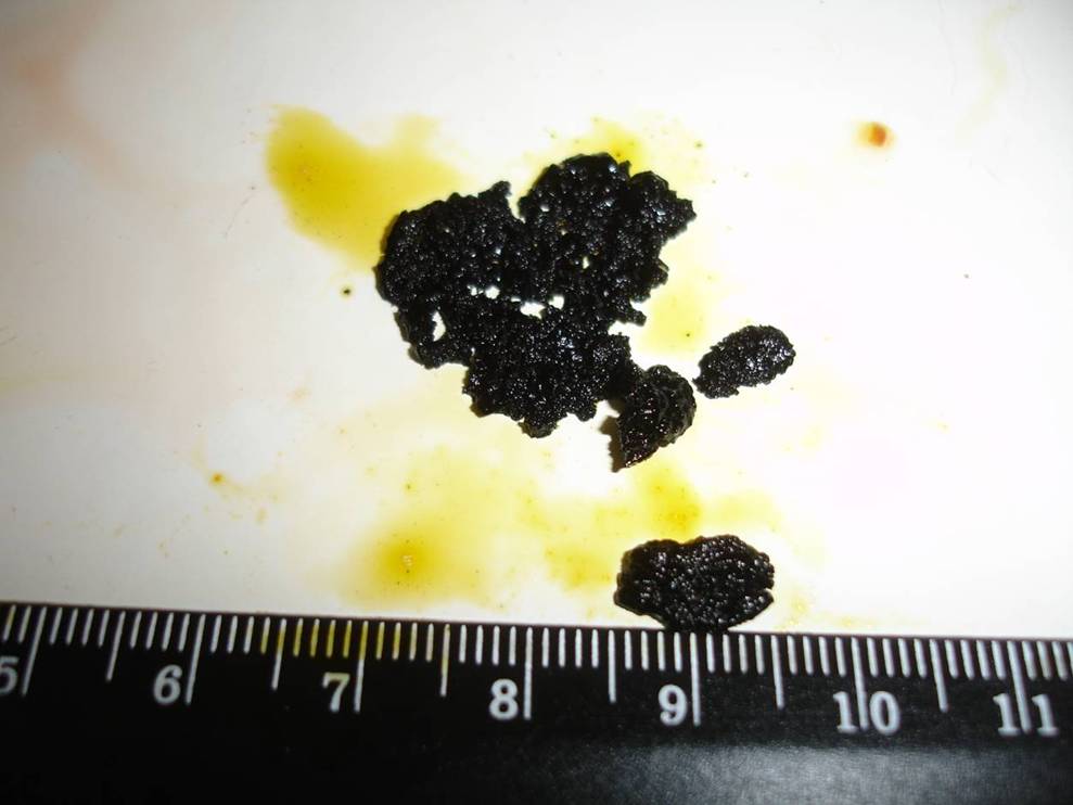
Rice. 10. Cholecystolithiasis. Pigment stones of the gallbladder in Fig. And are shown by arrows. The stones have a loose consistency and crumble under moderate pressure (Fig. B)
Discussion and conclusions
Cholelithiasis - rare disease dogs and cats, most often asymptomatic. In most cases, the pathology is concomitant with the development of the underlying disease. Only in one of the three clinical cases we described can we say that cholelithiasis was the main disease of the animal.
The main etiological factor of the pathology, both according to the veterinary literature and according to the above clinical cases, is liver pathology. Among the animals with cholelithiasis we studied, severe liver damage was confirmed (histologically) in all three cases. It represented like fatty degeneration, and hepatitis or perivascular cirrhosis.
Severe kidney pathologies (intermediate nephroso-nephritis, nephroso-nephritis with cirrhosis and polycystic disease, and nephrolithiasis, identified in each special case) and pancreas (fibrosis or cirrhosis of the organ, which we established in two out of three cases) may indicate a possible correlation of cholelithiasis with failure of these organs. It should be noted that in all three cases the disease was detected in females, and according to numerous medical literature data, the disease has a gender predisposition (in women, stones are 3-4 times more common).
Changes in hematological and biochemical parameters that appear during obstruction of the bile ducts with stones, leading to cholestasis, are more often manifested by leukocytosis and increased liver parameters.
The main instrumental method for studying the disease is ultrasound or CT, which makes it possible to identify the presence of stones, their size, quantity, location and, to a certain extent, structure.
In the presence of stones in the gallbladder, the main treatment method is cholecystotomy with removal of stones, and in case of serious pathology of the gallbladder, cholecystectomy. Restoring the outflow of bile by applying various anastomoses between the biliary system and duodenum(cholecystoduodenostomy).
Bibliography
1. Kaliteevsky P.F. Macroscopic differential diagnosis of pathological processes. Moscow, “Miklos”, 1993. p. 221-226.
2. Lyutinsky S.I. Pathological physiology of animals. M.: KolosS, 2005. p. 351-352.
3. Paltsev M.A. Pathology: course of lectures. Volume 2. M., “Medicine”, 2007. p. 287-289.
4. Savoysky A.G., Baimatov V.N., Meshkov V.M. Pathological physiology. M.: KolosS, 2008, p. 409-411.
5. Buote N.J. The surgical treatment of cholelithiasis in cats: a study of nine cases. J Am Anim Hosp Assoc. 2002, 38(3): 290-6.
9. Fahie M.A., Martin R.A. Extrahepatic biliary tract obstruction: A retrospective study of 45 cases (1983–1993). J Am Anim Hosp Assoc. 1995, 31: 478–481.
10. Heidner G.L., Campbell K.L. Cholelithiasis in a cat. J Am Vet Med Assoc. 1985, 15; 186(2): 176-7.
11. Kirpensteijn J., Fingland R.B., Ulrich T., Sikkema D.A., Allen S.W. Cholelithiasis in dogs: 29 cases. J Am Vet Med Assoc. 1993, 202: 1137–1142.
12. Neer M.T. A review of disorders of the gallbladder and extrahepatic biliary tract in the dog and cat. J Vet Intern Med 1992; 6: 186–192.
13. Rege R.V., Prystowsky J.B. Inflammatory properties of bile from dogs with pigment gallstones. Am J Surg. 1996; 171(1):197–201.
14. Strombeck D.R., Guilford W.G. Small Animal Gastroenterology, 2nd ed. Davis, California: Stonegate Publ, 1990, p. 686–689.
15. Wolf A.M. Obstructive jaundice in a cat resulting from choledocholithiasis. J Am Vet Med Assoc. 1984, 1; 185(1): 85-7.
Summary
D.E. Mitrushkin. Cholelithiasis in dogs and cats. The frequency of cholelithiasis in dogs and cats is rare and often is subclinical, but can result in clinical signs such as icterus, anorexia, vomiting, dehydration, abdominal pain, bradycardia, skin itch and acholia. Values for bilirubin total, alanine aminotransferase, alkaline phosphatase, cholesterol and white blood cells are higher than normal at obstructive cholelithiasis. In this article three cases of cholelithiasis were presented. In all three cases we expressed found histopathological changes of liver, pancreas and kidneys. It is suggested that the pathology of these organs might have contributed to gallstones formation. The main method of treatment of disease is cholecystotomy, however, cholecystectomy is indicated if damage of gall bladder is severe.
Unfortunately, many owners are faced with a disease such as cholecystitis in dogs. This pathology occurs when the normal function of the bile ducts is disrupted, which leads to the development of inflammation in the gallbladder.
Due to poor outflow, bile becomes denser and more caustic. In this case, there is a danger of injury to the walls of the bladder, which leads to the formation of ulcers on it. If the disease is not treated, bile will leak out into the abdominal cavity. This threatens to happen without urgent surgery, otherwise the pet will die.
There are several factors that provoke the development of cholecystitis.
Cholecystitis can develop in dogs that are frequently fed smoked meats.
These include:
An unbalanced diet is considered a provocateur of many diseases, including cholecystitis. The correct structure of the gastric mucosa is maintained due to the presence of a sufficient amount of carotene in the body. It is he who is responsible for the restoration of failed cells. Therefore, a lack of vitamin A in a pet’s diet negatively affects its health.
 An unbalanced diet is the main cause of cholecystitis in dogs.
 How to recognize the disease
It is almost impossible to find out about the presence of the disease until the first symptoms appear. If your dog begins to behave strangely, you should watch it. When the first clinical signs cholecystitis, the pet should be immediately referred to a veterinarian for further diagnostics.
Symptoms of cholecystitis
The chronic form differs in its manifestation from the acute one. It is characterized by slight deviations in the behavior of the animal.
It can be detected by the following symptoms:
- decreased activity, the animal sleeps a lot;
- lack of appetite, refusal of water;
- orange urine, light-colored feces, as bilirubin in the blood has increased sharply;
- intestinal disorders;
- severe hair loss ();
- The main position of the pet is on its stomach, with its back arched.
The acute form of cholecystitis occurs if measures are not taken in time and the disease starts.
When the disease worsens, there is real threat pet's life. When the bile ducts are completely blocked, the bladder stretches and bursts, causing peritonitis.
 With cholecystitis, dogs often develop jaundice.
 How is the disease diagnosed?
At the first visit, the doctor interviews the owner, finds out the symptoms and examines the pet. He assesses the condition of the skin, mucous membranes, and fur. Probes the abdominal area.
If there is a suspicion of cholecystitis, then the veterinarian prescribes the following diagnostic procedures:
- Ultrasound to identify pathologies that cause inflammation of the digestive system and gallbladder;
- x-rays to determine the presence or absence of stones;
- general blood test - identifying the content of leukocytes, increased level which indicate inflammation;
- urine and stool analysis to assess bilirubin levels;
- liver biopsy gives an idea of the viscosity of bile and its stagnation;
- Bile analysis allows you to determine the causative agent of the infection;
- diagnostic laparotomy - performed if there is a possibility of peritonitis.
Drug treatment
When starting treatment, first of all you should relieve dangerous symptoms and save your pet from dehydration. Glucose solution and calcium gluconate will help alleviate the general condition. Having normalized it, you can begin to directly eliminate the causes of the disease.
 Treatment of cholecystitis should only be prescribed by a veterinarian after examining the dog.
 Important. The doctor makes prescriptions and selects treatment depending on each specific case. In this case, the degree of neglect of the disease, its form and the cause of its occurrence play an important role. Also taken into account individual characteristics dogs, such as age, weight, general condition, accompanying illnesses and other factors.
The following medications are used to treat cholecystitis:
The last but not least important step in treatment is heat-based physical therapy. Such procedures relieve the effects of inflammation, improve blood flow and relieve pain.
Diet for cholecystitis
Special nutrition will help return the body to normal functioning. It is the basis effective treatment, as it puts minimal stress on the gastrointestinal tract.
 In case of cholecystitis, the dog is put on a special diet.
 Dog food for cholecystitis is selected by a veterinarian. Most often, specialized foods are prescribed aimed at restoring normal digestion. Dry cheap food is completely excluded.
If the pet also eats homemade food, then certain restrictions are introduced.
The dog's diet is based on the consumption of the following foods:
- lean meats, such as chicken or turkey;
- boiled cereals such as rice and buckwheat;
- calcium-rich foods - cottage cheese, any unsweetened sour milk with a low fat content;
- vegetables rich in vitamin A;
Having figured out what to feed your sick pet, you need to choose the right diet. It is best if there are at least five meals. Food must be fresh and served pureed. When the disease worsens, the animal needs to fast for about a day.
Disease prevention
 It is very important to monitor your dog’s weight and prevent obesity.
 Let's present the main ones:
It is necessary to monitor the dog’s health and follow preventive measures, then this disease can be avoided. If there is a suspicion of cholecystitis, then timely diagnosis and competent treatment will help avoid complications.
Experienced dog owners know that their four-legged friends can suffer from quite “human” diseases. For example, cholecystitis in dogs is a fairly common occurrence. Animals of irresponsible or simply inexperienced owners are especially often faced with this disease.
What do dogs look like and how can such a disease threaten the animal?
What is cholecystitis?
This is a disease that affects the biliary system. An inflamed gallbladder is unable to cope with the task assigned to it. As a result of poor outflow, bile becomes more caustic and quite dense. This condition contributes to injuries to the walls of the bladder and the formation of ulcers on it.
With prolonged inactivity of the owners, cholecystitis in a dog may well develop into an acute stage. This is fraught with the formation of gallstones. As a result of blockage of the ducts, acholia develops and bile completely stops flowing into the intestines.
Why is he dangerous?
If it gets into the blood a large number of enzyme causes weakness, high temperature, jaundice eyeballs and gums, itchy skin. If cholecystitis occurs in a dog, bile enters the peritoneum through the perforated walls and the animal may die from peritonitis.
Veterinarians distinguish between acute and chronic forms of this disease. Chronic cholecystitis in dogs is usually almost asymptomatic. This is where its danger lies. Often, when the dog is brought to the doctor, the disease is already quite advanced. An attentive owner may notice nausea, lethargy after eating, and signs of vomiting in the animal. In some cases, problems with stool begin: constipation alternates with diarrhea.
Acute cholecystitis in a dog is much easier to notice. The animal may experience fever, the sclera and gums turn yellow. The most severe situation occurs due to rupture of the gallbladder. Here the dog can only be saved by immediate assistance from a veterinary surgeon. No less dangerous is the formation of stones and other neoplasms.
Causes
What causes cholecystitis in dogs? In this matter, four-legged friends are incredibly similar to people. The causes of inflammation in the gallbladder may be as follows.

How can you tell if your pet is sick?
As you already understand, the danger will be much less if you start treatment immediately. Symptoms of cholecystitis in a dog are not always immediately noticeable. In humans, this disease is accompanied by a feeling of disgusting bitterness in the mouth, as well as pain in the right hypochondrium. In a dog, in principle, everything is exactly the same. Only she can't tell you about it.
Owners should be wary if:
- the animal has lost its appetite and refuses to eat;
- the dog often lies on its stomach and arches its back;
- the dog is lethargic and gets tired very quickly;
- vomiting often occurs with insufficiently digested food particles, and sometimes with bile;
- there are digestive disorders (constipation, diarrhea, flatulence, belching, bad smell from the mouth).
Have you noticed these symptoms of cholecystitis in your dog? Treatment should be started immediately.

More symptoms
With cholecystitis, other symptoms can be observed quite often:
- the dog drinks a lot and often;
- the animal is emaciated, there is significant weight loss;
- urine may turn orange;
- there is a fever or slight increase in temperature;
- the dog does not allow his belly to be touched and protests;
- the whites of the eyes and gums may have a yellowish tint;
- the animal's feces are very light;
- the fur becomes dull, brittle and begins to actively fall out.
Diagnosis of cholecystitis
If you notice symptoms of cholecystitis in a dog, treatment in Moscow, for example, can be carried out even at home. In the capital and others big cities Many veterinary clinics offer such a service as calling a doctor at home. In small populated areas You will have to go to the vet yourself.
During the initial examination, the doctor will listen to the owners’ complaints, examine the pet’s stomach, and check the gums and sclera. The veterinarian will also assess the condition of the coat, weight, elasticity and dryness of the skin, and assess the general condition of the dog.

However, a general inspection alone will not do the job. To establish an accurate diagnosis, you will need to carry out the following: laboratory research.
- Blood analysis. The disease will be indicated by an increase in the number of leukocytes, and if its origin is infectious, neutrophils will go off scale.
- Stool and urine analysis. It will tell about stagnation of bile high content bilirubin and bile acids.
- X-ray. Indicates the formation of stones and calcification of the walls of the diseased organ.
- Ultrasound. Will exclude diseases that have a similar clinical picture: peritonitis, enterocolitis, hepatitis and others. Ultrasound examination can also look at narrowing of the ducts, thickening of bile and other signs of cholecystitis.
- Fine needle biopsy. Studies of bile taken for analysis will identify and identify pathogenic microorganisms.
- Scintigraphy. One of the most modern methods checks. It is based on radionuclide scanning of the gallbladder.
How to treat?
Treatment of cholecystitis in dogs depends on the degree of neglect of the disease and the condition of the animal. To begin with, it is important to stop dehydration and reduce the level of toxins in the dog's body. To do this, in the hospital, a four-legged patient is injected with a saline solution (dropper), to which is often added:
- glucose (5%) - to maintain metabolic processes;
- hemodez - to reduce intoxication;
- calcium gluconate - to reduce the effect of toxins on the liver and improve heart function;
- reopolyglucin is an analogue of hemodez.

During treatment, the doctor may advise giving the animal a twelve-hour therapeutic fast. This will allow the body to rest a little. Further nutrition should contain little fat and a lot of carotene. It is very useful to give your dog pumpkin or carrots. The following products are also great:
- veal, lean beef, poultry;
- low-fat dairy products with minimal sugar content;
- skim cheese;
- eggs;
- rice or buckwheat;
- foods rich in vitamin A.
Owners need to ensure that meals are split: at least five times a day. The dog should eat little by little, in small portions. During treatment, it is best to completely abandon dry food, especially when its quality leaves much to be desired. For cholecystitis, dog food should be the most best quality. It is advisable that this be a therapeutic food specifically designed for dogs with digestive problems.

Preventive measures and diet
To ensure that your dog never finds out what cholecystitis is, canine nutritionists recommend following simple rules of behavior.
Following these simple rules will allow your pet to remain cheerful, active and in excellent health for a long time.
Cholestasis in dogs is not very common and cats are more susceptible to it, which makes this disease more dangerous. It is an insufficiency of bile outflow and secretion, which manifests itself as a result of disruption of the bile ducts. Cholestasis in dogs can be extagepatic and intrahepatic, and occurs due to viral, toxic and other damage to liver cells. Among the most striking symptoms of this disease are the following:
- Pale stool;
- Increased appetite;
Also, cholestasis in a dog, the symptoms of which will help determine the disease by early stages, can manifest itself through the presence of jaundice, severe itching and discoloration of stool.
Dog cholestasis - treatment and prevention.
It is possible to accurately determine whether your dog has cholestasis or not only under the careful supervision of an experienced veterinarian. To do this, he must conduct a number of tests, which will allow him to make a clear diagnosis. In addition to the above symptoms, cholestasis in a dog is characterized by increased levels of cholesterol, bile acids, direct bilirubin and alkaline phosphatase in the blood. For he may be the harbinger of many more serious illnesses, its treatment should be approached very responsibly. Advanced cholestasis can lead to the development of severe enteritis, cholangitis, tumor formation, and so on.
Treatment methods are selected individually, depending on the condition of the animal and the cause of the disease. Based on this, both conservative and surgical interventions can be used.
It is worth noting that failure to provide assistance in a timely manner will lead to problems with the functioning of the gallbladder and liver, so if you notice symptoms of cholestasis in your pet, immediately contact a specialist to carry out all the necessary tests.