6-09-2010, 10:19
Description
Human visual functions It is perception with photosensitive cells of the back of the outside world's eye by trapping reflected or emitted by light objects in the wave range from 380 to 760 nanometers (NM).
How is the act of vision?
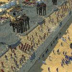
The rays of light pass through the horn shell, moisture anterior chamber, a lens, a glassy body and reach the retina. Horny shell and lens do not just miss the light, but also refract its rays, acting as a biconvecake glass. This allows them to collect them in a row and direct to the mesh shell so that it turns out a valid, but inverted (inverted) image of objects (Fig. 1).

Fig. 1. Scheme image of the subject in the eye
In molds and chopsticks, light energy is converted into nervous impulses, the latter are carried out by visual nerves, paths, paths into the visual centers of the brain, where the transformation of the nerve impulse energy in the visual perception (Fig. 2).

Fig. 4. Lights-crossing cells: A - sticks; B - Columns
As a result, there is a feeling of shape, the size and color of objects, the degree of their remoteness from the eye, etc. the ability of the organ of visionit was developed in the process of long-term evolutionary human development. Thus, in functionality, the eye consists of a light-conducting and light-crossing departments.
Depending on the illumination of the subjects under consideration, it should be distinguished day, twilightand night vision.
Day visionExercised by Kollovka with a high light intensity, is characterized by a high sharpness and good perception of color.
Twilight vision Provide sticks with a weak degree of illumination. It is characterized by low sharp and lack of perception of colors.
Night visionalso carried by chopsticks at a very low (so-called threshold and overpowing) light and comes down only to the sensation of light.
The dual nature of visual functions allows us to distinguish between central and peripheral vision.
Central vision
Central vision - This is the ability of a person to distinguish not only the shape and color of the objects under consideration, but also their small parts, which is provided by the central fossa of the yellow spat of the retina.
Central vision is characterizedits sharpness, that is, the ability human Eyes. Perceive separately points located apart at a minimum distance. For most people, the threshold angle view corresponds to one minute. In this principle, all tables are built to study visual acuity for Dali, including those adopted in our country Tables of Golovin-Sivzev and Orlova, which consist of 12 and 10 rows of letters or signs, respectively. So, the details of the largest letters are visible from a distance of 50, and the smallest - from 2.5 meters.
Normal visual acuity
Normal visual acuity in most people corresponds to one. This means that with such a visual sharpness, we can freely distinguish the letter or other images of the 10th row of table from a distance of 5 meters. If a person does not see the largest first line, he shows signs of one of the special tables.
For very low visual sharpness check light. If a person does not perceive the light, it is blind. Quite often meets the excess of the generally accepted norm of view. As shown by the Department of Adaptation of the Scientific Research Institute of Medical Problems of the North of the Siberian Branch of the Academy of Medical Sciences of the USSR, held under the leadership of the Doctor of Medical Sciences V. F. Bazarova, in the conditions of the Far North in children aged 5-6 years of visual acuity of the distance exceeds the generally accepted conditional The norm, reaches two units in some cases.
On condition central view A number of factors are influenced: the intensity of light, the ratio of the brightness and the background of the object under consideration, the exposure time, the degree of proportionality between the focal length of the refractive system and the length of the axis of the eye, the width of the pupil, etc., and also the general functional state of the central nervous system, The presence of various diseases.
Visual acuityeach eye is examined separately. Start with small signs, gradually go to the larger. There are also objective methods for determining visual acuity.
Color Wechness or Color Vision
One of important functions of the eye is an coloring - The ability to distinguish colors. A person is able to perceive about 180 color tones, and taking into account brightness and saturation - more than 13 thousand. This is due to the mixture in different combinations of red, green and blue flowers.
A person with the right sense of all three colors is considered a normal trichrome. If two or one component is functioning, the coloranomalia is observed. The lack of red-colored perception is called protanalia, green - deteransal and blue - tritomalia.
Known innate and acquired disorders color view. Congenital disorders are called daltonism named English scientist Dalton, who himself did not perceive the red color and first described this state.
For congenital violations of color vision There may be complete color blindness, and then all items seem to be gray. The reason for such a defect is underdevelopment or lack of mesh mesh.
Pretty common partial color blindnessespecially on red and green colorand which, as a rule, is inherited.
Blindness on the green color is twice as much as on red; On blue - relatively rarely. Partial color blindness is observed approximately every twelfth of a hundred men and one, out of two hundred women. As a rule, this phenomenon is not accompanied by a violation of other visual functions and is detected only with a special study.
Congenital color blindness is incurable. Often, people with abnormal color may not know about their condition, as they get used to distinguish between the color of the objects are not in color, but in brightness.
Acquired color assistance disorders are observed in the diseases of the retina and spectator nerveas well as during the disorders of the central nervous system. They can be both in one and both eyes and accompanied by disorders of other visual functions. Unlike congenital, acquired disorders may vary in the process of the disease and its treatment.
Coloring disorders Received with the help of special polychromatic tables and devices.
Peripheral vision
The possibility of visual work is determined not only by the state of acuteness of vision and at close range from the eyes. A big role in a person's life playing peripheral vision. It is provided by peripheral retinal departments and is determined by the magnitude and configuration of the field of view - space that is perceived by the eye with a fixed gaze. The peripheral vision is affected by the illumination, the size and color of the subject or object in question, the degree of contrast between the background and the object, as well as the general functional state of the nervous system.
The field of view of each eye has certain boundaries. Normally, the average borders on the white color of 90-50 °, including: the dust and the book-duck - 90 °, up-duck - 70 °; Book and Knutrice - at 60 °, upstairs and upstairs - 55 °, Book-Knutka - 50 °.
To accurately determine the boundaries of the field of view, they are projected into a spherical surface. In this method, a study on a special device - perimeter is founded. Each eye is examined separately in less than 6 meridians. The degree of the arc, on which the test for the first time saw the object, is noted in a special scheme.
The extreme peripherals of the retina, as a rule, does not perceive colors. Thus, the feeling of blue occurs only 70-40 ° from the center, red - 50 -25 °, green-at 30-20 °.
Forms of change peripheral vision Very multifaceted, and the causes are varied. First of all, these are tumors, hemorrhages and inflammatory diseases of the brain, the disease of the retina and the optic nerve, glaucoma, etc. It is not uncommon and so-called physiological seats (blind spots).
Example is blind spot - place of projection in the space of the optic nerve disk, the surface of which is deprived of photosensitive cells. The increase in the size of the blind spot is diagnosed, being an early sign of glaucoma and some diseases of the optic nerve.
Light
Light- This is the ability of the eye to perceive the light of various brightness, in other words, to distinguish light from darkness. It is carried out by the retinal chopstick apparatus and provides twilight and night vision.
Sensitivity of the eye A man to the light is very high. It happens absolute and distant. The first is characterized by the threshold of the perception of light, the second allows a person to distinguish objects from the surround background based on unequal brightness.
Absolute light sensitivity depends on the degree of illumination. Therefore, the change in this sensitivity in unequal illumination is called adaptation. There are two varieties of adaptation - light and dark. Eye adaptation to different light brightness occurs quite quickly, after 3-5 minutes. On the contrary, addictive to the dark is achieved only in 45-50 minutes. Twilight vision disorder is called hemoralopia, or "chicken blindness".
The hemoralopia is symptomatic and functional. The first is associated with the lesion of the photosensitive layer of the mesh shell and is one of the symptoms of the pattern of retina and optic nerve (glaucoma, pigment abodester of the retina, etc.). Functional hemoralopia develops due to vitamin A deficiency and is well treatment.
No matter how perfect vision with one eyeIt gives an idea of \u200b\u200bthe objects under consideration only in the same plane. Only in the vision at the same time in two eyes, it is possible to perceive the depth and the correct idea of \u200b\u200bthe mutual arrangement of objects considered by each eye. This ability to merge individual images; obtained in each eye, in a single whole ensures the so-called binocular vision.
Binocular vision in man
Binocular vision in man It is found at the fourth month of life, it is formed to two years, but its development and improvement ends only at 8-10 years. External manifestation It is a stereoscopic (volume) vision, without which the execution of driving, flight and a number of other works is difficult, as well as classes in many sports. The study of binocular vision is carried out on special devices.
To have a more complete picture of our visual functions, you should also know about such important properties of the eyes as accommodation and convergence.
Accommodation
Accommodation- This is the ability of a person to clearly see objects at various distances from the eye. It is realized due to the elasticity of the lens and the contractile ability of the ciliary muscle. Accommodation has its limits. So, a normal, commensurate eye, a person cannot clearly see the small details of the objects under consideration closer 6-7 cm from the eye. In myopia, even complete relaxation of the ciliary muscles does not allow clearly seeing items located away.
The volume of accommodation (space between the nearest and further points of clear vision) It will be the largest eye in the normal optical installation of the eye, the smallest - at the very high degree; The amount of accommodation will be reduced and with highly highness of a high degree. Accommodation is weakened and with age, and due to various diseases.
As already mentioned, the best vision is provided by the central fossa of the yellow stain. The straight line, conventionally connecting the object under consideration with the central fossa, is called a visual line, or a visual axis. If you manage to send both visual lines to the subject considered, the eyes acquire the ability to converge, that is, to change the position of the eyeballs by means of their inside. This property is called convergence. Normally, the closer the subject matter is, the more convergence.
There is a direct relationship between accommodation and convergence: The greater the accommodation voltage, the greater the convergence, and vice versa.
If a single eye acuity Much higher than the other, in the brain, the image of the object under consideration is only from a better-visible eye, the second eye can only provide peripheral vision. In this regard, the worse seeing eye periodically turns off from the visual act, which leads to ambulopia - a decrease in visual acuity.
In this way, summary functions Closely related to each other and constitute a single integer, called the act of view.
Now that you have become familiar with the device and the functions of the organ of vision, it is necessary to tell both about the main diseases of the eyes, their prevention, i.e. the prevention of disease.
An article from the book.
The main purpose of this function - serve the perception of small objects or their details. This vision is highest and characterized by the concept of "visual acuity".
Visual acuity - The ability of the eye to distinguish between two points separately with a minimum distance between them, which depends on the characteristics of the structure of the optical system and the light-crossing apparatus of the eye. Central vision provide Retinal columns, occupying its central pocket with a diameter of 0.3 mm in the field of yellow spots. As you remove from the center of the acuity of vision, it decreases sharply.
The diameter of the columble determines the magnitude of the maximum visual acuity. The smaller the diameter of the colums, the higher the visual acuity. Images of two points, if they fall into two neighboring columns, merge and will be perceived as a short line.
The angle of view is an angle formed by the points of the object under consideration and the nodal point of the eye.
To study visual acuity Use special tables containing letters, numbers or icons of various sizes, and for children - drawings (cup, christmas tree, etc.). They are called optotypes.
In physiological optics there are concepts The minimum visible, distinguishable and recognizable. The survey should see optotype, distinguish its details, recognize the presented sign or letter. All optotype corresponds to an angle view of 5 degrees.
Method for determining visual acuity on a head table - Sivseva. The lower edge of the table must be at a distance of 120 cm from the floor level. The patient sits at a distance of 5 m from the exhibited table. First determine the visual sharpness, then the left eye. The second eye is closed with a damper.
The table has 12 rows of letters or signs, the value of which gradually decreases from the top row to the bottom. In the construction of the table, a decimal system was used: when reading each subsequent lines of visual acuity increases by 0.1. So, under normal vision, adopted for 1.0, the upper line will be visible from a distance of 50 m, and the tenth - from a distance of 5 m.
There are people and with a higher urgency of view - 1.5; 2.0 or more. They read the eleventh or twelfth row of the table.
Under visual sharpness, it is necessary to bring closer to the table below 0.1 when it sees its first string. The calculation of visual acuity should be made according to the formula of Smelllen:
Where D is the distance from which the surveyed recognizes optotype; D is the distance from which this optotype is visible under normal visual sharpness.
The minimum sharpness of view is light With proper or irregular light. Lighting is determined by directions to the eye from different sides of the beam of light from an ophthalmoscope. In the absence of light, the visual acuity is zero and the eye is considered to be blind.
To determine visual acuity below 0.1 Optotypes developed by B. L. Pole, in the form of bar tests or rings of landslip, intended for presentation at a certain close distance, indicating the corresponding visual acuity.
There is also an objective (independent of the patient's testimony) A method for determining visual acuity based on octocinetic nystagme. With the help of special devices, the surveyed demonstrates moving objects in the form of strips or chessboard. The smallest value of the object, which caused the involuntary Nistagm (seen by the doctor), and corresponds to the severity of the examination
Peripheral vision, methods for its definition, field boundary in normal. Changes in sight. The impact of peripheral violations on the working capacity and the choice of profession. 26. Views and causes of peripheral violations. The value of the study of the field of view in the clinic of eye and nerve diseases.
Peripheral vision It is the function of a sticky and colummer apparatus of the entire optical retinal and is determined by the field of view.
line of sight - This is visible to the eye (eyes) space at a fixed gaze. Peripheral vision helps to navigate in space.
The field of view is examined by perimetry.
The easiest way - Control (approximate) Donders study. The examined and doctor are located face to each other at a distance of 50-60 cm, after which the doctor closes the right eye, and the surveyed is left. At the same time, the surveyed open right eye looks into the open left eye of the doctor and vice versa. The field of view of the left eye of the doctor serves as a control in determining the field of view of the examined. On the median distance between them, the doctor shows the fingers moving them away from the periphery to the center. In the coincidence of the boundaries of the detection of the fingers of the fingertips and the examined field of view, the latter is considered unchanged. During the mismatch, there is a narrowing of the field of view of the right eye being examined in the direction of the movement of the fingers (upwards, a book, with a nose or temporal side, as well as in radii between them). After checking the rod of the right eye, the field of view of the left eye is subject to closed right, while the doctor has a left eye.
The simplest device for studying the field of view It is a fermeter perimeter, which is a black arc (on a stand), which can be shifted in various meridians.
Perimetry on a universal projection perimeter (PPU) widely entered into practice (PPU) is also carried out monocular. The correctness of the centering of the eye is controlled by the eyepiece. First spend perimetry on white.
Modern perimeters are more complex, Including on a computer basis. On a hemispherical or any other screen in various meridians, white or colored labels are moving or flashed. The corresponding sensor captures the test indicators, indicating the boundaries of the field of view and the sections of the fallout in it on a special form or as a computer printout.
Normal boundaries of the field of view The white color is considered to be up to the top of 45-55 °, up the dust of 65 °, the dust of 90 °, the Book of 60-70 °, the book Knutrice is 45 °, Knutrice is 55 °, up to the top of 50 °. Changing the boundaries of the field of view can occur at various lesions of the retina, choroids and visual tract, in the pathology of the brain.
In recent years, the practice includes visa-protectoperimetrywhich is a method for evaluating spatial vision using black and white or colored strips of different spatial frequencies that are presented in the form of tables or on the computer display.
Local loss of internal sections of the field of view that are not related to its borders are called scotomes.
Castoms are Absolute (complete loss of the visual function) and relative (decrease in the perception of the object in the explored field of view). The presence of cattle indicates focal retinal lesions and visual paths. Scott can be positive and negative.
Positive cattle Sees the patient itself as a dark or gray spot in front of the eye. Such a fallout in the field of view occurs with the lesions of the retina and the optic nerve.
Negative cattle The patient itself does not discover, it is detected in the study. Usually, the presence of such a cattle indicates a damage to conductive paths.
Casual cattle - It is suddenly emerging short-term moving loss in sight. Even in the case when the patient closes his eyes, he sees bright, flickering zigzag lines leaving on the periphery. This symptom is a sign of a spasm of the brain vessels.
At the location of cattle In the field of view, peripheral, central and paraventional cattle are visible.
At removal of 12-18 ° from the center in the temporal half there is a blind spot. This is a physiological absolute scotoma. It corresponds to the projection of the optic nerve disk. The increase in the blind spot has an important diagnostic value.
Central and paraccentral cattle are detected at stoneimetry.
The central and paraccentral cattle appear in the damage to the papillomacular beam of the visual nerve, the retina and choroid. Central scotoma can be the first manifestation of multiple sclerosis
Binocular vision. Conditions for the implementation of binocular vision. The concept of identical and unidentic retinal points. Physiological branch. The value of the study of binocular vision during the trade unit.
Binocular vision - the perception of the surrounding items with two eyes - is ensured in the cortical department of the visual analyzer thanks to the most complex physiological mechanism vision - fusion, i.e., the merger of visual images arising separately in each eye (monocular image), into a single combined visual perception.
Single image of the subjectperceived by two eyes is possible only in the case of its images on the so-called identical, or corresponding, the retina points to which the central mesters of the retina are both eyes, as well as the retinal points located symmetrically with respect to the central pits. In central pits, individual points are combined, and at the rest of the retina, receptor fields related to one ganglion cell. In case of projecting an image of an object to asymmetrical, or so-called dispattors, the retinal points of both eyes arises for the image - diplopia.
For the formation of a normal (sustainable) binocular view, the following conditions are necessary:
Sufficient visual acuity of both eyes (at least 0.4), at which a clear image of the items on the retina is formed.
Free mobility of both eyeballs.
Equal sizes of images in both eyes - iseclony.
The normal functional ability of the retina, conductive paths and higher visual centers.
Location of two eyes in one frontal and horizontal plane.
Lecture 1 on the topic 2.04.01 Methods of study of the organ of vision. The system of organization of ophthalmological assistance.
PLAN:
The functions of the visual analyzer and the methods of their research
Clinical refraction and accommodation of the eye, research methods. Myopia and myopic disease
Brief historical essay of ophthalmology
Section of medicine studying etiology, pathogenesis and clinical picture Violations of vision and eye diseases are called ophthalmology (from Greek. ophtalmos.- eye I. logos.- the science). Doctors this specialty are called ophthalmologists.
Ophthalmology originated in ancient times. The primitive science on eye diseases existed in Egypt for 4400 years before the new era.
Up until the XVIII-XIX centuries, in many countries of the world, in the treatment of eye diseases, we used the recommendations of the greatest doctors of the ancient times of hippocrates and Galen. The famous Hippocrat (460-372 BC) in his treatises described the structure of the eye and the development of many of his diseases.
A large role in the development of ophthalmology also played "Medical Canon", written at the beginning of the second millennium of our era outstanding Tajik philosopher, scientist, poet and physician (Abu-Ali Ibn-blue). His unique labor for 600 years was considered the main benefit for the doctors of Europe and the countries of the East.
In 1805, the world's first specialized eye hospital was opened in Moscow.
The first eye department was established in 1818 at the Medical and Surgery Academy (now the Warnedic Academy in St. Petersburg), where in 1819, lectures on eye disease Readed N.I. Pies.
The founders of the Moscow Ophthalmological School were A.N. Maklakov and A.A. Hooks. The third large ophthalmological school in Russia was opened in Kazan under the leadership of E.V. Adamyuk.
Professor A.N. McLakov has a merit in creating a tonometer for measuring intraocular pressure (1884). A great contribution to the science of an outstanding ophthalmologist Academician M.I. Averbach (1872-1944). He organized the first in the Soviet Union the Scientifier Institute of Eye Diseases (Helmholts).
V.P. Filatov (1875-1956) created the world-famous Odessa Research Institute of Eye Diseases, which after the death of his founder began to wear his name.
In the second half of the XX century. Considerable attention was paid to the study of pathogenesis, diagnosis and methods of treatment of glaucoma (T.I. Yeroshevsky, M.M. Krasnov, A.P. Nesterov). MM Krasnov became a pioneer of the domestic micro and laser surgery of glaucoma. Student T.I. Yeroshevsky was S.N. Fedorov (1927-2000), whose name became world famous in connection with the development of a new refractive operation - anterior radial keratotomy.
In the XX century Many new diagnostic methods in ophthalmology were proposed: biomicroscopy, gonoscopy, fluorescent angiography, electrophysiological and ultrasound methods. Laser treatment has gradually become one of the inalienable components of the modern treatment of glaucoma and an ametropi. Lasers found their place in the diagnosis of diseases of the eye.
Methods for studying the organ of vision
Collecting Anamnese and Complaints
The patient's survey begin with the clarification of his complaints.
Building a conversation with a patient, you should always clarify a number of important provisions.
1. It is necessary to highlight the patient's priority problem, which caused it to apply for help to an ophthalmologist. To do this, ask the following questions:
What are you complaining about now?
When and how the disease began, specify the earliest symptoms.
Do you notice the progression of the disease or, on the contrary, the manifestation of it gradually pass?
Some complaints are very characteristic of a particular disease. For example, pain in the eye and headache, nausea, vomiting, the sudden loss of vision of the eye is characteristic of the acute attack of glaucoma; Redness of the edges of the eyelids and itching in this area testify to bluffy; The feeling of eye clogging, the feeling of sand, the severity of the eyelids - signs of chronic conjunctivitis. Inflammation of the cornea, iris or ciliary bodies cause pain in the eye, light-friendly, tear. A sudden loss of vision without pain in the eye may occur during the damage to the retina, the optic nerve, with an acute impaired blood circulation in the retinal vessels (thrombosis, embolism).
2. Specify as a patient assesses the state of his vision, After listening to the answers to your questions:
Is the vision reduced, and if so, then on one eye or both?
Reduced vision only in the distance or only near, or even then together?
How did the vision reduced? Quickly or gradually?
Reducing vision keeps stably or is the vision periodically improving?
What, according to the patient, was the reason for reducing vision?
Does patient difficulty in fulfilling their daily duties due to reducing vision?
Be sure to find out what means of the correction of vision used the patient (glasses, contact lenses) and how long ago.
So, the patient glaucoma can be considered the beginning of the disease a sudden loss of vision, and asking him, it is possible to find out that it was long before that there was a periodic vision of vision in the morning, when looking at the electric light bulbs around them, rainbow circles appeared. These are the early signs of one of the forms of glaucoma, and they define the beginning of the disease.
Finding out when the patient got sick, you need to know how the illness began: suddenly, gradually, at what time of day, the time of year than at that time did, where he worked, there was no common disease, injuries or the actions of harmful external factors. In case of injuries of the organ of vision, it is important to find out in detail how they have occurred, what kind of help was the patient.
3. Discover if the patient has similar problems (diseases) before And, if so, then what treatment was carried out.
4. It is necessary to clarify how the disease proceeded, were there previously any diseases of the eye or an operation on the eye. Reverse special attention For the presence of glaucoma, cataracts, eye complications of diabetes.
5. Evaluating the patient's complaints, a medium medical worker should know that a number of vision complaints may be reflected not only eye, but also common diseases man. So, bumping ripening, "flyers" before your eyes
they can be observed both with the eye (glaucoma, the damage of the retina and the optic nerve) and in common diseases (diabetes, hypertensive disease, osteochondrosis of the cervical spine, etc.). Therefore, it is very important to collect anamnesis in more detail in this patient.
Get information about the presence of concomitant common diseases, as well as about the nature and timing of their treatment. Information on hypertension, diabetes mellitus, chronic diseases of lungs and kidney, on the reception of oral contraceptive means (they can cause circulatory disorders in the retina vessels) are especially important.
7. Collect anamnesis of the disease, go to the history of life. Find out what previous eye diseases suffered a patient what common diseases suffering. It is important to know whether it was sick with tuberculosis, venereal diseases.
Check out S. family history: Does not have the nearest blood relatives of glaucoma, cataracts, myopia, strabismus, congenital reducing vision or blindness.
8. Find out whether allergic reactions for drugs, food products, pollen plants and so on.
9. Ask what conditions living Your patient, about the nature of its nutrition, features professional activities (visual loads, harm) whether there are familiar inxication (alcohol, nicotine, etc.).
Having finished the collection of complaints and anamnesis of the disease of the patient, go to the inspection of the organ of vision. Before the inspection you need to thoroughly wash your hands.
10. Sequence of an inspection of the organ of sight
Inspection eyeball And its appendages are produced, firstly, using bright lighting and, if possible, magnifying optical devices (for this purpose, ordinary glasses with plus glasses can serve).
Secondly, it is necessary to inspect very carefully, not losing parts, consistently moving from superficial to deeper structures.
Thirdly, it is necessary to compare the state of the studied departments of both eyes.
In the survey process, inspect: century - Rate the color and appearance skin, shape, position, eyelash growth, configuration and the size of the eye slit;
conjunctiva The eyelids of the eyelid apple: its color, surface, transparency, separated by the conjunctiva. In the norm of conjunctiva pink, smooth, transparent, without separated;
tear organs The position of the tear points (in the normal one they are not visible without pulling out the eyelid from the eyeball), the condition of the skin in the inner corner of the eye in the projection of the laction of the lacrimal bag. It is necessary to check whether there is a separated from the tears when pressing on this area (there is no normal);
the position of the eyeball in orbit, the volume of its movements; cornea - transparency, surface, sensitivity. Normally, it is transparent, mirror-shiny, spherical, very sensitive;
sclera - Color, presence of foci. Norma it is white and smooth;
front camera - Depth, moisture transparency. Normally, the front camera is uniform, the moisture is transparent;
iris - color, drawing. Healthy heights of both eyes are painted equally, the drawing is clear;
pupil - position, quantity, color, shape, reaction. Normally, it is located in the center, has a round shape, black, the diameter is about 3-4 mm, alive responds to light, accommodation and convergence;
ciliary body - Palpal soreness. Normally palpation of the eyeball is painless;
crystalik. - vitreous body (transparency). Normally, they are transparent, so the study in the transmitted light, the pupil has a bright pink glow (reflex);
ocular fundus - examined by a doctor. When it is inspected, it is estimated: the optic nerve disk (form, color, borders, level); the state of the vessels of the retina (stroke, caliber); region of yellow stain, the periphery of the eye dna;
intraocular pressure It is estimated palpafact. When registering received data, the following notation was taken:
OD. (Oculus Dexter) - right eye;
OS. (Oculus Sinister) - left eye;
Ou. (Oculi Utriusque) - both eyes (each of two).
Ehoofthalmoscopy
Echoofthalmoscopy - ultrasound procedure Anatomical structures of the eye with a special device - echoofthalmoscope. This method allows you to define the form, dimensions and structure of the eye.
Fluorescent angiography
Fluorescent angiography - 5-10% solution of sodium salt of fluorescein intravenously introduced, the solution is distributed over the vascular channel, which allows to determine the pathology of the retina and the vascular shell. In pathology, the solution of fluorescein accumulates in inflammatory foci, serous population, tumors. The passage of the solution is blocked in the presence of inflammatory foci in the retina and obstruction of the vascular vascular bottom.
Ophthalmotonometry
Ophthalmotonometry is made with palp born determination of the level of intraocular pressure, method of tonometry on Maclakov and tonometria by shiottsu.
Central vision
Central vision(uniform) is measured by urgency. Under the sharpness of vision (Visus)understand the ability of the eye to perceive separately 2 points located apart at a minimum distance, i.e. The ability of the eye to separate perception of 2 points under the smallest angle of view.
Most people have the smallest angle of view, under which the eye can distinguish 2 points, equal to 1 "(1 angular minute). The highest visual acuity is provided only by the central zone of the retina (spot area and central fox), where there is a maximum flange density.
In the event that the eye sees separately 2 points, the angle between which is at least 1 ", the visual sharpness is considered normal and determine it equal to one unit. Some people have visual acuity 2 units and more.
With the age of acuity vision changes. Subsection appears at the age of 2-3 months. The acuity of vision in children aged 4 months is about 0.01. By the year, visual acuity reaches 0.1-0.3. The acuity of view equal to 1.0 is formed by 5-15 years.
For definition of visual acuityspecial tables containing letters, numbers or signs (for children use drawings - machine, Christmas tree, etc.) of various sizes. These signs are called optotypes. The basis of the creation of optotypes is the international agreement on the magnitude of their parts constituting the angle of 1 ", while all optotype corresponds to the corner of 5" from a distance of 5 m.
In young children, visual sharpness is determined approximately, assessing the fixation of bright items of various sizes. Starting from three years, visual sharpness is estimated with special tables.
In our country, the Golovin-Sivzian table was most common, which is placed in the Rota apparatus - a box with mirror walls, providing uniform lighting of the table. The table consists of 12 lines (Fig. 3-1). The table is calculated to test the visual acuity from 5 m.
First determine the sharpness of the right eye, the left eye is covered with an opaque damper. Then check the visual sharpness of the left eye. Only complete acuity of vision is taken into account. The first 6 lines of the table (VIS \u003d 0.1-0.6) are considered read if all signs are recognized in them. In the 7-10th lines (VIS \u003d 0.7-1.0) an error is permissible in one sign.
The study can be simplified if you show the patient from different distances of your hand. With this method of measuring 1 m, equivalent to 0.02 visual sharpness. From here it follows, for example, that, with the right account of the fingers at a distance of 1 m, the visual acuity is 0.02, 2 m - 0.04, by 2.5 m - 0.05, etc.
When the vision is so little that the eye does not distinguish between objects, but only light perceives, the visual sharpness is considered to be equal to the light
Fig. 3-1.Table of Golovin-Sivzieva to determine visual acuity
If the surveyed sees the light and correctly determines its direction, the visual sharpness is considered to be equal to the light with the correct projection of light. If the eye of the surveyed incorrectly determines the projection of light at least on the one hand, the visual sharpness is evaluated as lightness with incorrect projection of light.
Peripheral vision
Peripheral vision is determined by field of view.
line of sight- space visible to the eye at a fixed gaze. The dimensions of the field of view are determined by the boundary of the optically active part of the retina and the protruding parts of the face: the back of the nose, the top edge of the orders, cheeks.
Changes in vision fields occur in pathological processes in various sections of the visual analyzer.
Unilateral changes in the field of view (only in one eye on the side of the lesion) are due to damage to the retina or optic nerve.
Bilateral vision changes are detected during localization pathological process in Hiazme and above.
Highlight three types of field changes:
Focal defects in sight (cattle) 1;
Narrowing peripheral boundaries of the field of view;
Loss of half of the field of view (hemianopsy).
The field of view is investigated using the control method and special instruments - perimeters and campmers.
Control methodused in outpatient practice and in seriously ill, especially lying. The patient and the doctor are opposite each other at a distance of 1 m and closed over one variety of eye, and open eyes serve as a fixed point of fixation. The doctor begins to slowly move from the periphery of the field of view a brush of his hand by moving to the center of the field of view. Research is repeated from all sides. If the study sees the hand at the same moment when the doctor can be said that the boundaries of the field of view in the patient are normal. Prerequisite It is a normal field of view from a doctor.
Perimetry- Method for studying the field of view on a spherical surface. Currently there are two main methods perimetry: kinetic and static.
1 scotoma is a limited defect in sight. In a normal field of view, physiological scotoma is always there - the projection of the optic nerve disk.
Kinetic perimetry is carried out on hemispherical perimeters. The chosen diameter object (from 1 to 5 mm) is slowly moving along the perimeter arouse from the periphery to the center, the patient who fixes the central mark of the perimeter under study, should determine the appearance of an object in the field of view.
The field of view is examined by 8 or 12 meridians (after 45 or 30 °). The boundaries of the field of view are expressed in degrees. Normally, the average borders for a white label with a size of 5 mM Knod 90 °, the Book of the Source - 90 °, the book - 60 °, the book Knutka - 50 °, knutut - 60 °, upstairs knutut - 55 °, upstairs - 55 ° and upstairs - 55 ° and upstairs - 55 ° 70 °. In children of preschool age, the boundary of the field of view is 10% already than in adults, and expand to normal school age. Fields of view on colors significantly already than white. A particularly narrow field of view on green, somewhat wider on red and even wider on blue.
With static perimetry, the surveyed alternately impose fixed test objects. Static perimeter is carried out on the automatic computer perimeters of the domestic production "Peric".
Campimetry- Study of the central and paracentral departments of the field of view on a flat surface (campimeter) or on the computer monitor screen.
Light adaptation
The ability of the visual analyzer to perceive light and different degrees of its brightness is called lightweight.This is the earliest and basic function of the organ of vision. The light is due to the function of the sticks, they are many times more sensitive to light than kolkovka. When exposed to the eye of strong light, visual substances are destroyed faster and, despite their periodic recovery, the sensitivity of the eye towards the light decreases. In the dark, the disintegration of visual substances does not occur as fast as in the light, and, therefore, the sensitivity of the eye towards the light increases in the dark.
Eye adaptation process various conditions Illumination is called adaptation.Adaptometers serve to study light sensitivity.
Hemeraralopia- Weakening of the adaptation of the eye to the dark. Hemeraralopia is manifested by a sharp decline in twilight, while day vision is usually saved. Eliminate symptomatic, essential and congenital hemoralopia:
Symptomatic hemerlopia accompanies various ophthalmological diseases: retinal pigment abiotrophy, sideriosis, high degree, with pronounced changes in the eye dna;
Essential hemoralopia is due to hypovitaminosis A. Retinol serves as a substrate for the synthesis of rhodopcin, which is disturbed by exo- and endogenous vitamin deficit;
Congenital hemerlopia - genetic disease. Ophthalmoscopic changes are not detected.
Central vision
The acuity of vision is the ability of the eye to perceive separately points located apart at a minimum distance, which depends on the characteristics of the structure of the optical system and the light-crossing device of the eye. The angle formed by the extreme points of the object under consideration and the nodal point of the eye is called an angle of view.
Determination of visual acuity (visometry). Under normal urgency, the ability of the eye is understood to distinguish separately two glowing points at an angle of view in 1 min. It is much more convenient to measure the visual sharpness of vision by not visual angles, but the values \u200b\u200bof the ring value, i.e. in relative units. For normal visual acuity, equal to one, the inverse angle of view is adopted in 1 min. The acuity of vision is inversely proportional to the angle of view: the less angle of view, the higher the acuteness of sight. Based on this dependence, tables are calculated to measure visual acuity. There are many options for tables to determine visual acuity that differ in test objects, or optotypes.
In physiological optics there are the concepts of minimally visible, distinguishable and recognizable. The survey should see optotype, distinguish its details, recognize the presented sign or letter. Optotypes can be projected onto the screen or display of the computer. As optotypes use letters, numbers, drawings, stripes. Optotypes are constructed so that from certain distances of the optotype details (the thickness of the lines and gaps between them were visible at an angle of view of 1 min, and the entire optotype - at an angle of view in 5 minutes. International optotype is taken by a broken ring of the landslon. In domestic ophthalmology, the table is most common. Golovin - Sivtsieva, containing the letters of the Russian alphabet and rings of the Landold. The table has 12 rows of optotypes. In each row, the size of the optotypes is the same, but they gradually decrease from the upper row to the bottom. The magnitude of the optotypes changes in arithmetic regression. Within the first 10 Rows Each series differs from the previous one by 0.1 units of visual acuity, in the last two rows by 0.5 units. Thus, if the three row of letters reads, then the visual acuity is 0.3; fifth - 0.5, and t. d.
When using a head table - Sivzsev, visual sharpness is determined from 5 m. The bottom edge of the table must be at a distance of 120 cm from the floor level.
First, the sharpness of the view of one eye (right) is determined, then the left eye. The second eye is closed with a damper. From a distance of 5 m at an angle of view of 1 min, parts of the optics of the tenth row of the table are visible. If the patient sees this number of tables, its visual acuity is 1.0. At the end of each row of optotypes, the symbol of V shows the visual acuity corresponding to the reading of this series from the distance of 5 m. To the left of each row, the distance from which the optotypes of this row differ in visual sharpness of 1.0 are indicated. So, the first row of tables in visual sharpness of 1.0 can be seen from 50 m.
To determine the severity of vision, you can use the formula of Sielenna - Diders U181 1 8 \u003d FTE, where th - the distance from which the studied sees this series of tables (the distance from which the study is carried out), m; ^ - The distance from which the studied should see this series, m.
Using the above formula, it is possible to determine the visual sharpness in cases where the study will cause a length in a length, for example, in 4.5 m, 4 m, etc. If the patient sees the fifth row of tables from a distance of 4 m, then its visual acuity is equal to: 4/10 \u003d 0.4.
There are people and with a higher urgency of view - 1.5; 2.0 or more. They read the eleventh or twelfth row of the table. The case of visual acuity is described: the examined differed satellites of Jupiter, which are visible from the ground at an angle of 1 sec. Under visual sharpness, it is necessary to bring closer to the table below 0.1 when it sees its first string.
Since the thickness of the fingers of the hand approximately corresponds to the width of the prototypes of the first line of the table, can be demonstrated to the expired fingers (preferably on a dark background) from different distances and, accordingly, determine visual acuity below 0.1 also according to the above formula. If visual acuity is below 0.01, but the surveyed fingers at a distance of 10 cm (or 20, 30 cm), then the visual acuity is equal to the account of the fingers at a distance of 10 cm (or 20, 30 cm). The patient may not be able to count the fingers, but determines the movement of the hand in the person, this is considered the next gradation of visual acuity. The minimum sharpness of view is the light supply (U18 \u003d 1 / -) with the correct or incorrect lighting. Lighting is determined by directions to the eye from different sides of the beam of light from an ophthalmoscope. In the absence of light, visual acuity is zero (U18 \u003d 0), and the eye is considered to be blind.
To determine the visual acuity, children use the E. M. Orlova table. It uses pictures of the objects of them and animals as optotypes. And yet at the beginning of the study of visual acuity, the child is recommended to bring it close to the table and ask to name the optotypes.
The table for the study of visual acuity is placed in an open front wooden box, the walls of which from the inside are lined with mirrors. The table contains an electrical lamp, closed behind the screen for constant and uniform illumination (Rota Device - Roslavsetva). The optimal is the coverage of the table, which gives an ordinary incandescent lamp in 40 W. Illuminator with tables strengthen on the wall opposite to the windows. The lower edge of the illuminator is placed at a distance of 120 cm from the floor. The room where patients expect a reception, and the eye office should be well lit. Currently, test marks are increasingly used to study the visual signs. On the screen from a distance of 5 m, optodypes of various sizes are projected. Screens are made of matte glass, which reduces the contrast between optotypes and the surrounding background. It is believed that such a threshold definition more contributes to real severity.
To determine the acuteness of vision below 0.1, optodypes developed by B. L. Poles are used in the form of bar tests and rings of the landslide intended for presentation at a certain close distance with an indication of the corresponding acuity of vision. These optotypes are specially created for military-medical and medical - social expertise, conducted in determining the shelf life military service or a group of disability.
There is also an objective (independent of the patient's testimony) a method for determining visual acuity based on a nutritional nystagm. With the help of special devices, the surveyed demonstrates moving objects in the form of strips or chessboard. The smallest value of the object, which caused the involuntary nastagm (seen by the doctor), and corresponds to the severity of the examination of the eye.
When determining the visual acuity, it is necessary to fulfill certain rules.
1. To explore the visual sharpness (separately) in each eye, starting with the right.
2. When checking both eyes should be open, one of them is observed by a palate from an opaque material. If it is not, then the eye can be closed with palm (but not with your fingers) of the subject. It is important that he does not press over the eyelids on the covered eye, as this can lead to a temporary decrease in vision. The shield or palm is kept vertically in front of the eye, so that the possibility of intentional or unintentional peeping is excluded, and that the light fall into the open eye slot.
3. The study must be carried out with the right position of the head, eyelids and gaze. There should be no head slopes to one or another shoulder, turns of the head to the right or left, the slopes of her kleedi or the stop. It is unacceptable to pushed. In myopia, this leads to an increase in visual acuity.
4. In the study, the time factor should be taken into account. In conventional clinical operation, the exposure time is 2-3 s, with control and experimental studies - 4-5 s.
5. Optotypes in the table should be shown by pointer; Its end should be well distinguishable, it is installed exactly under an optotype exposed at some distance from the sign.
6. Start research is necessary from displaying the progress of the tenth row of the table, gradually moving to rows with larger signs. In children and people with a deliberately reduced urgency vision, checking the visual acuity is permissible to start from the top line, showing from the top down one sign in the row to a row in which the patient is mistaken, after which it should return to the previous row.
Vision sharpness should be assessed by a number in which all signs were correctly called. One error is allowed in the third - sixth rows and two errors in the seventh - tenth rows, but then they are registered in the recording of visual acuity. The visual sharpness is determined by a special table, which is designed for a distance of 33 cm from the eye. If the patient does not see the top row of the head table - Sivzev, i.e., visual acuity is less than 0.1, then determine the distance from which it distinguishes the optotypes of the first row. For this, the studied will be closer to the table until it sees the first row, and the distance from which he distinguishes the optotypes of this series is noted. Sometimes we use split tables with the optotes of the first row that approach the patient.
On the presence of vision in a newborn can be judged in direct and friendly reactions of pupils into light, with a sudden illumination of the eyes - on the total motor reaction and the occasion of the eyelid. Since the second week, the newborn reacts to the appearance in the field of vision of bright items by turning the eyes in their direction and can be briefly monitored by their movement. In 1-2 months, the baby fixes the moving item for both eyes long enough. From 3-5 months, shaped vision can be checked with a bright red ball with a diameter of 4 cm, and from 6-12 months - the ball of the same color, but with a diameter of 0.7 cm. Having it at various distances and attracting the ball on the ball by swinging the ball, Determine the visual sharpness. The blind child reacts only on sounds and smells.
It can be approximately checking the visual sharpness, which is crucial in a virtue, labor and military examination.
A visual acuity may decrease depending on many reasons. They can be divided into three groups. SAMI frequent cause - This is an abnormal refraction (myopia, hyperopia, astigmatism). In most cases, visual acuity increases or is completely corrected using spectacular glasses. The second reason for the reduction of vision is the turbidity of the refractive transparent structures of the eye. The third cause is the diseases of the retina and the optic nerve, conducting paths and visual centers.
It should also be noted that during the life of visual acuity changes, reaching a maximum (normal values) by 5-15 years and then gradually decreased after 40-50 years.
From book Full Son. by Dipak Chopra.Vision visual stimuli is powerful causative agents of the nervous system. As I said earlier, try not to watch TV late in the evening, especially avoid bright and colorful shows and movies. Any excessive excitement in your life has
From the book listen to your body, your best friend on earth by Liz BurboVision You allow yourself to worry about some things or events that you have to see at present. The body tells you: "What is worried about, has nothing to do with you - this is not your business ..." or: "Instead of worrying, do
From the book the beginning of your child's life AuthorVision to see as adults, newborns, of course, can not. All of its reactions are limited to the fact that he is buried from very bright light and sometimes turns to the light source. Already by monthly age, the baby may not be detained for a short time and
From the book Homeopathic Directory Author Sergey Aleksandrovich NikitinVision defeat vision - the appearance of haze or fog in front of the eyes -
From the book Health of the child and common sense His relatives Author Evgeny Olegovich Komarovsky1.7.1. Vision to see as adults, newborns, of course, can not. All of its reactions are limited to the fact that he is buried from very bright light and sometimes turns to the light source. By monthly age, the baby can briefly detain on any glance
From the book Lost and Returned World Author Alexander Romanovich LuriaSomething happened to him with him, something never happened before. He looks around - and what is it? He cannot see any things at once: the world is fragmented into pieces, and pieces do not fold into entire objects, whole pictures. The right side of what he is trying to watch
From the book Introduction to the psychological theory of autism by the author Francesca AppeCentral coordination and representation of internal representations Central coordination may be useful to explain the peculiarities of the daily activity of autists, which still remain unexplained, as well as to understand the hull
From the book Brain, Mind and Behavior Author Floyd E. BloomThe central coordination and function of programming and control of the hypothesis of central agreement, which matter of the case with the peculiarities of cognitive style, rather than with a violation as such, is distinguished by this not only on the theory of representation of internal representations, but
From the adventure of another boy. Autism and not only Author Elizabeth Zavarzina Mammy From Book Beauty and Health Women Author Vladislav Gennadevich Liflyandsky From the book How to improve your eyesight when you ... Author Gennady Mikhailovich KybardinVision Vision W. different people Very varies in sharpness, color perception and other parameters. The acuity of sight is changing when the light is changed. At the same time, it changes with age and can be different for each eye, as a result of hereditary features or
From the book of the authorGlasses and vision reader, you did not in vain in my hands this book. Perhaps the acuity of your view deteriorated due to changes in the parameters of the lens and the visual eye system included in the work of the eye muscles to compensate for the anomaly. In such a situation, you need to train eye
From the book of the authorBreathing and vision There is a close relationship between the degree of focus of human focus with the frequency and its breathing depth. The more closely we look into anything, the less deep breathe. In this situation, all sorts of external sounds, extraneous muscular
From the book of the authorPalming and Vision With the help of the Palming Exercise Millions of people on Earth seeks to effectively relax eye muscles. Palming is very simple performed and is very effective for relaxation (relaxation) of the eyes. Palming was first developed in the 20s of the last century
From the book of the authorGlacers and vision from the blink by moving to glims. According to the efficiency of increasing visual acuity, the so-called glimpses are located next to the effect of exercises for blinking. To understand what is the essence of exercises on glimpses, right now
From the book of the authorAnalytical vision Analytical vision lies in fast and short movements of eyes and attention from point to point throughout the object of view. Consider as an example of a typical house. Go outside and start contemplating the nearest house. pay attention to
Central vision - Central section of visible space. The main purpose of this function is the perception of small objects or their details. This vision is highest and characterized by the concept of "visual acuity". Central vision is provided by the metalls with a central yam in the field of yellow spots.
As you remove from the center of the acuity of vision, it decreases sharply. This is due to a change in the density of the location of neuroelements and the peculiarities of the transfer of momentum. The pulse from each tip of the central fifth passes through separate nervous fibers through all the separation paths.
Visual Acid (Visus) - The ability of the eye to distinguish between two points separately with a minimum distance between them, which depends on the characteristics of the structure of the optical system and the light-crossing device of the eye.
Point A and B will be perceived separately if their image on the retina b and A will be separated by one non-excited flap with. This creates a minimum lighting gap between two separate lying columns. The diameter of the columble C determines the magnitude of the maximum visual acuity. The smaller the diameter of the colums, the higher the visual acuity. The image of two points, if they fall into two neighboring columns, are alive and will be perceived as a short line.
Vision angle - angle formed by the extreme points of the object under consideration (A and B) and the nodal point of the eye (O). Nodal point - The point of optical system, through which the rays pass without refraarting (are located at the rear pole of the lens). Eye only in the event sees separately two points, if their image on the retina is not less than the arc in 1 ', i.e. The angle of view should be at least one minute.
Methods of research of central vision:
1) using Special Golovin-Sivzian Tables - Optotypes - contain 12 rows of specially selected signs (numbers, letters, unclosed rings, pictures) of different values. The basis of the creation of optotypes is the international agreement on the magnitude of their parts, distinguished at an angle of view of 1 minute, while all optotype corresponds to an angle of view for 5 minutes. The table is calculated on the study of visual acuity from a distance of 5 m. At this distance, the details of the optotypes of the tenth row are visible at an angle of view 1 ', therefore the acuity of vision of the distinguishing optotypes of this row will be equal to 1. If the acuity of sight is different, then it is determined in which a number of the table. The examinable distinces signs . At the same time, visual sharpness is calculated according to the formula Smelllen: Visus \u003d d / d, where D is the distance from which the study is made, D is the distance from which the normal eye distinguishes the signs of this series (they have been laid in each row to the left of optotypes). For example, the first row is examined from the distance of 5 m, the normal eye distinguishes the signs of this series from 50 m, which means Visus \u003d 5/50 \u003d 0.1. In the construction of the table, a decimal system was used: with reading each subsequent line of visual acuity increases by 0.1 (except for the last two lines).
If the acuity of visual is less than 0.1, then determine the distance from which it spills the optotypes of the first row, and then calculate the visual sharpness according to the formula of Smelllen. If the acuity of the view is below 0.005, then it is indicated for its characterization, from which distance he consider fingers. For example, Visus \u003d 10 cm bill account.
When the vision is so little that the eye does not distinguish between objects, but perceives only light, visual sharpness is considered to be equal to the light: Visus \u003d 1 / ¥ with proper (Proctia Lucis Certa) or with incorrect (Proectia Lucis Incerta) light production. Lighting is determined by directions to the eye from different sides of the beam of light from an ophthalmoscope.