It is one of the diseases that pose the greatest threat to human life. According to statistics, up to 50% of pathology cases end in death. Some patients do not have time to get to the hospital to receive fast qualified assistance. And even if the person who suffered the disease survived, his lifestyle changes dramatically, since traces of the defeat remain forever on the heart.
According to statistics, people over the age of forty are most susceptible to the disease. Patients who immediately seek help after the onset of severe pressing pain in the chest have the highest chances. Primary diagnosis is made by ambulance workers. Later, the patient is admitted to the hospital and a full examination is performed in the hospital.
The similarity of the manifestation of myocardial infarction with other diseases of the cardiovascular system, for example, angina pectoris, complicates its definition. For this reason, the term "acute coronary syndrome" is often used before diagnosis.
Methods for diagnosing myocardial infarction include:
- physical examination;
- differential diagnosis;
- laboratory research;
- instrumental diagnostics.
Diagnostically significant data at this stage are revealed by collecting complaints, studying the medical record, examining the patient, palpation, listening to the heart and lungs. The purpose of this stage of the study is not so much the diagnosis of MI, which will be carried out by other methods, but the analysis of the current state of the patient.
The patient finds out:
- how long the attack lasted;
- what was the effect of taking the medication;
- how many attacks the patient has experienced, and with what regularity they appeared;
- how the pain syndrome changed with a change in body position.
Complaints
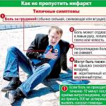
The main complaint of patients is prolonged pain syndrome. The pain can be described by patients in different ways: as burning, pressing, bursting, cruel.
The pain is usually felt in the chest, but can also be felt in both arms, back, neck, and jaw. The duration of the pain syndrome is 20-60 minutes, and sometimes persists for hours. The use of nitroglycerin in case of MI does not bring relief.
Pain syndrome may not be observed.
Concomitant complaints inherent in the disease are dizziness, arrhythmia, weakness, sweating. Cold limbs. Partial loss of contractile function by the myocardium or damage to the valvular apparatus leads to shortness of breath. Elderly and diabetic patients may experience a sudden brief loss of consciousness. Syndromes are often accompanied by fear of death.
During the survey, the reaction of patients to the pain syndrome is clarified. Patients are characterized by agitation, anxiety. In an attempt to relieve pain, they are constantly moving, writhing in bed. They try to induce vomiting. This is in contrast to the response to chest discomfort in angina patients who tend to freeze to stop the pain.
Anamnesis
The vast majority of patients with acute MI have a history of risk factors such as arterial hypertension, diabetes mellitus, overweight, poor heredity, and smoking. In addition, there are indications of manifested coronary heart disease (angina pectoris).
The relatives of the patient can learn about the period preceding the onset of MI, and the factors that provoked it (heavy physical activity, strong emotional stress).
Inspection
Patients with MI are characterized by pronounced excitation. They move restlessly, change positions, can walk around the office. Everything indicates that they are uncomfortable and are trying to change this state. The excited state passes when the pain syndrome is removed.
The exception is patients with left ventricular failure. They are characterized by shortness of breath, hoarseness, bluish coloration of the lips, coldness of the skin.

Auscultation
Deterioration of myocardial contractility leads to muffled tone I. II tone may be slightly weakened or not changed at all. However, if the blood flow is disturbed, then the II tone splits over the pulmonary artery. At a part of patients the III tone is listened.
If the subject has shortness of breath or the lungs are swollen, then wheezing is heard in the lower sections.
Blood pressure and pulse
Blood pressure rises due to stress, fears of the patient, pain. Blood pressure drops when circulatory failure develops. The frequency of contractions immediately after MI is 50-60 per minute.
Differential Studies
Differential diagnosis of myocardial infarction distinguishes it from pathologies with a similar clinic by comparing their signs. Excluding illnesses that are not suitable for symptoms, the diagnosis is reduced to the only correct decision. This technique allows the most efficient conduction of the myocardium. Diagnosis should be based on comparison of MI with diseases such as angina pectoris, acute coronary insufficiency, aortic aneurysm, acute pancreatitis, pericarditis, hepatic colic, and others.
Laboratory diagnosis of myocardial infarction includes a complete blood count and a biochemical study.
Biochemical diagnosis of myocardial infarction takes into account the level of biochemical markers, that is, proteins that make up the heart muscle, which, with irreversible cell damage, begin to be released into the blood. The proteins under study include:
- troponin;
- myoglobin;
- MW-fraction of creatine phosphokinase.
Tests for myocardial infarction show an excess of these enzymes in the blood.
Troponin is the most specific biomarker that can be used to diagnose myocardial infarction. Normally, the level of troponin in the blood is minimal and often not detected. However, 2 hours after MI, its concentration increases sharply and remains at a high level for 1-2 weeks, after which it begins to gradually decrease. The peak concentration of troponin falls on the second day after a heart attack.
Another marker is the level of myoglobin. Its growth is diagnosed 2-4 hours after a heart attack and persists for two days. However, a high concentration of the enzyme can also be caused by other circumstances, for example, kidney pathologies, physical overexertion.

The CPK MB-fraction begins to rise 6-8 hours after CPB and tends to normal on the third day. The more the heart muscle is affected, the more active are the CPK MB-fractions. A number of other circumstances can also lead to the growth of this protein. These can be damage to brain tissue, the consequences of surgery, physical exertion, and others.
A blood test for myocardial infarction also reveals other laboratory signs of the disease:
- increased erythrocyte sedimentation rate;
- increased leukocytosis;
The severity of these symptoms depends on the size of the focus, so they may be absent in mild infarcts.
Electrocardiogram
Electrocardiography is an extremely valuable source of information in determining a heart attack. The cardiogram should be recorded in dynamics.
In the normal state, the cardiac cycle opens with atrial activation. It is shown on the cardiogram by the R wave. A certain time interval passes before the onset of excitation of the ventricles. It corresponds to the P-Q section. The process of involvement of all ventricles is reflected by the QRST complex, while R reflects the maximum amplitude. At the T point, ventricular repolarization is fixed.
When the heart muscle is damaged, its electrical potential decreases relative to healthy tissues. This allows you to establish the exact localization of myocardial infarction on the ECG.
The result of blood flow disorders in MI is the creation of pathological zones. All of them take part in the ECG in myocardial infarction:
- zone of necrosis. Central, marked by the QRS complex. ECG signs of myocardial infarction in this area are the registration of an infarcted Q wave and a sharp decrease in the amplitude of the QR wave;
- damage zone. It is located around the area of necrosis. A heart attack on the ECG is manifested by a rise in the ST segment and its fixation above the isoline;
- ischemia zone. It is located at the border with unchanged tissues. Changes on the ECG are manifested in the fact that the T wave changes its polarity and amplitude.
ECG diagnosis of myocardial infarction also allows you to set the depth of necrosis:
- with a transmural form of MI, the R wave falls out;
- subpericardial MI is characterized by ST depression and T wave transformation
- the intramural form of MI is expressed by merging ST with the T wave and raising this segment.
ECG diagnostics is standardly carried out in 12 departments. This method allows you to accurately detect infarction, localization of necrosis, the degree of damage and temporal changes. In some cases, additional compartments are used, for example, in the case of an abnormal location of the heart.
Echocardiography is used as an additional source of information. According to its results, necrosis is visualized, the scale of the lesion is determined, complications and the effectiveness of therapy are evaluated.
Myocardial infarction is getting younger every year, the number of cases is growing. And, despite the fact that the percentage of deaths is decreasing every year, the overall mortality is still extremely high. A third of the sick die before hospitalization, a tenth - within a year. Therefore, timely diagnosis of the disease is of great importance and is the basis of effective therapy.