1. Definition, classification. 2. Etiology and pathogenesis of leukemia. 3. Acute undifferentiated leukemia. 4. Pathological anatomy of acute myeloblastic leukemia.
5. Pathological anatomy of acute lymphoblastic leukemia.
Chronic leukemia.
1. Chronic myeloid leukemia, its stages. 2. Pathological anatomy of chronic myeloid leukemia. 3. Pathological anatomy of chronic lymphocytic leukemia. 4. Determination of paraproteinemic leukemia. 5. Pathomorphology of multiple myeloma.
Malignant lymphomas. Lymphogranulomatosis.
1. Definition, etiology, pathogenesis. 2. Pathological anatomy of lymphosarcoma. 3. Burkitt's tumor. 4. Lymphogranulomatosis - microscopic characteristics. 5. Clinical and morphological classification of lymphogranulomatosis.
Anemia.
1. Definition, classification. 2. Posthemorrhagic anemia, pathomorphology. 3. Iron-deficiency anemia. 4. Pernicious anemia. Pathomorphology. 5. Hemolytic anemia.
Atherosclerosis.
1. Definition of disease. 2. Etiology of atherosclerosis. 3. Macroscopic views atherosclerotic changes. 4. Stages of morphogenesis of atherosclerosis. 5. Signs of undulating course and progression of atherosclerosis.
Clinical and morphological forms of atherosclerosis.
1. Typical complications of atherosclerosis. 2. Atherosclerosis of the aorta, types of aneurysms.
3. Atherosclerosis of cerebral vessels. 4. Atherosclerosis of the renal arteries.
5. Atherosclerosis of intestinal vessels and limbs.
Hypertonic disease.
1. Definition. The concept of symptomatic hypertension. 2. Pathogenesis of hypertension. 3. Morphological characteristics of the stages of the disease, vascular changes. 4. Clinical and morphological forms of the disease. Organ changes.
5. Causes of death in patients with hypertension.
Ischemic disease hearts. Myocardial infarction.
1. Definition of disease. 2. Etiology and pathogenesis, risk factors. 3. Ischemic myocardial dystrophy. 4. Acute myocardial infarction, morphology. 5. Complications of myocardial infarction.
Rheumatic diseases. Rheumatism.
1. Definition of rheumatic diseases. 2. Rheumatism, etiology, pathogenesis.
3. Morphogenesis of changes connective tissue. 4. Morphology of rheumatic granuloma of Ashoff-Talalaev. 5. Nonspecific cellular reactions.
Pathological anatomy of rheumatism.
1. Morphology of endocardial damage, types of endocarditis. 2. Rheumatic myocarditis, forms. 3. Rheumatic pericarditis. 4. Clinical and anatomical forms of rheumatism. 5. Features of rheumatism in children.
Rheumatoid arthritis. Systemic lupus erythematosus.
1. Rheumatoid arthritis, etiology and pathogenesis. 2. Rheumatoid arthritis - changes in periarticular connective tissue. 3. Stages and morphology of rheumatoid arthritis. 4. Systemic lupus erythematosus, etiology and pathogenesis. 5. Systemic lupus erythematosus - main morphological changes.
Acute bronchitis. Bronchopneumonia.
1. Acute bronchitis - etiology, pathogenesis and pathological anatomy. 2. Definition of bronchopneumonia, etiology and pathogenesis. 3. Pathological anatomy of bronchopneumonia. 4. Morphological features of bronchopneumonia depending on the infectious agent. 5. Legionnaires' disease.
Lobar (lobar) pneumonia.
1. Definition, etiology and pathogenesis. 2. Pathological anatomy of the stages of the disease.
3. Outcomes. 4. Pulmonary complications. 5. Extrapulmonary complications.
54. Chronic nonspecific lung diseases. Chronic bronchitis, bronchiectasis, pneumofibrosis.
1. Definition. 2. Chronic bronchitis. 3. Bronchiectasis, mechanism of development, morphology. 4. Pneumofibrosis, classification. 5. Atelectasis, types.
Emphysema.
1. Definition. 2. Etiology and pathogenesis. 3. Classification. 4. Pathological anatomy of emphysema. 5. " Pulmonary heart", causes and mechanisms of development, morphology.
Acute gastritis. Chronic gastritis.
1. Etiology and pathogenesis. 2. Forms acute gastritis, morphology. 3. Classification chronic gastritis. 4. Morphology of forms of chronic gastritis. 5. Complications. Outcomes.
Peptic ulcer stomach and duodenum.
1. Definition. 2. Etiology and pathogenesis. 3. Acute stomach ulcers. 4. Morphology of a chronic ulcer during the period of exacerbation and healing. 5. Complications of peptic ulcer.
Acute appendicitis. Chronic appendicitis.
1. Definition, etiology and pathogenesis acute appendicitis. 2. Morphological forms of acute appendicitis. 3. Pathological anatomy of forms of acute appendicitis. 4. Definition of chronic appendicitis, morphology. 5. Complications of acute and chronic appendicitis.
Liver diseases. Hepatosis. Toxic liver dystrophy. Fatty hepatosis.
1. general characteristics liver diseases, definition of hepatosis. 2. Toxic liver dystrophy, etiology, pathogenesis. 3. Pathological anatomy of toxic liver dystrophy. Outcomes. 4. Fatty hepatosis, etiology and pathogenesis, the role of alcohol. 5. Pathological anatomy of fatty hepatosis. Outcomes.
Viral hepatitis. Alcoholic hepatitis.
1. Etiology. Pathogenesis. Types of viral hepatitis. 2. Clinical and morphological forms of viral hepatitis. 3. Microscopic characteristics of acute viral and alcoholic hepatitis. 4. Pathomorphology chronic hepatitis. 5. Outcomes and complications of hepatitis
Cirrhosis of the liver.
1. Definition. Classification. 2. Morphogenesis. 3. Pathological anatomy of portal cirrhosis (macro-micro). 4. Pathological anatomy of postnecrotic cirrhosis (macro-micro). 5. Complications.
Glomerulonephritis.
1. Definition of disease. 2. Etiology and pathogenesis. 3. Topography of inflammation in the glomeruli. 4. Pathomorphology of acute and subacute glomerulonephritis. 5. Chronic glomerulonephritis, morphology of its types.
Acute renal failure.
1. Definition of the syndrome, etiology. 2. Pathogenesis. 3. Stages of the syndrome, pathomorphology of the kidneys. 4. Complications. 5. Outcomes.
Pyelonephritis.
1. Definition. 2. Etiology. 3. Pathogenesis. 4. Pathomorphology of acute and chronic pyelonephritis. 5. Complications, outcome.
Nephrosclerosis.
1. Definition, etiology, classification. 2. Pathological anatomy of nephrosclerosis. 3. Definition of chronic renal failure. 4. Pathological anatomy of uremia. 5. Chronic suburemia.
Diseases of the endocrine glands.
1. Addison's disease. 2. Goiter (struma) - micro- and macroscopic types. 3. Endemic and sporadic goiter. 4. Diffuse toxic goiter ( Graves' disease), pathomorphology. 5. Thyroiditis.
Diabetes.
1. Definition. Classification. 2. Etiology and pathogenesis. 3. Pathological anatomy diabetes mellitus, angiopathy. 4. Kidney damage due to diabetes. 5. Complications of diabetes.
Viral diseases. Flu.
1. Features viral infection. 2. The relationship of the virus with the target cell. 3. Influenza - etiology, pathogenesis. 4. Pathomorphology of influenza forms.
5. Brief characteristics of parainfluenza, adenoviral infection.
Measles.
1. Definition. 2. Etiology and pathogenesis. 3. Enanthema. 4. Exanthema. 5. Complications.
Pathological anatomy of AIDS.
1. Etiology and pathogenesis. 2. Periods of AIDS. 3. Lesions of the lungs and brain. 4. Defeats gastrointestinal tract. 5. Skin changes in AIDS. Causes of death of patients with AIDS.
Diphtheria.
1. Definition. 2. Etiology and pathogenesis. 3. Pathomorphology of local changes.
4. Morphology of general toxic changes. 5. Complications and causes of death.
Scarlet fever.
1. Etiology and pathogenesis. 2. Morphology of the first period of the disease. 3. Morphology of the second period. 4. Forms of the disease. 5. Complications and causes of death.
Meningococcal infection.
1. Etiology and pathogenesis. 2. Classification. 3. The role of immune complexes in pathogenesis. 4. Pathomorphology of the main forms. 5. Complications.
Typhoid fever.
1. Definition, etiology. 2. Pathogenesis. 3. Local changes in the intestines. 4. Pathomorphology of the stages of the disease. 5. General changes. Complications of typhoid fever.
Dysentery.
1. Definition of disease. 2. Etiology. 3. Epidemiology and pathogenesis. 4. Pathological anatomy of the stages of dysentery. 5. Complications.
Tuberculosis. Primary tuberculosis. Flow options. Forms of progression.
1. Definition, etiology, pathogenesis. 2. Morphology of the primary tuberculosis complex in the lungs. 3. Morphology of the primary complex in the intestine. 4. Healing of the primary complex. 5. Forms of progression of primary tuberculosis.
Hematogenous tuberculosis.
1. Definition. Differences between hematogenous tuberculosis and primary tuberculosis. 2. Types of hematogenous tuberculosis. 3. Morphology of generalized hematogenous tuberculosis. 4. Hematogenous pulmonary tuberculosis. 5. Hematogenous extrapulmonary (organ) tuberculosis - damage to bones, joints, and other organs.
Secondary tuberculosis.
1. Definition, distinctive features. 2. Pathogenesis, forms. 3. Acute focal tuberculosis. 4. Fibrous focal tuberculosis. 5. Infiltrative tuberculosis.
Secondary tuberculosis.
1. Tuberculoma. 2. Caseous pneumonia. 3. Pathomorphology of fibrous-cavernous tuberculosis. 4. The structure of the wall of acute and chronic cavity.
5. Morphology of cirrhotic tuberculosis.
Sepsis.
1. Sepsis - definition, characteristics of infection. 2. Pathogenesis. 3. Pathological changes - local, general. 4. Classification. 5. Morphology of septicemia and septicopyemia.
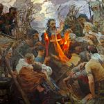
General characteristics of pathological changes. Leukemia is characterized by the proliferation of hematopoietic tissue cells, which differ from normal ones by the loss of their ability to mature. The leukemic process begins with damage to the hematopoietic organs (bone marrow, lymphatic tissue). It also involves those organs that were hematopoietic in the prenatal period (liver, spleen). Gradually, the process becomes generalized and leukemic infiltrates are found almost everywhere. They arise autochthonously from undifferentiated reticular cells located in the walls of blood vessels, the interstitium of glands and parenchymal organs, in the membranes of nervous tissue, and serous leaves. Leukemic infiltrates either diffusely infiltrate the organ, or have the appearance of small or larger nodes. In adults, in 8-10% of cases of acute leukemia (N.A. Kraevsky and M.P. Khokhlova), in children much more often, leukemic infiltrates can be tumor-like in nature, they grow, fuse and compress neighboring organs and tissues.
Rice. 5. Tumor-like growths in thymus gland and mediastinal lymph nodes in hemocytoblastosis.
In children, there is a tumor-like form of acute leukemia with localization and onset of the process in the area thymus gland[J. Cook; color table, fig. 5].
At chronic forms leukemia, tumor-like forms can also be observed, and tumor growths are localized in organs that, in this form of leukemia, are most involved in the process (N. A. Kraevsky and M. P. Khokhlova). For example, in chronic lymphocytic leukemia, tumor-like growths are observed in the retroperitoneal lymph nodes or in the mediastinal lymph nodes.
Secondary changes in leukemia depend on the failure of the organs of the blood system as a whole and on local circulatory disorders and tissue trophism that develop in connection with the growth of leukemic infiltrates. Due to anemia (leukanemia), there is pronounced pallor of the skin, mucous membranes and internal organs.
The skin may show focal leukemic infiltrates in the form of bluish-red raised nodules or diffuse leukemic infiltration, such as facies leonina (see below Skin manifestations leukemia). In the area of leukemic infiltrates and beyond them on skin hemorrhages, necrosis, gangrene, and secondary suppuration often develop. Necrosis is localized at the corners of the mouth, the openings of the nose, on the back, buttocks (places of bedsores).

Rice. 4. Tumor-like nodular leukemic infiltrates in the gastric mucosa with hemocytoblastosis.
Leukemic infiltration of mucous membranes is more often observed in digestive tract. Tonsils can be sharply enlarged, especially when acute forms ah leukemia, with symptoms of gangrenous decay. Diffuse leukemic infiltration of the gums (leukemic gingivitis) may occur. Diffuse leukemic infiltration of the gastric mucosa leads to its sharp thickening with emphasized folding (hirus-like leukemic infiltration of the stomach). In the stomach area, nodular plaque-like leukemic infiltration is also observed (color table, Fig. 4). Peyer's patches and solitary intestinal follicles, increasing in volume, can morphologically simulate the picture of medullary swelling with typhoid fever; This similarity is enhanced by the presence of necrosis and ulceration. Ulcerative-necrotic changes can occur in the mucous membrane of the larynx and epiglottis; in the mucous membranes of the urinary and genital tracts they are less common.
Diffuse leukemic infiltration of serous leaves leads to their opacity and thickening, which can simulate the picture chronic inflammation, such as the meninges. Nodular or plaque-like flat leukemic infiltrates are found on the pleura, pericardium, and dura mater.
Leukemic infiltration of the bone marrow, the detection of which is fundamental for the diagnosis of leukemia, is usually diffuse. Fatty bone marrow is replaced by cellular marrow and when cutting long tubular bones, for example the diaphysis of the femur, it can be removed in the form of a “sausage”; The color of the bone marrow depends on the form of leukemia and may be reddish, reddish-gray, greenish, or blackish from areas of hemorrhage.
Bone tissue - in most cases with symptoms of osteoporosis, noticeable even on macroscopic examination. In severe cases of osteoporosis, the bone can be cut with a knife. Osteosclerosis is less common.
Lymph nodes in the vast majority of cases are diffusely infiltrated, whitish, tumor-like on section, with reddish and blackish lesions.
In parenchymal organs (liver, kidneys) and glands, diffuse leukemic infiltration leads to a significant increase in their volume and weight. The parenchyma of the organ undergoes atrophy; circulatory disorders lead to hemorrhages, necrosis, infarction, sometimes with rupture of the organ capsule. Leukemic infiltrates in parenchymal organs have the appearance of multiple, identical in volume, nodes or nodules of a whitish color, homogeneous appearance, soft consistency and thus resemble metastases malignant tumors(color table, Fig. 1).

Rice. 1. Tumor-like leukemic infiltrates that developed along the liver vessels in hemocytoblastosis.
In the lungs, leukemic infiltration, spreading along the peribronchial tissue and along the alveolar septa, is detected in most cases by histological examination. Circulatory disorders lead to the appearance of effusion in the cavity of the alveoli. The addition of autoinfection is accompanied by the development of pneumonia, abscesses and gangrene.

Rice. 2. Leukemic infiltration of the kidney with massive hemorrhage into the pelvis in acute hemocytoblastosis.
The leukemic process is usually accompanied by hemorrhagic diathesis, which is often the direct cause of death of patients. Brain hemorrhages with massive foci of red softening or in the form of hemorrhagic purpura are typical. Characteristic hemorrhages are in the epicardium, pleura, peritoneum, in the renal pelvis (color table, Fig. 2), in the lungs, and in the cavity of the gastrointestinal tract.
Autoinfection in tissues affected by the leukemic process can lead to the development of coccal or mycotic sepsis.
Modern methods of treatment with hormones and antimetabolites significantly change the picture of leukemia. Therapeutically caused pathomorphosis (see) of leukemia leads to a change in the quality of the leukemic cells themselves, to degeneration and necrosis of leukemic infiltrates. Qualitative changes in the nature of leukemic infiltrates are expressed in the appearance of atypical reticular cells and plasmatization. Particularly significant are dystrophies and necrosis of leukemic cells in the bone marrow, which in some cases leads to panmyelophthisis. In rare cases, massive disintegration of leukemic cells and their nuclei leads to stone formation in the kidneys and uric acid infarctions, up to the development of anuria. IN bone tissue, especially with hormone therapy in children, osteoporosis intensifies with the development of brevispondyly (subsidence of the vertebrae in the craniocaudal direction), a violation of statics and the appearance of calcareous metastases in organs. Less commonly, there is an increase in osteosclerosis up to eburnation. Modern complex therapy leukemia may also be accompanied by the appearance of Itsenko-Cushing syndrome, atrophy of the adrenal cortex, hyperplasia of the anterior pituitary gland, plasmorrhagia and arteriolonecrosis, focal necrosis of parenchymal organs, and an increase in septic processes, despite the use of antibiotics.
Leukemia is characterized by a systemic progressive proliferation of leukemic cells, first in the bone marrow, spleen and lymph nodes, then they move to other organs and tissues, forming leukemic formations around the vessels and in their walls, and leukemic cells appear in the blood. In leukemia, blast cells suppress the differentiation of normal stem cells. Leukemia cells, like all tumor cells, are characterized by atypia varying degrees expressiveness. The use of chromosomal analysis has made it possible to establish that with any leukemia, a clone of tumor leukemia cells, the descendants of one initially mutated cell, spreads throughout the body.
The group of acute leukemias is united by common feature: The tumor substrate consists of young, blast cells. Their course is malignant. The substrate of chronic leukemia is made up of mature cells. Their course is relatively benign.
In acute myeloid leukemia, the bone marrow becomes red, grayish, or greenish. The spleen, liver, and lymph nodes are enlarged. Necrosis in the oral cavity, pharynx, tonsils, stomach. In the kidneys, widespread and focal tumor formations are found. In a third of cases, leukemic infiltration of the lungs develops - leukemic pneumonitis, in a quarter of cases - leukemic infiltration of the meninges - leukemic meningitis. Hemorrhages are detected in the mucous and serous membranes and internal organs.
In acute lymphoblastic leukemia, leukemic infiltration is most pronounced in the bone marrow, spleen, lymph nodes, lymphatic system of the gastrointestinal tract, kidneys, and thymus. Bone marrow is raspberry-red and juicy. The spleen enlarges sharply. The mediastinal and mesenteric lymph nodes are significantly enlarged. The thymus gland reaches gigantic size. Often, the leukemic formation extends beyond the thymus gland and grows into the tissue of the anterior mediastinum, compressing the organs of the thoracic cavity.
Chronic myeloid leukemia develops as a result of malignant transformation of a hematopoietic stem cell, which retains the ability to specialize and mature into mature cellular elements. Chronic myeloid leukemia is characterized by primary damage to the bone marrow with a gradual increase in tumor mass, which is accompanied by an increase in the number of leukemic myelokaryocytes, replacement of fatty bone marrow and infiltration of organs and tissues by leukemic cells. Displacement of normal hematopoiesis, ineffective erythropoiesis (formation of red blood cells), and the appearance of autoantibodies to erythrokaryocytes and platelets lead to the development of anemia and thrombocytopenia. A large tumor mass, especially during cytotactic therapy, is accompanied by a rapid increase in its size, which contributes to the development of severe complications.
As the disease progresses, additional mutations arise in the tumor clone, which lead to the development of new cell clones with high proliferative activity. Their appearance indicates the clonal evolution of the disease, its transition to the final stage. Tumor cells lose their ability to mature.
In this form of the disease, the bone marrow is juicy, gray-red, gray-yellow, and contains young and blast cells. Sometimes osteosclerosis occurs in the bones. The blood is gray-red, the organs are anemic, the spleen is sharply enlarged, sometimes occupying almost the entire abdominal cavity. Its weight reaches 6-8 kg. The liver is enlarged to 5-6 kg. Lymph nodes are significantly enlarged. Leukemia cells form blood clots in blood vessels, penetrate into vascular wall. In this regard, heart attacks and hemorrhages are common. Autoinfection occurs quite often.
Subleukemic myelosis is characterized by a violation of bone marrow hematopoiesis. Myelofibrosis develops in various bones skeleton and extends to all bones. In this case, leukocytosis and thrombocytosis develop. Anemia in myelofibrosis is a consequence of ineffective formation of red blood cells and their autoimmune breakdown, reduction of erythropoiesis, or failure of bone marrow hematopoiesis. Sometimes activation of erythropoiesis is noted.
The subleukemic (aleukemic) nature of leukemia is explained by a violation of coordination between bone and hematopoietic tissue and the elimination (removal) of leukocytes to the periphery due to the development of fibrosis.
In addition to leukemic transformation of hematopoietic tissue, the disease is characterized by the presence of leukemic infiltration in other organs and primarily in the spleen, as well as in the lymph nodes and liver, dysplasia (impaired formation) of bone tissue in the form of pathological bone formation. The enlargement of the hematopoietic organs occurs not only as a result of myeloid metaplasia (impaired formation of bone marrow cells), but also due to the growth of connective tissue in them.
With erythremia, complete hyperplasia (increased formation) of three myelopoiesis lineages, predominantly erythrokaryocytes, is observed in the bone marrow. Cellular elements retain the ability to specialize and mature. The accumulation of tumor mass leads to an increase in the number of red blood cells both in the vascular bed and in the sinuses of the bone marrow, spleen and other organs, causing a violation of the rheology (flow movement) of the blood and, as a consequence, oxygen deficiency of tissues and thrombotic complications. Erythremia is characterized by a certain staged process. As a result of failure of bone marrow hematopoiesis, the disease is accompanied by sudden structural changes. All organs are sharply full of blood. Blood clots often form in arteries and veins. The yellow bone marrow of the long bones turns red. The spleen enlarges sharply. There is an enlargement of the myocardium, especially the left ventricle.
Chronic monocytic leukemia develops as a result of tumor transformation and is manifested by the proliferation of monocytoid cellular elements in the bone marrow, an increase in their content in the blood and their infiltration of the spleen and liver.
Chronic myelomonocytic leukemia occurs as a result of tumor transformation. The mechanisms of tumor development, which are basically characteristic of all leukemias, lead to the suppression of erythro- and thrombocytopoiesis, and to insufficiency of bone marrow hematopoiesis.
Chronic lymphocytic leukemia is benign tumor lymphatic tissue. Tumor cells are predominantly mature lymphocytes. The number of lymphocytes in the lymph nodes, spleen, and liver increases.
The bone marrow is red with areas of yellow. The lymph nodes of all areas of the body are sharply enlarged, merging into huge soft or dense packets. The size of the tonsils and intestinal lymphatic follicles increase. The liver, kidneys and spleen are enlarged. Leukemic infiltration is observed in many organs of the mediastinum, mesentery, myocardium, serous and mucous membranes.
Paraproteinemic leukemia. The group of paraproteinemic leukemias includes tumors from B-lymphocytes:
- multiple myeloma;
- primary macroglobulinemia (Wandelstroem's disease); heavy chain disease (Franklin disease).
Tumor cells synthesize homogeneous immunoglobulins or their fragments, the so-called pathological immunoglobulins. Highest value has multiple myeloma. Most often, myeloma develops at the age of 45-60 years. Men and women get sick equally often. Myeloma metastases are observed in the spleen, liver, kidneys, lungs, and lymph nodes. Nephrosis and sclerosis develop in the kidneys. Edema of the myocardium and lungs is noted. Inflammatory changes in the form of pneumonia and pyelonephritis are found in the lungs and kidneys. Lime is deposited in the organs, as well as an altered protein - amyloid.
Various forms of leukemia differ in the uniqueness of their morphological manifestations, but they are characterized by common features. All forms of leukemia are characterized by systemic tumor growths arising from hematopoietic cells. Leukemic growths (infiltrates, proliferates) are constantly observed in the bone marrow, often in the spleen, lymph nodes, liver, and other organs and tissues.
Leukemic infiltration often causes an increase in the size and weight of organs, and the formation of extensive nodular growths is possible. The leukemic process is naturally accompanied by signs of general anemia, hemorrhage, dystrophic and necrotic-ulcerative changes, and complications of an infectious nature.
The listed specific and nonspecific changes, closely related to each other, determine the pathological picture.
Depending on the stage of the disease, the form of leukemia, the treatment performed, the volume of leukemic growths and their prevalence in the body, the intensity of the accompanying changes can be expressed to varying degrees. When treated with cytostatic agents, changes often differ from the typical pathological manifestations of the disease.
Pathoanatomical diagnosis of leukemia and its particular forms is based on the analysis of sectional data and intravital examination of hematopoietic organs, mainly bone marrow, using trepanobiopsy. Less commonly, the objects of lifetime study are:
- enlarged lymph nodes removed for diagnostic purposes;
- nodular growths various localizations;
- liver biopsy material;
- removed spleen.
Acute leukemia is characterized by the proliferation of poorly differentiated leukemic (blast) cells.
During pathological examination, the manifestations of various forms of acute leukemia are similar to each other, and therefore their differentiation into sections is not possible. When autopsying the dead, signs of leukemia can be expressed to varying degrees depending on the prevalence of the process. With a detailed picture of acute leukemia, the bone marrow of flat bones is dark red, pink-red in color, and the juicy, fatty bone marrow in the tubular bones is replaced by leukemic growths.
In some cases, an enlargement of the spleen and lymph nodes is detected, however, which, in the liver, is not as pronounced as in chronic leukemia.
Spleen
The weight of the spleen in most cases increases 2-3 times compared to the age norm, less often - the size and weight of the spleen remain normal even in the presence of leukemic infiltration, sometimes it can significantly increase (up to 700-1000 g).
The relationship between the degree of splenomegaly and the duration of the disease (according to clinical data), as well as the form of acute leukemia, has not been established. The spleen tissue on the section is red, with an erased pattern of the structure, sometimes there are infarctions, the pulp gives copious scrapings. Capsular ruptures, even with massive leukemic infiltration, rarely occur.
The lymph nodes
Enlargement of the lymph nodes can be expressed to varying degrees and is usually systemic; sometimes the lymph nodes of one anatomical region are enlarged. In some cases, the lymph nodes are not enlarged. The affected lymph nodes have a soft consistency, are not fused to each other, and their tissue is pink-red in color.
Liver
The liver, as a rule, is slightly enlarged; small grayish-white stripes are sometimes visible on the section, corresponding to areas of leukemic infiltration. Specific growths are often detected in the gastrointestinal tract. In such cases, swelling of the gums, enlargement of the tonsils, group follicles (Peyer's patches) are observed. small intestine and solitary follicles of the colon, areas of diffuse thickening of the wall of the stomach and intestines.
Characterized by extensive foci of necrosis of the mucous membrane of the gastrointestinal tract with ulceration. Occasionally, perforation of the intestinal wall and peritonitis are observed. With a significant prevalence of the process, nodular or diffuse whitish growths of leukemic tissue are detected in the skin, heart, kidneys, pleura, epicardium, genitals, and in children - in the thymus gland. Against the background of systemic leukemic infiltration in children in approximately 50% of cases and in adults in 10% of cases of acute leukemia, untreated or resistant to therapy, extensive nodular growths of various localization are observed.
Nervous system
In acute leukemia, central lesions are often detected. nervous system– neuroleukemia – in the form of infiltration of the meninges and substance of the brain and spinal cord, cranial nerves. This type of damage occurs when various forms acute leukemia most often with acute lymphoblastic leukemia in children. Leukemic infiltration of the meninges can be isolated or combined with pathological changes brain substances.
Focal or diffuse thickening of the arachnoid membrane and extensive hemorrhages in it are detected; the dura mater is less often affected. With leukemic infiltration of the brain, multiple dark red foci are detected - from pinpoint to several centimeters in diameter, mainly in the white matter. Sometimes large areas of red softening of the brain matter appear with a breakthrough into the ventricles of the brain and under the dura mater.
In acute leukemia, concomitant changes are expressed due to insufficiency of hematopoiesis and leukemic infiltration:
- multiple hemorrhages;
- extensive necrotic-ulcerative processes;
- complications of an infectious nature.
In some cases, these changes come to the fore during autopsy, and the signs of leukemia are weak or even absent (organs are not enlarged in size; in adults, fatty bone marrow remains in the diaphysis of the tubular bones). This picture can occur not only under the influence of treatment, but sometimes also in patients who have not been treated. In such cases, diagnosis of acute leukemia at autopsy is difficult and is possible only after microscopy.
At microscopic examination differential diagnosis forms of acute leukemia is based on the cytochemical characteristics of blast cells that form leukemic growths. The structure of blast cells, the frequency of specific damage to various internal organs, the nature of the location of leukemic infiltrates in them differ quite relatively in different forms, and therefore cannot serve as the main criteria for establishing the form of acute leukemia.
IN practical work pathologists, when examining biopsy material obtained from hematopoietic organs and sectional material, the form of acute leukemia is usually established taking into account the data of a cytochemical study of intravital peripheral blood smears and bone marrow punctures.
Identification of the form of acute leukemia in the study of tissue sections is carried out using histochemical techniques, including methods for determining peroxidase, glycogen based on the PAS reaction, the activity of nonspecific esterase (with the substrate α-naphthyl acetate and incubation of control sections in a medium with the addition of sodium fluoride). For control and more detailed differentiation, lipids with black sudan, chloroacetate esterase activity, and acid phosphatase are usually also detected.
Tissues taken only in the first hours after death are subject to histochemical examination. The results should be assessed on thin sections (up to 5 µm) at high microscope magnification. Due to the fact that in tissue sections it is difficult to detect a positive PAS reaction in granular form and the degree of acid phosphatase activity, especially with a small number of positively reacting blasts, histological examination It is advisable to combine it with a cytochemical study of cells in prints from hematopoietic organs. In patients at the treatment stage, determining the form of acute leukemia is difficult due to frequent changes in the cytochemical properties of blasts under the influence of cytostatic agents. In such cases, when examining biopsy and sectional material, the form of acute leukemia is diagnosed based on the results of a cytochemical study of leukemic cells in the peripheral blood and bone marrow before treatment.
In the bone marrow, according to trepanobiopsy, in the first stages of the disease, accumulations of blast cells are focal, and elements of active bone marrow are found in sufficient quantities. Acute circulatory disorders and areas of hypoplasia are often observed, and necrosis may be detected.
The progression of the disease is characterized by an increase in the number and size of leukemic growths, their fusion, and then diffuse infiltration of bone marrow tissue. The number of normal hematopoietic cells decreases markedly. There is often infiltration of the periosteum, and the process may spread to the surrounding soft tissue.
The development of leukemic growths is accompanied by pronounced resorption of bone tissue, mainly by the type of smooth resorption. Very rarely, osteoclasts take part in this process. Due to partial, and in some places complete resorption of the bone beams, a significant expansion of the bone marrow cavities, thinning, and in some areas destruction of the cortical layer occurs.
In the early childhood sharp changes are observed in the zone of enchondral ossification.
In acute leukemia, focal new formation of atypical bone tissue rich in osteoid and proliferation of fibrous tissue are rarely observed. Along with small foci of necrosis, extensive infarct-like coagulation necrosis sometimes occurs, defined as yellow-white dry areas; There are hemorrhages in the circumference, and there may be an accumulation of macrophages. In the area of leukemic growths, thinning and partial disintegration of the reticular fibers are often detected.
Observations with focal or diffuse myelofibrosis, which is more often detected in myeloblastic leukemia, are described. In such cases, puncture usually fails to obtain bone marrow, and to establish a diagnosis it is necessary to examine the trepanobiopsy material.
Leukemic infiltration of the tissue of the spleen and lymph nodes is accompanied by a decrease in the size and number of follicles until their complete disappearance. In the spleen, infiltration of trabeculae is often observed with disintegration of the walls of trabecular vessels, with deformation and narrowing of their lumen, and there are fields of hemorrhages. Focal infiltrates initially appear in the lymph nodes, which, as the disease progresses, increase in size and the lesion becomes diffuse.
II. PRIVATE PATHOLOGICAL ANATOMY. CHAPTER 12. DISEASES OF THE HEAT-POIZING ORGANS AND LYMPHOID TISSUE: ANEMIA, LEUKEMIA, LYMPHOMA
II. PRIVATE PATHOLOGICAL ANATOMY. CHAPTER 12. DISEASES OF THE HEAT-POIZING ORGANS AND LYMPHOID TISSUE: ANEMIA, LEUKEMIA, LYMPHOMA
Anemia (general anemia) is a condition characterized by a decrease in the concentration of hemoglobin per unit volume of blood, which is often accompanied by a decrease in the number of red blood cells.
Principles of classification of anemia: By etiology And pathogenesis(anemia due to blood loss, due to increased destruction erythrocytes due to insufficiency of erythropoiesis), according tomorphological characteristics of erythrocytes, the average hemoglobin content in an erythrocyte, the nature of the course, functional status bone marrow, peculiarities of iron metabolism.
Types of anemia:posthemorrhagic (acute and chronic), hemolytic, dyserythropoietic, iron deficiency, megaloblastic (Addison-Beermer, pernicious, B 12-deficient and the like), aplastic (hypoplastic).
Tumors of hematopoietic and lymphoid tissues are clonal growths of transformed (as a result of mutation) cells of hemopoiesis or lymphopoiesis. All tumors of hematopoietic and lymphoid tissues are divided into 2 large groups - leukemia (leukemia) And lymphomas. In leukemia, the bone marrow is primarily affected. By histogenesis leukemias are divided into myeloid and lymphoid; according to the degree of cell differentiation (cell maturation ability) - acute and chronic.
Classification of leukemia: acute (blastic) - lymphoblastic B- and T-leukemias and myeloblastic. Acute myeloblastic leukemia: with minimal differentiation, without maturation, with partial maturation, myelomonoblastic, monoblastic, erythroblastic and megakaryoblastic. A separate subgroup is formed by acute myeloid leukemia with persistent cytogenetic abnormalities (promyelocytic and a number of others).
Chronic leukemias of myeloid origin(chronic myeloproliferative diseases): chronic myeloid (myelocytic)
leukemia (myeloid leukemia), polycythemia vera(Vaquez-Osler), chronic (idiopathic) myelofibrosis, essential thrombocythemia.
Chronic leukemia of lymphoid origin:B-cell - chronic lymphocytic leukemia(lymphocytic leukemia), prolymphocytic, hairy cell; chronic T-cell.
Plasma cell tumors (myeloma, etc.) consist of terminally differentiated B cells (plasmocytes), with focal or diffuse growth in the bone marrow, but without the release of tumor cells into the peripheral blood.
Lymphomas are primary regional tumors of lymphoid tissue. The tumor can be localized in the lymph nodes, spleen, thymus, mucosa-associated lymphoid tissue (MALT), and less often in any other tissues and organs (extranodal lymphomas). As the tumor progresses, tumor infiltration of the bone marrow and other organs develops, and tumor cells enter the peripheral blood (leukemia).
Classification of lymphomas:Hodgkin's lymphoma (Hodgkin's disease, lymphogranulomatosis), B-cell lymphomas, T- and NK-cell lymphomas. The last 2 groups of lymphomas are traditionally designated by the term"non-Hodgkin's lymphomas".

Rice. 12-1. Macropreparations (a-d). Spleen in chronic myeloid leukemia: the spleen is sharply enlarged in size (splenomegaly, weight up to several kilograms), in cross-section it has a juicy appearance, dark red color, with yellowish-white and reddish-brown dense foci under the capsule (ischemic infarctions of varying duration) (a, d - preparations by L.V. Kaktursky; b, c - preparations by I.N. Shestakova)


Rice. 12-1. Ending

Rice. 12-2. Microslides (a, b). Liver in chronic myeloid leukemia: pronounced infiltration of parenchymal lobules along the sinusoids by tumor myelocytes, fatty degeneration and lipofuscinosis of hepatocytes. In the portal tracts, infiltration is not expressed. Staining with hematoxylin and eosin: a, b - x 200

Rice. 12-3. Macropreparations (a, b). Lymph nodes in chronic lymphocytic leukemia: the lymph nodes of the mesentery of the intestine are sharply enlarged, compacted, merge into dense packets, on the section they are presented as a homogeneous juicy white-pink tissue, in places with small hemorrhages (preparations by I.N. Shestakova)

Rice. 12-3. Ending

Rice. 12-4. Microslides (a, b). Liver in chronic lymphocytic leukemia: pronounced infiltration of portal tracts by tumor lymphocytes of varying degrees of maturity (but not blasts). In parenchymal lobules there is practically no infiltration. Hepatocytes in a state of fatty degeneration and lipofuscinosis. Staining with hematoxylin and eosin: a - x 100, b - x 400

Rice. 12-5. Microslides (a, b). Vertebral bone marrow in acute undifferentiated leukemia: the bone marrow is infiltrated with homogeneous undifferentiated hematopoietic cells. Staining with hematoxylin and eosin: a - x 100, b - x 400

Rice. 12-6. Electron diffraction pattern. Leukemia cell in acute undifferentiated leukemia: pathological mitosis with uneven chromatin distribution (from)


Rice. 12-7. Macropreparations (a, b). A fragment of the parietal bone in multiple myeloma: multiple round, up to 2 cm in size (“stamped”) foci of destruction of the spongy substance of the flat bone of the calvarium, especially clearly visible in the light (1); (b - specimen from the museum of the Department of Pathological Anatomy of Moscow State Medical University)

Rice. 12-8. Electron diffraction pattern. Myeloma cell: in the cytoplasm there are sharply dilated tubules of the endoplasmic reticulum filled with paraprotein (1). Eccentrically located core (2); (from )

Rice. 12-9. Macropreparation. Spleen in Hodgkin's lymphoma (lymphogranulomatosis): the spleen is sharply enlarged (splenomegaly), dense in consistency, with a smooth surface, when cut, its tissue has a variegated appearance: with multiple, different-sized foci of white, yellow, dark red and brownish color (“porphyry spleen” ); (preparation from the museum of the Department of Pathological Anatomy of the Moscow State Medical University)

Rice. 12-10. Microslides (a-d). Cervical lymph node in Hodgkin lymphoma (lymphogranulomatosis), mixed-cell variant: structural pattern lymph node erased, the lymphoid tissue is replaced by cells, including large mononuclear Hodgkin cells (1), multinucleated Berezovsky-Reed-Sternberg cells (2); lymphocytes, plasma cells, eosinophilic and neutrophilic leukocytes are also present (foci of necrosis and sclerosis may occur in the tumor).
a, b - staining with hematoxylin and eosin; c, d - immunohistochemical method; c - diagnostic cells with CD15 expression; d - with CD30 expression; x 400 (c, d - preparations by G.A. Frank)

Rice. 12-10. Ending

Rice. 12-11. Microslide. Extramedullary (extramedullary) hematopoiesis in the liver in chronic anemia: multiple islands of hematopoietic tissue are detected in the liver tissue, represented by different cells hematopoietic systems, including erythroblasts, etc. 1 - cells of hematopoietic tissue, 2 - fatty degeneration of hepatocytes (from)


Rice. 12-12. Macropreparations (a-c). Pioid (pus-like) bone marrow of the diaphysis femur(a), vertebral bodies (b) and a pronounced increase in the size and weight of the liver (hepatomegaly) (c) in chronic myeloid leukemia (b - preparation by L.V. Kaktursky)

Rice. 12-13. Hemorrhagic syndrome (purpura) in acute leukemia (a-d): multiple petechial and confluent hemorrhages in the skin (a), gastric mucosa (b), epicardium (c), soft meninges (d) (preparations by I.N. Shestakova )

Rice. 12-13. Ending

Rice. 12-14. Microslide. Bone marrow trephine biopsy for acute myeloblastic leukemia: positive immunohistochemical reaction for myeloperoxidase in blast cells: x 200 (preparation by G.A. Frank)

Rice. 12-15. Macropreparations (a-c). Malignant lymphoma of the anterior mediastinum (a-b) and mesenteric lymph nodes (c) (c - preparation by I.N. Shestakova)

Rice. 12-15. Ending

Rice. 12-16. Microslides (a, b). Lymph node biopsy for T-cell lymphoblastic lymphoma (lim-
fosarcoma): a - diffuse proliferation of lymphoblasts, b - expression of CD10 in lymphoblasts.
a - hematoxylin and eosin staining, b - immunohistochemical method; a - x200; b - x400 (preparations by G.A. Frank)

Rice. 12-17. Microslide. Lymph node biopsy. Burkitt's lymphoma: a picture of a “starry sky”. Staining with hematoxylin and eosin: x 100 (preparation by G.A. Frank)

Rice. 12-18. Microslide. Trephine biopsy of bone marrow for megaloblastic anemia: hyperplasia of erythrokaryocytes.
Hematoxylin and eosin staining: x400 (G.A. Frank’s preparation)

Rice. 12-19. Macropreparations (a-d). Hemorrhagic shock in acute posthemorrhagic anemia: a - “shock” kidney (necrotic nephrosis, multiple confluent hemorrhages in the mucous membrane of the pelvis and calyces); b - acute anemia of the spleen - reduced, flabby consistency of the spleen, with moderate scraping on the incision; c - petechial and confluent hemorrhages under the endocardium of the left ventricle of the heart; d - “shock” lung - pronounced edema, foci of hemorrhage, atelectasis, increased volume and weight of the lungs

Rice. 12-19. Ending

Rice. 12-20. Microslides (a-b). Acute posthemorrhagic anemia. Hemorrhagic shock. DIC syndrome: fibrin microthrombi (red): a - in the lumen of the pulmonary veins; b - in the sinusoids of the red pulp of the spleen. Staining according to Marcius-Scarlet-Blue (MSB), a - x 100, b - x 200 (preparations by A.V. Dobryakova)