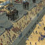
Fig. 27. Microscopic Cala: A - native drug normal; b - muscular fibers of varying degrees of digestibility; in - neutral fat; g - crystals of fatty acids and soap; d - potato cells, starch grains, ozofyl flora; E - digestible and unsecured fiber
For a complete microscopic study of Cala, several drugs are prepared. In most cases, wet native drugs are used, but sometimes for the study of cellular elements and differentiation of simplest, fixed painted drugs are prepared. To prepare native drugs, a piece of feces are placed in a porcelain mortar and rub in a small amount of isotonic solution to the consistency of liquid cashem, then the prepared emulsion is placed on the slide glass.
Typically prepare four drugs.
Native unpainted drug. In the native preparation, most elements of feces are differentiated: muscle fibers, vegetable fiber, neutral fat, fatty acids, soaps, cellular elements, mucus, helminth eggs, simplest crystals.
With a solution of ligol (1 g of iodine, 2 g of potassium iodide, 50 ml of distilled water). The emulsion is prepared in analogously to the native drug, but the feces are rubbed not with isotonic solution, but with a solution of a double fortress. In these drugs, it is possible to detect starch, iodofilic flora, as well as differentiate the cysts of the simplest.
The third drug is prepared in the form of a thick water emulsion, to which a drop of a solution of methylene blue (0.5% solution) is added. These drugs make it possible to detect fat and products of its splitting.
Native drug with glycerin. Glycerin is used to enlighten the eggs of helminths and helps to detect them. Preparations are covered with coating glass and are considered at first under small (8 x 10), and then under large (40 x 10) magnification.
Elements of food origin
Dyrette is the main background with normal feces microscopy. It represents the remains of the foodstuffs of microorganisms that have broken cell elements. Petriton has the form of amorphous formations of small sizes, mostly a grainy form. According to the grains of Derita, it is impossible to determine the source of their education. Petriton in large quantities is contained in the crash. The fat feces, the less Derita. When making data from microscopic research, child is not marked.
The mucus during microscopic examination is defined as a unstructured substance with single cells of the cylindrical epithelium. The mucus, detected only by microscopically, occurs from those intestinal departments, where the wheel weights are still so liquid that it mixes with them during the peristaltics.
In the case of the decorated feces, the origin of only microscopically detectable mucus should be attributed to the subtle intestine or blind intestine.
With a cascidious or liquid chair, the origin of small mucus particles is more difficult to determine, but the absence of a mucus simultaneously visible to the naked eye speaks rather against its origin from the colonist. In general, the smaller the lumps of the mucus and the more closely they are mixed with the feet, the higher the place of their allocation. The mucus lumps visible to the naked eye should be microscopic examination. Pumps of mucus are first gently washed in a physiological solution, freeing from feces. Under a small increase in the microscope, the mucus has a form of light lumps or seasures with fuzzy, incorrect outlines, linked into the main brown or yellow mass.
To distinguish the mucus from the elements of the feces, you can apply painting on GEHTU: mix before use equal volumes of 1% neutral red and 0.2% diamond green. 1-2 drops of reagent are applied to the solder of Kala. After a few minutes, the mucus is painted in a light red color, the rest of the mass is in green, except for shells and nuclei of plant cells, acquiring a lilovo-red color.
Muscular fibers in feces in a healthy person, located on the usual diet, are not detected or detected in the form of single yellowish boulders. Muscle fiber detection in large quantities (creative) indicates deficiency of protein digestion. Microscopically distinguishes untrained, weakly edged and scraps well-doused muscle fibers. Unpained muscle fibers have a more elongated cylindrical shape with well-preserved straight corners and pronounced transverse aperture. Specify the failure of the digestion of protein food in the stomach.
Weakly digested fibers have a cylindrical shape with slightly smoothed corners. They can see the longitudinal, and sometimes weakly-wave cross-allocated. Indicate a violation of the function of the pancreas. Scraps of well-doused muscle fibers have the form of small homogeneous lumps, more than oval shape with rounded edges, bright yellow. Indicate the absence of peptidases produced in the intestine.
Small scraps of muscle fibers that have lost allocated and have acquired an irregular shape, to accurately determine with simple microscopy it is not possible. To identify the protein nature of such unformed boulders or particles, simple chemical samples can be used - Biuret and Xanthoprotein. At the first rocket, the feces are stirred on the subject glass with a 10% solution of caustic potassium and add 1-2 drops of 1% of the solution of copper sulphate. Protein particles acquire purple staining. To carry out xanthoprotein sample, the feces are mixed with a strong nitric acid and heated. Protein particles are painted in yellow.
Connectual fibers have the kind of grayish, refracting light fibers, sometimes similar to the mucus. However, in contrast to the latter, they swell under the influence of acetic acid and lose the fibrous structure. Normal feces are not detected. It is detected in violation of gastric digestion, poor feeding of food, with poorly roasted meat.
Fat and products of its splitting. Normally, the fat arrived with food in moderate quantity is absorbed almost completely. Therefore, a small amount of soap can meet in Kale with the absence of neutral fat. The detection of a significant amount of neutral fat and products of its splitting indicates a violation of digestion and suction of fat. Neutral fat in the native preparations of Cala has the form of colorless droplets.
Fatty acids and soaps are found in the form of boulders, drops and crystals. Crystals have the shape of thin needles pointed from two ends, often fold into small beams, are sometimes located radially, surrounding the glybank of fatty acids. Detection in the native preparation of colorless droplets, boulders and needle crystals suggests the steamer. To confirm this assumption, the drug is painted by Sudan III.
The drops of neutral fats and drops of fatty acids are painted in red, and crystals and bumps of fatty acids and soaps are not stained.
For differentiation of droplets of neutral fat and drops of fatty acids, it is mixed with methylene blue: 1 drop of 0.5% methylene blue solution is mixed with 1 drop of a carte emulsion, covered with coating glass, a preparation is viewed under a microscope. Neutral fat is not painted, drops of fatty acids are painted in blue.
Fatty acids with soaps are differentiated using a sample with heating. When the native preparation is heated to 80-90 ° C, the crystals and bolbs of fatty acids are melted in drops (re-turning into the locks during cooling), the locks and crystals of the soap in the drops are not melted. Their fusion occurs after the addition of 30% acetic acid and the subsequent heating to the boil.
Violation of the absorption of fat is associated in most cases with the insufficient activity of the lipase or with an insufficient entry into the intestine of bile. However, if the fat is concluded in the connecting tissue (fatty tissue), then for its release, sufficient digestion is necessary in the stomach of the connective tissue, so the violation of the specified process can also lead to the steamer. Plant fiber and starch are the residues of the carbohydrate component of food. There are two types of fiber: digested and unsecured. The unsecured fiber is a support fiber (vegetable peel, fruit, vessels and hairs of plants, etc.), the intestine does not split and completely distinguishes with the feet. With microscopy of native unpainted drugs, it has a variety of sharp outlines, the correct pattern in the form of thick dual-circuit cellulose shells of brown, yellow and gray color.
The digestible fiber is meal, parenchymal cells of vegetables and fruits and consists of rounded cells with a thin shell and a cellular structure. In microscopy, the tissue is digested by the fiber differs from unsecured gentle contours, the presence of starch grains or coloring pigments. The digestible tissue includes large oval potato cells. They stand out in a native preparation in the form of colorless ovals on a yellow or brown dedrete background. They are located either by one by one or small groups of 2-3-4 cells.
Their difference from mucus is that the outlines of potato cells are clear, rounded, while the outlines of the lumps of mucus are vague and their shape is uncertain.
Starch grains in a native non-colored preparation have the appearance of oval colorless formations located extracellular or inside the cells of the tissue of fiber. The presence of starch grains is better detected in the preparation painted with a solution of lugol. Under the influence of iodine, unchanged starch is painted in a blue-black color, the products of its partial cleavage - in purple (amylodextrin) and in red-brown (erytrodextrin). Almost completely digested grains of starch remain colorless. The abundance of the cockle of the starch and the digestible fiber is accompanied by the usually rich iodophilic flora. The microbes belonging to it, feeding at the expense of carbohydrates split by them, put the granules inside the granules, staining with iodine. The fervor caused by this flora leads to the formation of organic acids that give to the calves with an acidic reaction.
Normally, only unsecured fiber and single potato cells are found in the feces, there is no starch. The presence of starch in Calais (amylorius) indicates disadvantage of digestion or accelerated evacuation of food chimus.
The pancreas lesions significantly reflected on the digestion of fats and proteins, relatively little affect the absorption of starch, if they are not accompanied by diarrhea. The lack of amylase is compensated by amylolytic enzymes of other departments of the digestive tract and bacteria.
Elements separated by the intestinal wall
The elements separated by the intestinal wall make up a second group of microscopic research objects. In addition to the mucus, these are red blood cells, leukocytes, tissue macrophages, cell epithelium cells, cages of malignant tumors.
A flat epithelium, captured occasionally when passed by dense feed masses through an anal hole, does not have a diagnostic value. Cell elements are detected in a cage containing mucus. When preparing a drug for microscopy from the feces, mucous membranes, bloody lumps are isolated, washed with isotonic solution and applied to the slide.
Investigate native drugs and drugs painted by Romanovsky - GIMZE.
The cells of the intestinal epithelium are usually found in lumps of mucus. Sometimes cells are well preserved, more often they are deformed due to soaping them with soaps or the digestion started.
Single cell epithelium cells can be found in normal feces as a consequence of physiological lunches. Large groups of similar cells should be regarded as a sign of inflammation of the intestinal mucosa.
Leukocytes, located in the mucus in a significant amount (accumulation), indicate an inflammatory process in the thick intestine. Leukocytes in the mucus, coming from the small intestine, have time to collapse.
Unchanged red blood cells are found in feces in bleeding from a large intestine and rectum (ulcerative processes, hemorrhoids, etc. and); When bleeding from higher underlying intestinal departments, erythrocytes are either completely destroyed, or acquire the character of the shadows, and it is very difficult to recognize them.
Macrophages are found in some inflammatory processes, especially with bacterial dysentery. When painting on Romanovsky - GIMZE, these cells are larger than leukocytes, contain a circular or oval kernel and various cytoplasmic inclusions. To distinguish macrophages from the ciste of the simplest, it should be resorted to the color of the Lugol solution, in which a dark-colored shell is noticeable in cysts, as opposed to macrophages.
Cells of malignant tumors can get into the feces when the tumor is located in the rectum. At a higher localization of the cell tumor are subject to changes that make it difficult to recognize them. The diagnostic value is not the foundation of non-single cells, but fragments of tissue, cell groups that differ in characteristic atipsis. If the tumor is suspected, a cytological examination of the material obtained with a suspicious area should be carried out.
A copron is a study of the roasting masses to determine their properties, chemical and physical composition, the presence of anomalous inclusions to confirm the diagnosis of one or other disease, as well as to track the dynamics of diseases and therapy efficiency.
Calurate content is formed while promoting Himus (lump of food) through the digestive tract from entering the oral cavity to the anal channel. And therefore, the results of the coprogram are a valuable diagnostic criterion for determining the diseases of the gastrointestinal tract.
When a coprogram is assigned
In Kale, it is possible to detect various in terms of quantity and type of microorganisms, felling pigments, particles of untapped food products, epithelial cells from various intestinal areas.
In accordance with the peculiar features of the capabilities, an experimental laboratory will easily find pathological processes that are localized in one or another intestinal department.
Coprogram is assigned at:
- Glice invasion.
- Acute, chronic stomach diseases.
- Neoplasms.
- Diseases of the duodenum.
- Various infections.
- Pathologies of the pancreas, liver, gallbladder and ducts.
- Pathological processes in the small intestine.
- To assess the effectiveness of the therapy and the need to correlate treatment.
With the help of coprological analysis, you can identify dysbacteriosis (the condition when the ratio of normal and pathogenic microorganisms is disturbed and recent reproduction of the latter).
The coprogram is rarely used as an isolated diagnostic method, more often its purpose is combined with other studies. However, despite this, the diagnostic value of coprological analysis is very large.
Rules for handing analysis
There are several simple rules that need to be considered when delivering the material for this analysis:

Calm mass collection

It is necessary to collect material in the morning and to get enough to the laboratory as quickly as possible (the accuracy of the data obtained) depends on this).
If necessary, the filled container can be stored in the refrigerator no more than eight hours (the temperature is not higher than five degrees).
Calate's study is carried out within two or three days, sometimes it takes a little longer (five to six days).
Value fees in infants

In infants, the feces are assembled with the help of a linen or diaper (in the case of a liquid chair).
In the presence of constipation, the stimulation of defecation is carried out with the help of a belly massage, sometimes they put a gas pipe.
Hands before collecting material necessarily wash.
Collecting material from diapers is undesirable.
Cales Coprogram: Decryption in older children and adults (research order)
Initially, macroscopy is carried out, while evaluating:
- Appearance Cala.
- His density.
- Tint (normal or pathological).
- Smell.
- The presence of blood, untapped food, mucus, pus.
- The presence of helminths.
- The presence of pancreatic or bile stones.
After carrying a mating microscopy, allowing to evaluate the digestion function of food.
Cales Coprogram (decrypt in children): Table
Color
Calla color is normal (a variety of shades) due to the presence of Sterkobilo in it. Shades of feces depend on nutrition and drugs taken. So, a vegetable diet can give Kala greenish tint, coffee and blueberries - black, dairy products - light yellow, beets - red, and antibiotics - golden.
With some pathologies, the color of the carts also changes:
- Red-brown cal - bleeding from the lower parts of the intestine.
- Black - bleeding with duodenal ulcers or stomach.
- Green - the presence of enteritis, dysbacteriosis.
- - Diseases of the biliary tract, liver.
When deciphering the core coprogram in children (see the photo below), which are on breast (natural) feeding is determined by yellow, green-yellow, golden yellow color of the cart. The artificials feces can be light brown or pale yellow.

In kids, a bilirubin, giving out the feces, a greenish color can be released to six months. That is, if, besides the green chair, there are no other symptoms, then such a state of treatment does not require.
Microscopy
The presence of a protein indicates inflammation in the gastrointestinal bodies, polyps, ulcers and neoplasms. There is no protein in a normal coprogram.

Blood appears in the root masses due to bleeding, which can be caused by helminths, tumors, ulcers, polyps. Changed blood points to bleeding in the upper gastrointestinal departments, and unchanged - from the lower.
The increase in the level of sterkobilo appears in feces in hemolytic anemia. A decrease in this indicator is a sign of blockage of bile ducts.
The appearance of bilirubin indicates dysbacteriosis and acute inflammatory processes.
The presence of mucus is a sign of intestinal infections (dysentery, salmonellosis, colitis). However, it is necessary to take into account the age of the child, because when Cala Coprogram (deciphering in children), the mucus can be an option for the norm (children up to year).
The presence of pathological flora is a sign of dysbiosis.
In the Cala coprogram (when deciphering in children), child, if its number is below the appropriate normal age, may indicate violations in the digestive process.
The presence of a large amount is a violation of the selection or suction of bile.
Unchanged - pancreatic pathology.
The presence of starch grains - Malabsorption syndrome, chronic pancreatitis.
Soap (which is normal there must be a small amount) - the problems of the duodenum intestine, pancreatitis, the stones of the gallbladder.
In the Cala coprogram (deciphering in children), leukocytes in large numbers are talking about the presence of inflammatory gastrointestinal processes.
Fatty acid. Normally not defined. If they are presented in the cartoons, it is worth susceptible to the lack of enzymes, accelerating the intestinal activities and disturbance of the outflow of bile.
Plant fiber. If the fibers are insoluble (for example, vegetable peel and so on) is an option of the norm, if soluble fibers are present in the feces - this indicates a lack of hydrochloric acid in the stomach.
There are no connectual fibers in the norm. Their appearance is a sign of pancreatitis.
Increased ammonia content in Kale - a sign of the intestinal inflammation.
The presence of pathogenic bacteria - intestinal diseases and dysbacteriosis.
the pH of the caval masses may be different (weakly acid, weakly, neutral). This indicator depends on human nutrition.
Features of the results obtained
When Cala Coprogram (decrypting in children up to 1 year old and infants), the main data of the coprological study are similar to the adult data, but there are some features in the children's coprogram.

In children, before the year, the presence of leukocytes in feces can even be observed in absolutely healthy kids. If a child is normally gaining weight, parents do not complaints, the presence of leukocytes (as well as mucus) is one of the options for the norm.
The main mass of kallets has a neutral or a weakly alkaline reaction (pH from 6 to 7.6). However, it is worth remembering that the feces are more often sour, which depends on the nature of the nutrition.
If, when deciphering the core coprogram in children, caol masses have an alkaline reaction, in this case it is worth suspecting imperfect absorption or the development of rotary processes in the intestine.
For children up to three months, which are on breastfeeding, present in the Calais Bilirubin - option of the norm. When deciphering the core coprogram in older children, only Sterkobilin should be present in the norm.
Studies include several stages of study.:
- Physical properties of feces;
- Chemical study;
- Microscopic examination;
- Bacteriological research;
Physical properties.
Chemical study Cala.
Includes determining the content in the fence of blood, which is not visible not by an armed view, bilirubin, sterkobilo, and other substances.
Bacteriological research Cala.
If the calm acquires a black color, a tar-shaped consistency (melan), then these are signs or duodenal. This occurs as a result of the breaking of the blood vessel at the bottom of the ulcer. Varicose extension of the veins of the esophagus, occurs in people with. If the blood from the veins of the esophagus got into the stomach, then a black, tar-shaped chair appears.
Fresh blood appearance.
If the visual inspection looks through fresh blood fragments, this indicates such diseases as, the cracks of the anal hole ,.
Changing the smell of Cala.
A sharp, unpleasant scent of the feces is a consequence of the flow of extensive reactions of rotting or fermentation. It is found in such a disease as chronic pancreatitis. The disease is characterized by insufficient production of the pancreas juice, which is involved in digesting fats, proteins and carbohydrates. Insufficiently digested food contributes to an increase in grinding bacteria that excrete sall substances. In addition to the rotor odor, the feces contains many visible fragments of non-digestible food.
Dysbacteriosis, a disease in which the ratio of normal and pathological microflora of the intestines is disturbed. Cal becomes a piercing, with a sharp unpleasant odor, and a high content of leukocytes.
Availability of protein in feces.
Availability of muscle fibers.
Under the muscle fibers are meant the elements of meat food that did not digest in the digestive tract, and got into the feces. If the presence of muscle fibers exceed the norm, then this phenomenon is called a creature. It is found in case of such diseases as: chronic atrophic gastritis is a decrease in the acidity of the stomach. At the same time, the extraction of hydrochloric acid is disturbed, and meat food elements are not subjected to the necessary processing, which further reduces their quality digestion in the lower digestive tract.
Normally, in the study of the feces, the result must be negative. This indicates that eggs, cysts, larvae of worms are absent. With a positive result, it is indicated which kind of helminths is detected.
The presence of giardia in feces.
In children for up to one year, driving hard food, increased content in the feces of muscle fibers, fats, carbohydrates, allowed. As food, food begins to digest almost completely, the digestion comes back to normal.
To study the feces are collected in a clean, dry, colorless dishes.
Cal, obtained in the morning defecation, is sent immediately to the laboratory.
It is impossible to direct the material after taking medicines (belladonna, castor oil, vaseline oil, iron, bismuth, sodium sulfate), the introduction of candles, enema.
Cal should not contain urine impurities.
When preparing for analysis, drinking regime (diet) with a dosage amount of proteins, fats and carbohydrates is observed.
Clinical analysis of Cala provides:
macroscopic
chemical
microscopic, and in some
cases I.
bacterioscopic examination.
In macroscopic examination of the feces, the following properties define:
quantity
consistency
visible impurities.
number
Reducing the daily qualance
Hyperdefection
Normally, the feces have a weak unpleasant fecal smell, which depends on the presence of aromatic substances.
The stencil smell happens with putrid processes in the intestines, especially with a rotary dyspepsia.
In the ferment dyspepsia, Cal acquires a sour smell.
In a healthy person, Cala color varies from yellow (dairy-vegetable food), to dark brown (meat). Change the colors of the feces can use vegetables (when used beets - reddish, black currant - black), medicinal substances (carbollane, bismuth, Vicalin, iron - give to Kala black color).
In cases of bleeding from the stomach or subtle intestine, Cal acquires black (tar-shaped) color.
When bleeding from the lower departments, the nickname marks the red color of the fakes.
Bleeding from the distal ileum department can paint steels in brown.
The gray color of feces (aholic) is due to the absence of biliary pigments (mechanical or parenchymal jaundice).
With the pancreas lesions, the colors of Cala Gray, it has a large amount of fat.
With cholera Cal - inflammatory exudate of gray with fibrin flakes and slices of the gum of the colon (rice decoction).
Diesening is accompanied by the release of mucus, pus and scarlet blood.
With abdominal title in feces, there are many mucus and pus, which give feces yellowish - grayish color ("pea soup").
The intestinal separated by Amebiaz may have a jelly character of a rich-pink ("crimson jelly").
Form and consistency
A healthy person has a cylindrical (sausage shape) and a homogeneous dense consistency.
With permanent constipation due to excessive water absorption, Cal becomes dense ("sheep feces"), indicates a spastic state of the intestine, maybe in neural, starvation, ulcer of the stomach of the and12-rosewood, constipation, due to the intestinal anomaly, intestinal anomaly, etc.
When the peristaltics is enhanced by unformed, casket or liquid, especially if there is a large amount of mucus and exudate.
Maceous conservation feces most often occurs during pancreatitis due to the presence of a large amount of fat in it.
The linto shape ("pencil") of the feces can take in the spa and stenosis of the sphincter of the rectum, anal sphincter, hemorrhoidal nodes, spastic colitis, tumors of a sigmoid or rectum, polypose.
The consistency depends on the predominance of plant food (abundance makes it cascidious), from the content of water, mucus and fat. The water is normally 80-85% of the mass of the feces, during constipation-70-75%, with diary-90-95%.
Liquid consistency is due to the presence of mucus hypersection, an increased motor function of the intestine or inflammatory exudation of the mucous membranes (watery feces).
The frothy nature of feces is acquired with enhanced fermentation processes in the colon and carbon dioxide formation; The mucosa is a large amount (hypersecretion) of mucus (O.Entherocolitis, inflammatory exudate, or transudate when resorption of edema).
Mazevoid, in the presence of more unchanged or split fat (O.Pancreatitis, pancreatic necrosis, fiber opticosis, o. And xp. Enteritis, etc.).
With a long stay of the roam masses in the colon, they irritate the mucous membrane, causing the formation of mucus and the transduction of water, followed by a disclosure of the decorated path masses (shut-off diarins).
Visible impurities:
Fat, mucus, pus, blood, etc.
Appish of food origin.
Large chips of undigested food (connecting tissue, fat, untapped meat) are found in the insufficiency of gastric and pancreatic digestion (Lientoria).
The presence in the feces of undigested meat is called creators, fat - steamer.
Impurities mucus
When evaluating impurities of non-invisible origin (mucus, blood, pus, etc.) draw attention, first of all, on its location relative to the roaming masses.
If the mucus is mixed with the feces, it comes from the upper intestinal department.
If it is located on the surface of the roaming masses or is allocated separately from them - from the lower sections of the large intestine.
Blood impurities
Normally, via the gastrointestinal tract is lost up to 1 ml of blood per day, which is practically not diagnosed with modern chemical methods and is not reflected in the coloration of the feces.
When bleeding from the distal division of thick and rectum, blood is located in the form of residences, bulls and clots on the decorated feces. Alay blood occurs when bleeding from the lower sections of the sigmoid and rectum (hemorrhoidal nodes, cracks, ulcers, tumors) and in profuse bleeding from more distant sections of the intestine.
Blood from the proximal department of PS (stomach), mixing with the feet, paints it in black (melena).
Impurities Mouth
Pump is destroyed leukocytes. Petina is distinguished by inflammation and ulceration of the chip mucosa (tuberculosis, dysentery, ulcerative colitis, tumor disintegration), often with blood and mucus.
The scarce amount of pus is detected only with microscopy. Pump occurring from the upper intestinal departments is quickly destroyed.
Cal, consisting of pus or mucus with blood, indicates the defeat of the sigmoid and rectum.
Microscopic examination of carts
Preparations for microscopy are prepared from water-mounted feces and from visible impurities (preparation of the emulsion by Vishnyakov method). A drop of material is placed on slide glass.
Usually 4 drugs are prepared:
native unpainted (for review study),
painted Sudan III (for the presence of fat),
lugal solution (for the presence of starch and iodophilic flora grains),
Native drug
Detriton is a polymorphic, fine-grained mass from the residues of digested food, a living and dead bacterial flora.
With constractions, the amount of detritus increases, with digestion violation - decreases.
Connecting tissue - the remains of untapped vessels, ligaments, fascia, cartilage are found in the form of convulsion, shiny, homogeneous fibers, uniform thickness, isolated in bunches of elastic fibers.
Muscular fibers
Muscle fibers in a healthy person, which is not detected on the usual food diet.
With insufficient digestion of meat food (the damage of the stomach, pancreas, the small intestine), muscle fibers are found in large quantities.
Microscopically, they have a cylindrical shape with transverse or longitudinal allocations. If there is a well-pronounced transverse aperture - untapped muscle fibers, if there is only longitudinal allocations - weakly ordered.
The emergence of weakly or undequired muscle fibers is characteristic of the insufficiency of gastric and pancreatic digestion.
If there is a large number of muscle fibers in the feces with preserved connective tissue shells, you can think about the combined insufficiency of gastric and pancreatic digestion.
Vegetable fiber
Vegetable food is digested throughout the gastrointestinal tract.
Microscopically distinguishes two types of fiber: digestible and undevelopable.
Usually referenced fiber include the support fiber (vegetable peel, fruit) - this fiber is not digested under any conditions, it looks very diverse (in the form of hairs, plants vessels, spirals of different colors with clear contours).
Normally, it is not digested and stands out in large quantities.
The digestible fiber is parenchymal cells of vegetables and fruits. The appearance of the latter in feces is called amylorreys and is a pathological sign indicating the damage to the gastrointestinal tract. They are found with reservoirs for ahlorohydria, with accelerated evacuation against the background of the ferodyl dyspepsia.
Fat and products splitting
A healthy person in feces allowed a small amount of soap.
The remains of fat in the native preparation may be in three morphological forms: drops, needles, chucks. In the native preparation, droplets are convex, rounded, well refracted light.
Steatheree - the appearance of a large amount of fat.
Disruption and fat absorption disorders are most often associated with a decrease in the admission of bile in the intestine (cholecystitis).
With a lack of bile, resulting in the intestines, fat splitting products - fatty acids cannot be absorbed and highlighted in large quantities.
With pancreatic diseases (pancreatitis) in feces appear in large numbers neutral fats due to the lack of action of lipase.
With the diseases of the small intestine, a large amount of fatty acids and soaps are found in feces.
DEPARTMENT WITH LUGOL
Starch grains (in color - blue-black color) can be found only in minor quantities.
The greatest severity of the amylorrea is observed with the defeats of the small intestine, accompanied by an accelerated peristaltic.
Microflora (iodophilic flora) - Klostridia are in normal. A large number of clostrides is regarded as fermentation dysbiosis and is observed in the overdose of carbohydrates.
Cellular elements and epithelium.
Cylindrical epithelium can be detected as individual cells, clusters or layers. In a state of fatty dystrophy and vakolization, it is rounded.
Leukocytes - briefly neutrophils, are present in mucus by clusters or small groups.
Eosinophils indicate an allergic condition.
Erythrocytes unchanged are detected in the mucosy-numerous-bloody masses (pH 7.0-8.0); At acidic reaction (pH 5.0-6.0) is destroyed and are in the mucus in the form of rings.
Normally, single cells of cylindrical epithelium and single neutrophils are revealed in the mucus covering the operational cells.
A large number of leukocytes, cylindrical epithelium, erythrocytes, mucus in the mucous-purulent masses take place when about. and XP. The colitis of various etiologies, ulcerative-necotic lesions of the mucous membrane of the colon, polypose and malignant formations.
|
Forms. Elements |
Pathogenesis |
Clinical situation |
|
Muscular fibers: Undigested (many) Upsed (a lot) |
Akhlorhydria, Achilia, SSKO- food Evacuation. Accelerated Evacuation of Foods |
Xp Atrophic gastritis, malice celed tumor, yab. Failure deficiency. glands |
|
Radiant fiber (detection) |
Achilia, hypersecretion |
Lack of gastric digestion |
|
Unsecured fiber (increased) |
Changes in the composition of the microflora of the colon (dysbacteriosis) |
Permanent or periodic dyspeptic disorders |
|
Unchecked (a lot) In the digestion stage |
N and raise stomach. secretion, Accelerated. Evacuation of food, root naya dyspepsia. Ahlorhydria |
Xp Gastritis, enteritis, colitis, pancreatic failure |
|
Yodophilic Flora (abundant) |
Carbohydrate diet, dysbacteriosis |
Enteritis, colitis, fermentation dyspepsia, chronic gastritis |
|
Neutral (increased) Fatty acid salts (many) |
Akholia lipase deficiency due to blockage or intrahepatic cholestasis |
O. and XP Pancreatitis, Obstructive jaundice, infectious hepatitis, enteritis, colitis, gastroenteritis, sigmoiditis |
|
Mucus (increased) |
Inflammation of the intestinal mucosa |
Enteritis, enterocolitis, gastroenteritis, sigmoiditis |
|
Yeast Cells and Mycelium (Mushrooms) |
Pathogenic flora dysbacteriosis |
Xp Colitis, Candidiasis, etc. Mycoses, in the treatment of AB, constant or periodic dyspeptic disorders |
The food consumed by a person is pre-crushed in the oral cavity, wetting saliva and passing through the digestive system, in the colon is converted into steels. Various gastrointestinal departments are responsible for gradual digestion and absorption of nutrients.
The composition of the wheel masses can not only speak, but also to report exactly what the GTS department stopped functioning normally. Therefore, in order to diagnose some diseases, the doctor resorts to the appointment of Kala analysis - coprograms.
Muscular fibers in feces are not found in the norm
In order to assign a coprogram, the doctor must have certain bases. It can be shown in the following situations:
- when diagnosing the pathologies of the gastrointestinal tract
- with suspected
- in order to evaluate the effectiveness of the therapy
Comprehensive preventive examinations also suggest the analysis of key masses. With the help of a coprogram, you can identify various violations in the digestive system of the child:
In order for the coprogram to bring reliable results, when collecting a feast, it is necessary to adhere to some rules. A couple of days before the analysis, it should be abandoned from the use of dishes containing meat and affecting the dyeing of the carts.
These include various green vegetables, tomatoes, red fish. They are able to distort the result of the Coprogram, when searching for hidden blood in the patient's feces. Sometimes, the doctor independently prescribes a special diet for the patient. In the products prescribed by it, proteins, carbohydrates and fats in a certain amount are contained.
This is how the maximum load of the digestive system is created, as a result of which the feces analysis helps to detect any, even the slightest deviation in digestive processes. Before analysis, taking various and drugs that affect the intestinal peristalsis should be avoided. Reception of antibiotics, preparations, which include iron and bismuth, as well as anti-inflammatory means it is also necessary to postpone.
People who passed an x-ray examination with Barium, or you need to wait a few days with the analysis. Women are not desirable to hand over a coprogram during menstruation. People suffering from hemorrhoids need to postpone the study until the problem is eliminated if hemorrhoids bleeds.
The analysis for analysis must be obtained naturally. It is recommended to hand over the feces, which is obtained as a result of the morning defecation. Evening samples can be stored in the refrigerator for ten o'clock. Material for analysis is collected in a special sterile container. It will be enough to collect 15g material for analysis.
Coprogram is a feast analysis, held to confirm various diseases of the gastrointestinal tract. It can also be used in comprehensive preventive examinations.
What can a microscopic part of Kala

Coprogram: Decoding
The assimilation of food is a complex mechanism for the interaction of various organs of the human digestive system. It begins in the oral cavity and flows throughout the digestive tract, right up to the anal hole. Processing of food occurs not only at the mechanical level, but also on a chemical - as a result of the effects of gastric juice and various enzymes on nutrients.
With the help of microscopic research of carts, you can determine which products eaten by the patient, poorly digested. Based on the information received, the specialist can determine which person.
Cal in normal is a homogeneous mixture of various substances, which consists of products obtained as a result of secretion and excretion of the gastrointestinal organs, remnants of undigested or poorly digested food, particles of the upper bowel tissues and it. When conducting a coprogram, the homogeneity of the carts is defined as deriters. With the normal functioning of the gastrointestinal tract, food is processed well and deriters has a more homogeneous appearance.
In the case of the development of any violations in the patient's digestive system, food is not digested to the fullest, therefore undigested residues of consumed products begin to appear in the cartoons. So, among the remains of animal products, fats and muscle fibers can be found in feces.
Vegetable food is presented in the form of fiber and starch. All these components, to varying degrees present in the analysis material, can tell about the specific diseases of the patient's digestive system. The quality of human life depends on the efficiency of the organism of the organism's digestive system. Food is the main source of various nutrients that are necessary for the body to meet all its needs.
Microscopic examination of carts can tell about how effective the digestive system copes with its work. Depending on the presence of various components in the cartoons, the doctor diagnoses this or that deviation from the norm and determines its cause.
Causes of muscle fibers in feces

Elements of animal products submitted in the analysis in the form of muscle fibers can be divided into three types:
- changed fibers (digested food)
- low-changed fibers (weakly digested food)
- unchanged fibers (untrained food)
The fibers of various species have characteristic features of the form. Fibers that are fully digested, do not have a clear allocated and are presented in the form of small lumps.
Unpained fibers are distinguished by an elongated cylindrical form, in which you can clearly define their transverse allocations and acute angles. Low-hearted fibers are also distinguished by a cylindrical form, but have longitudinal allocations, and their corners have a more smoothed view.
Gastric juice, affecting the fibers in the process of digestion, violates their structure, longitudinal and transverse allocations. The final digestion of fibers occurs in where pancreatic juice produced by pancreas is affected.
The feces of a healthy person, which feeds on the products of both vegetable and animal origin, is not marked by the presence of fibers at all, or they can be detected in a very small amount. The appearance of muscle fibers in feces is called creature and may indicate various pathologies of the stomach and pancreas.
In cases where the stomach does not produce sufficient hydrochloric acid, or does not produce gastric juice, muscle fibers can be detected with a clear allocated. Often, the cause of this deviation is various forms. If muscle fibers are found in the feces without a allocated, this is most likely talking about the violations of the pancreatic functions or about too fast movement of food on the gastrointestinal tract, as a result of which the enzymes of the organ properly affect the nutritional mass.
Muscular fibers in the norm should not be present in the driving feces.
The emergence of muscle fibers of various shapes can talk about bad stomach operation, or in any case, the doctor will assign additional analyzes, if necessary, and confirms the diagnosis.
Coprogram - feces analysis, in order to obtain certain information about the state of the organism's digestive system. As a result of the study, various undigested food components can be discovered in feces, the presence of which speaks of various disorders of the digestion process. Reliable allows the doctor to determine the disease and prescribe its treatment.
What will tell a common feast analysis, you can learn from the video.
Tell your friends! Tell us about this article to your friends in your favorite social network with the help of social buttons. Thank you!
Telegram
Together with this article read:
 Feed analysis on Helicobacter Pilari: Features of the ...