Diseases of the nervous system in the modern world are increasingly common. Brain pathologies in a large percentage of cases are severe progressive processes leading to the loss of not only the ability to work, but also the quality of life and the ability to communicate with others. Such diseases include bulbar syndrome.
Definition of the concept
The human nervous system consists of a central section, which includes the brain and spinal cord, and peripheral - motor, sensory, autonomic fibers.
Any order of the brain in the form of an electrical impulse begins its journey in the convolutions of the cortex, then along the pathways comes to the second point - the accumulation of motor nuclei of nerve cells. Through the fibers of these structures that form the nerves, the impulse arrives at the destination - skeletal muscles.
Most of the centers of the cranial nerves are located in the medulla oblongata.
The muscles of the trunk and limbs have their own motor nuclei at different levels of the spinal cord. The musculature of the head and neck is controlled by clusters of nerve cells that lie in the most ancient formation of the central nervous system - the medulla oblongata. On a small area, paired motor centers are concentrated that control hearing, facial and oculomotor muscles, muscles of the tongue, pharynx, neck and upper shoulder girdle. These nerve cells make up the nuclei of the cranial nerves and are denoted by Roman numerals from one to twelve. In the immediate vicinity there are centers that control breathing and blood flow through the vessels.
The cranial nerves control the muscles of the face, neck, eyes, tongue
Bulbar syndrome is a medical term for a combined lesion of the nuclei of the ninth, tenth, twelfth pair of cranial nerves and their motor pathways.
Synonyms for the disease: bulbar palsy, bulbar paresis, bulbar disorder syndrome.
In bulbar syndrome, peripheral paralysis occurs, in which a nerve impulse is blocked at the level of the cranial nuclei or subsequent motor nerve fibers. When the source of impulses in the cortex or its pathways is damaged, a largely excellent state is formed - central paralysis of the ninth, tenth and twelfth cranial nerves, called pseudobulbar syndrome. The clinical picture in this situation will be very different.
Differences between bulbar and pseudobulbar syndrome - table
| Type of paralysis | Pseudobulbar \u003d central paralysis | Bulbar \u003d peripheral paralysis |
| Defeat level | Central motor neuron:
| Peripheral motor neuron:
|
| Symptoms | A triad of symptoms:
|
|
| Atrophy of the muscles of the tongue, drooping of the soft palate | Are characteristic | Not typical |
| Reflex raising of the soft palate in response to stimulus | Strengthened | Weakened |
| Violent laugh, cry | Are characteristic | Not typical |
| Reflex contraction of the mouth muscles in response to a stimulus | Characteristically | Not typical |
Classification
Bulbar palsy is classified into three main types:

Causes and factors of development of paralysis
In bulbar syndrome, the pathological process affects the nuclei of three pairs of cranial nerves. The ninth pair (glossopharyngeal) is responsible for conducting a nerve signal to the muscles of the tongue and pharynx, and also provides the perception of taste from the posterior third of the tongue. The tenth pair (vagus nerve) transmits signals to the muscles of the larynx, salivary glands and other internal organs located in the chest and abdominal cavity. The twelfth pair (hypoglossal nerve) controls the muscles of the tongue.
Peripheral paralysis of these nerves is characterized by a triad of signs: a decrease in muscle strength and tone in the form of paresis or paralysis, muscle atrophy, and convulsive contractions of individual muscle fibers (fascicular twitching).
The nuclei of the cranial nerves of the bulbar group can suffer from various pathological processes.
Bulbar syndrome often occurs as a result of cerebrovascular accident. Violation of blood flow in the central nervous system is, as a rule, in the nature of hemorrhage or ischemia due to blockage of blood vessels by atherosclerotic plaques. A large volume of blood that has flowed out into the cranial cavity inevitably leads to compression of adjacent structures in a confined space. Nerve cells are also very sensitive to a lack of oxygen, so even a short episode of reduced blood flow in the vessels can damage them.
Stroke is a common cause of bulbar syndrome
Clinical aspects of hemorrhagic stroke - video
The tumor process is also capable of affecting the bulbar group of the nuclei of the cranial nerves. A malignant neoplasm may be primary in nature and have nerve cells of the medulla oblongata as a source. In another case, the tumor is primarily located in another organ, and the nuclei of the cranial nerves are affected by the secondary foci of the process - metastases.
A brain tumor can cause destruction or compression of the bulbar centers
The formation of edema of gray and white matter against the background of various pathological processes can lead to the wedging of the medulla oblongata into the lumen of the adjacent round bone formation - the foramen magnum. This event is extremely dangerous by damage not only to the nuclei of the cranial nerves, but also to the respiratory and vasomotor centers.
Traumatic brain injury can also cause the development of bulbar syndrome. A fracture of the bones of the base of the skull leads to swelling, compression and damage by fragments of fragile nerve fibers of the cranial nuclei.
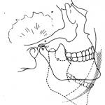
Fracture of the base of the skull - a traumatic cause of the development of bulbar syndrome
The cause of bulbar syndrome can also be inflammation of the substance and the meninges of the brain of an infectious nature. This process inevitably leads to edema of the structures of the brain, including the nuclei of the cranial nerves. These diseases include tick-borne encephalitis, poliomyelitis, meningococcal meningitis, enteroviral meningitis, diphtheria, syphilis, HIV infection. In addition, nerve cells suffer from the toxins of the pathogen released into the bloodstream. One of these substances is botulinum toxin secreted by the bacterium C. botulinum.
An infectious agent can cause damage to the bulbar nuclei
Degenerative processes of the brain substance can also cause bulbar syndrome. These diseases either lead to diffuse damage to nerve cells, or they affect a specific substance - myelin, which forms their surface membrane. Such pathologies include amyotrophic lateral sclerosis, Kennedy bulbar amyotrophy, Werdnig-Hoffmann spinal amyotrophy, and motor neuron disease.
Chronic progressive diseases of the nervous system can cause the development of bulbar syndrome
Clinical picture
Bulbar paralysis is characterized by a triad of clinical signs in the form of a disorder of articulation, voice and swallowing.
Voice disorder (dysphonia) is characterized by a change in the timbre of the voice and the appearance of nasal sound. The reason for the first symptom is non-closure of the glottis due to paresis of the muscles of the larynx. Snuffiness (rhinolalia) is caused by the immobility of the soft palate.
Articulation disorder (diazarthria) is a consequence of damage to the motor activity of the muscles of the tongue. The patient's speech in this situation becomes slurred or impossible.
Swallowing disorder (dysphagia) is a consequence of damage to the glossopharyngeal nerve. The brain is unable to command the act of swallowing. The consequence of this process is choking when taking liquid food and getting it into the nasal cavity.
The patient's appearance is also very characteristic: there is no live facial expressions on the face, the mouth is slightly open, saliva flows from the corner of the mouth, since the brain is not able to control salivation through the vagus nerve. The tongue, as a rule, is markedly reduced in volume from the affected side and deviated from the midline.
Deviation of the tongue from the midline is a typical sign of bulbar syndrome
The most difficult consequence of the pathological process in the group of bulbar nuclei of the cranial nerves is respiratory failure and cardiac disorder.
Diagnostic methods
To establish the correct diagnosis, it is necessary to carry out the following measures:
- careful questioning of the patient with the identification of all the details of the disease;
- neurological examination reveals impaired speech, swallowing, voice formation and concomitant sensory and movement disorders of the muscles of the trunk and limbs, as well as
Neurological examination is the basis for the diagnosis of bulbar syndrome
weakening of reflex reactions;
- examination of the larynx with a special mirror (laryngoscopy) reveals sagging of the vocal cord on the affected side;
- electroneuromyography allows you to determine the peripheral nature of paralysis, as well as graphically reflect the movement of a nerve impulse along the pathways;
Electroneuromyography - a method of graphically recording the passage of a nerve impulse
- x-ray of the skull reveals a violation of the integrity of the bones (fractures);
X-ray of the skull reveals violations of the integrity of the bones
- computer (magnetic resonance) tomography allows you to localize the pathological focus with great accuracy and study the anatomical structure of the brain and its departments;
Computed tomography is a way to reliably study the anatomical structures of the cranial cavity
- lumbar puncture allows you to identify signs of an infectious process or hemorrhage in the brain;
Lumbar puncture is used to diagnose an infectious brain injury
- a general blood test allows you to detect signs of an inflammatory process in the body in the form of an increase in the number of white cells (leukocytes) and an acceleration of the erythrocyte sedimentation rate (ESR);
- determination of specific proteins-antibodies in the blood allows to confirm the infectious nature of the process.
A blood test for antibodies is used when an infectious nature of bulbar syndrome is suspected
Differential diagnosis is carried out with the following diseases:
- mental disorders;
- pseudobulbar paralysis;
- hereditary neuromuscular diseases;
- encephalopathy against the background of impaired liver and kidney function;
- poisoning with neurotropic poisons.
Treatment methods for bulbar syndrome
Treatment of bulbar syndrome (the underlying disease and symptoms) is carried out under the guidance of a neurologist, if necessary, a neurosurgeon and an infectious disease specialist. In severe cases, therapy is carried out in a specialized department of a hospital or neuroreanimation.
Medication
For the treatment of bulbar syndrome and the main pathological process, the following drugs are used:
- antibacterial agents that have a detrimental effect on infectious agents: Ceftriaxone, Azithromycin, Clarithromycin, Metronidazole, Meronem, Tienam;
- means that relieve symptoms of cerebral edema and reduce intracranial pressure: Lasix, Furosemide, Diacarb;
- hormonal anti-inflammatory drugs: Prednisolone; Hydrocortisone;
- drugs that improve the metabolism in the nervous tissue: Cortexin, Actovegin, ATP;
- drugs that improve the functioning of the neurological system: Mexidol, Piracetam, Phezam;
- drugs that eliminate increased salivation: Atropine;
- b vitamins, which improve metabolic processes in the nervous tissue: Thiamin, Riboflavin, Pyridoxine, Cyanocobalamin;
- antineoplastic drugs that kill malignant cells: doxorubicin, cisplatin, methotrexate.
Pharmacological agents for the treatment of bulbar syndrome - gallery
 Diacarb is used to reduce intracranial pressure
  Actovegin has an active metabolic effect
  Cortexin is an active metabolic drug
  Prednisolone - hormonal anti-inflammatory drug
  Thiamin contains vitamin B1
  Riboflavin belongs to the B vitamins
  Azithromycin is an antibiotic that is active against many strains of bacteria
  Klacid is used for bacterial meningitis and encephalitis
  Lasix is \u200b\u200ban effective decongestant drug
  Doxorubicin belongs to anticancer drugs
  Cisplatin is used for tumor chemotherapy
  Neuromultivit - a combined vitamin preparation to improve the function of the nervous system
  Pyridoxine has a beneficial effect on the functioning of nerve fibers
  Tienam is a broad-spectrum antibacterial drug from the carbapenem group
  Mexidol contributes to the normal functioning of nerve cells
Surgical
Surgical intervention is performed by a neurosurgeon in the following cases:

Non-drug
Recommendations for changing the diet are given by a specialist depending on the type of underlying disease. Physiotherapy measures are an important component of the treatment of bulbar syndrome. The following methods are used in the treatment of the disease:

In children under two years of age, bulbar syndrome is an integral component of infantile cerebral palsy after birth injury and is additionally manifested by motor and sensory disorders, impaired sucking reflex, and frequent regurgitation. In other cases, the clinical picture in children is similar.
Complications and prognosis
The prognosis for the treatment of bulbar syndrome largely depends on the underlying disease that caused the pathological condition. With the infectious nature of the lesion of the nuclei of the cranial nerves, complete recovery is possible. The prognosis for hemorrhage in half of the cases is unfavorable. In degenerative diseases of the nervous system, bulbar paralysis is progressive.
In severe cases, the following complications may develop:
- edema of the brain;
- cerebral coma;
- breathing disorders, including those requiring artificial ventilation of the lungs;
- traumatic epilepsy;
- complete impossibility of independent swallowing of food and the use of food through a tube.
Prevention
Prevention of bulbar syndrome includes the following measures:

With damage to some parts of the brain, serious pathological processes can appear that reduce the standard of living of a person, and in some cases, threatening death.
Bulbar and pseudobulbar syndrome are disorders of the central nervous system, the symptoms of which are similar to each other, but their etiology is different.
Bulbar arises from damage to the medulla oblongata - the nuclei of the glossopharyngeal, vagus and hypoglossal nerves that are located in it.
Pseudobulbar syndrome (paralysis) is manifested as a result of impaired conduction of the cortical-nuclear pathways.
The clinical picture of bulbar syndrome
The main diseases during or after which bulbar paralysis occurs:
- a stroke that affects the medulla oblongata;
- infections (tick-borne borreliosis, acute polyradiculoneuritis);
- trunk glioma;
- botulism;
- displacement of brain structures with damage to the medulla oblongata;
- genetic disorders (porphyrin disease, Kennedy bulbospinal amyotrophy);
- syringomyelia.
Porphyria is a genetic disorder in which bulbar palsy is common. The unofficial name - the disease of vampires - is given because of a person's fear of the sun and exposure to light on the skin, which begins to burst, become covered with ulcers and scars. Due to the involvement in the inflammatory process of cartilage and deformation of the nose, ears, as well as exposure of teeth, the patient becomes like a vampire. There is no specific treatment for this pathology.
Isolated bulbar paralysis occurs infrequently due to the involvement of the nuclei of the nearby structures of the medulla oblongata during the lesion.
The main symptoms that arise in the patient:
- speech disorders (dysarthria);
- swallowing disorders (dysphagia);
- voice changes (dysphonia).

Patients speak with difficulty, indistinctly, their voice becomes weak, to the point that it becomes impossible to pronounce a sound. The patient begins to pronounce sounds in the nose, his speech is blurry, slowed down. Vowel sounds become indistinguishable from each other. Not only paresis of the muscles of the tongue can occur, but their complete paralysis.
Patients choke on food, often cannot swallow it. Liquid food enters the nose, aphagia may occur (complete inability to make swallowing movements).
The neurologist diagnoses the disappearance of reflexes of the soft palate and pharyngeal and notes the appearance of twitching of individual muscle fibers, muscle degeneration.
With severe damage, when the cardiovascular and respiratory centers are involved in the medulla oblongata, disturbances in the rhythm of respiration and heart activity occur, which threatens with death.
Manifestations and causes of pseudobulbar syndrome
Diseases after or during which pseudobulbar paralysis develops:
- vascular disorders affecting both hemispheres (vasculitis, atherosclerosis, hypertensive lacunar cerebral infarctions);
- traumatic brain injury;
- brain damage due to severe hypoxia;
- epileptoform syndrome in children (a single episode of paralysis may occur);
- demyelinating disorders;
- pick's disease;
- bilateral perisylvian syndrome;
- multisystem atrophy;
- intrauterine pathology or birth trauma in newborns;
- genetic disorders (amyotrophic lateral sclerosis, olivopontocerebellar degeneration, Creutzfeldt-Jakob disease, familial spastic paraplegia, etc.);
- parkinson's disease;
- glioma;
- neurological conditions after inflammation of the brain and its membranes.

Creutzfeldt-Jakob disease, in which not only pseudobulbar syndrome is observed, but also symptoms of rapidly progressing dementia, is a serious disease, the predisposition to which is genetically determined. It develops due to the ingestion of abnormal tertiary proteins, similar in their action to viruses. In most cases, death occurs within a year or two from the onset of the disease. There is no treatment to eliminate the cause.
The symptoms that accompany pseudobulbar paralysis, like bulbar paralysis, are expressed in dysphonia, dysphagia and dysarthria (in a lighter version). But these two lesions of the nervous system are different.
If atrophy and degeneration of muscles occurs with bulbar paralysis, then these phenomena are absent with pseudobulbar paralysis. Defibrillary reflexes also do not occur.
Pseudobulbar syndrome is characterized by uniform paresis of the facial muscles, which are spastic in nature: disorders of differentiated and voluntary movements are observed.
Since disorders in pseudobulbar paralysis occur above the medulla oblongata, there is no threat to life due to a stop of the respiratory or cardiovascular systems.
The main symptoms, which indicate that pseudobulbar paralysis has developed, and not bulbar paralysis, are expressed in violent crying or laughter, as well as reflexes of oral automatism, which are normally characteristic of children, and in adults indicate the development of pathology. This can be, for example, the proboscis reflex, when the patient pulls out the lips with a tube, if you make light tapping around the mouth. The same action is performed by the patient if an object is brought to his lips. Facial contractions can be triggered by tapping the bridge of the nose or pressing the palm under the thumb.
Pseudobulbar paralysis leads to multiple softened foci of the brain substance, so the patient has a decrease in motor activity, disorders and weakening of memory and attention, a decrease in intelligence and the development of dementia.
Patients may develop hemiparesis, a condition in which the muscles on one side of the body become paralyzed. Paresis of all limbs may occur.
With severe brain damage, pseudobulbar paralysis can appear together with bulbar.
Therapeutic effects
Since pseudobulbar syndrome and bulbar syndrome are secondary diseases, treatment should be directed to the underlying cause, if possible. When the symptoms of the primary disease decrease, the signs of paralysis can be smoothed out.

The main goal of treatment for severe bulbar paralysis is to maintain vital body functions. To do this, appoint:
- artificial ventilation of the lungs;
- tube feeding;
- proserin (with its help, the swallowing reflex is restored);
- atropine with profuse salivation.
After resuscitation measures, complex treatment should be prescribed, which can affect the primary and secondary diseases. This ensures the preservation of life and the improvement of its quality, the relief of the patient's condition.
The question of the treatment of bulbar and pseudobulbar syndromes through the introduction of stem cells remains controversial: supporters believe that these cells can produce the effect of physical replacement of myelin and restore the function of neurons, opponents point out that the effectiveness of using stem cells has not been proven and, on the contrary, increases the risk of developing cancerous tumors.
The restoration of reflexes in a newborn begins in the first 2 - 3 weeks of life. In addition to medication, he is given massage and physiotherapy, which should have a tonic effect. Doctors give an uncertain prognosis, since complete recovery does not occur even with adequately selected treatment, and the underlying disease may progress.
Bulbar and pseudobulbar syndrome are severe secondary lesions of the nervous system. Their treatment should be comprehensive and must be directed to the underlying disease. In severe cases of bulbar paralysis, respiratory arrest and cardiac arrest can occur. The prognosis is unclear and depends on the course of the underlying disease.
Bulbar syndrome is a neurological pathology caused by dysfunction of three pairs of cranial nerves at the same time: IX, X and XII. Disorder of the motor innervation of the muscles of the head and neck is manifested by a violation of the swallowing process, throwing food into the respiratory system, speech abnormalities, hoarseness, pathological changes in taste sensations and autonomic symptoms.
Bulbar syndrome is characterized by blockage of nerve impulses at the level of the cranial nuclei or motor fibers. A mild form of pathology develops with unilateral damage to the IX, X and XII nerves. Bilateral damage to the same nerves leads to the development of a severe degree of the disease.

Bulbar syndrome, in contrast, has a more severe course and is manifested by life-threatening dysfunctions: arrhythmia, atrophy of paralyzed muscles and respiratory arrest. A triad of symptoms is characteristic: dysphonia, dysphagia, dysarthria. Some patients are not even able to take food on their own. Diagnosis of the syndrome is based on patient examination data and the results of additional examinations. Usually, treatment begins with urgent measures, and then moves on to etiotropic, pathogenetic and symptomatic therapy.
Bulbar syndrome is a severe progressive process that leads to disability and a deterioration in the quality of life. A rapidly emerging syndrome with a rapid increase in clinical symptoms is deadly and requires emergency medical care and hospitalization of patients in the intensive care unit.
Classification
Bulbar syndrome is acute, progressive, alternating with unilateral or bilateral lesions.
- Acute paralysis is characterized by sudden onset and rapid development. Its main causes are strokes, encephalitis and neuroinfections.
- Progressive paralysis is a less critical condition characterized by a gradual increase in clinical symptoms. It develops in chronic degenerative diseases of the nervous system.
- Alternating syndrome - damage to the nuclei of the bulbar zone with unilateral damage to the muscles of the trunk.
Etiology

The etiopathogenetic factors of paralysis are very diverse: impaired blood supply to the brain, TBI, acute infections, neoplasms, cerebral edema, inflammation, exposure to neurotoxins.
Bulbar syndrome is a manifestation of various mental and somatic diseases, which by origin can be conditionally divided into the following groups:
- genetic - acute intermittent porphyria, Kennedy's disease, Chiari malformation, paroxysmal myoplegia;
- vascular - ischemic and hemorrhagic cerebral stroke, hypertensive crisis, venous sinus thrombosis, discirculatory encephalopathy;
- degenerative - syringobulbia, Guillain-Barré syndrome, myasthenia gravis, dystrophic myotonia, Alzheimer's disease;
- infectious - encephalitis, tick-borne borreliosis, poliomyelitis, neurosyphilis, Lyme disease, diphtheria polyneuropathy, botulism, meningitis, encephalitis;
- oncological - tumors of the cerebellum, gliomas, ependymomas, tuberculomas, cysts;
- demyelinating - multiple sclerosis;
- endocrine - hyperthyroidism;
- traumatic - fractures of the base of the skull.
Factors provoking the development of the syndrome:
- abuse of salty foods,
- frequent inclusion in the diet of high-carbohydrate and fatty foods and dishes,
- chronic stress, frequent conflict situations,
- excessive physical stress.
Pathogenesis
Electrical impulses from the brain go to the cortex, and then to the motor nuclei of the bulbar zone. Nerve fibers originate from them, along which signals are sent to the skeletal muscles of the upper body. The centers of the medulla oblongata in healthy people are responsible for hearing, facial expressions, the processes of swallowing and sound production. All cranial nerves are structural components of the central nervous system.

- The vagus nerve has many branches that span the entire body. The tenth pair of nerves starts from the bulbar nuclei and reaches the abdominal organs. Thanks to its correct work, the respiratory organs, stomach, and heart function at an optimal level. The vagus nerve provides swallowing, coughing, vomiting, and speech.
- The glossopharyngeal nerve innervates the muscles of the pharynx and the parotid salivary gland, providing its secretory function.
- The hypoglossal nerve innervates the muscles of the tongue and facilitates swallowing, chewing, sucking and licking.
Under the influence of the etiological factor, synaptic transmission of nerve impulses is disrupted and the nuclei of the IX, X and XII pairs of cranial nerves are simultaneously destroyed.
The etiopathogenetic factor can exert its negative impact at one of three levels:
- in the nuclei of the medulla oblongata,
- in the roots and trunks inside the cranial cavity,
- in fully formed nerve fibers outside the cranial cavity.
As a result of damage to the nuclei and fibers of these nerves, the trophism of muscle tissue is disturbed. Muscles decrease in volume, become thinner, and their number is reduced to complete disappearance. Bulbar paralysis is accompanied by hypo- or areflexia, hypo- or atony, hypo- or atrophy of the paralyzed muscles. When the nerves that innervate the respiratory muscles are involved in the process, patients die of suffocation.
Symptoms

The clinic of the syndrome is caused by a violation of the innervation of the muscles of the throat and tongue, as well as dysfunction of these organs. Patients have a specific symptom complex - dysphagia, dysarthria, dysphonia.
- Impaired swallowing is manifested by frequent choking, salivation from the corners of the mouth, and the inability to swallow even liquid food.
- Bulbar dysarthria and dysphonia are characterized by a weak and deaf voice, nasal and "blurred" sounds. Consonant sounds become the same type, vowels become difficult to distinguish from each other, speech becomes slow, tedious, indistinct, impossible. Nasiness and slurred speech are associated with the immobility of the soft palate.
- The voice of patients becomes weak, deaf, depleting up to complete aphonia - a violation of the sound of speech. The cause of the altered timbre of the voice is incomplete closure of the glottis due to paresis of the laryngeal muscles.
- Violations of mimic activity or its complete absence. Mimic functions lose their specificity, their general weakening occurs, and normal coordination is impaired. The patient's facial features become expressionless - the mouth is half-open, profuse salivation and loss of chewed food.
- Decrease and gradual extinction of palatine and pharyngeal reflexes.
- Weakness of the chewing muscles due to paralysis of the corresponding nerves. Violation of complete chewing of food.
- Atrophy of the muscles of the tongue and its immobility.
- Ingestion of liquid and solid food in the nasopharynx.
- Twitching of the tongue and drooping of the palatine curtain.
- In severe cases - disruption of the heart, vascular tone, breathing rhythm.
When examining patients, specialists find a deviation of the tongue towards the lesion, its hypotension and inactivity, and single fasciculations. In severe cases, glossoplegia is noted, which sooner or later ends with pathological thinning or folding of the tongue. Immobility and weakness of the palatine arches, uvula, and pharyngeal muscles lead to dysphagia. The constant throwing of food into the respiratory tract can result in aspiration and the development of inflammation. Violation of the autonomic innervation of the salivary glands is manifested by hypersalivation and requires constant use of a scarf.
In newborns, bulbar syndrome is a manifestation of cerebral palsy caused by birth trauma. Babies develop motor and sensory disorders, the sucking process is disrupted, and they often regurgitate. In children over 2 years of age, the symptoms of pathology are similar to those in adults.
Diagnostics
The diagnosis and treatment of bulbar palsy is carried out by specialists in the field of neurology. Diagnostic measures are aimed at identifying the immediate cause of the pathology and consist in examining the patient, identifying all the symptoms of the disease and conducting electromyography. The obtained clinical data and research results allow determining the severity of paralysis and prescribing treatment. These are mandatory diagnostic techniques, which are complemented by a general analysis of blood and urine, brain tomography, esophagoscopy, CSF examination, electrocardiography, and an ophthalmologist's consultation.

During the first neurological examination, the neurological status of the patient is determined: intelligibility of speech, timbre of voice, salivation, swallowing reflex. Be sure to study the appearance of the tongue, identify atrophies and fasciculations, assess its mobility. An assessment of the respiratory rate and heart rate is of great diagnostic value.
Then the patient is referred for additional diagnostic examination.
- Using a laryngoscope, the larynx is examined and a sagging of the vocal cord on the affected side is found.
- X-ray of the skull - determination of bone structure, the presence of fractures, injuries, neoplasms, foci of hemorrhage.
- Electromyography is a research method that evaluates the bioelectrical activity of muscles and allows you to determine the peripheral nature of paralysis.
- Computed tomography - the most accurate images of any part of the body and internal organs, made using X-rays.
- Esophagoscopy - determining the work of the muscles of the pharynx and vocal cords by examining their inner surface using an esophagoscope.
- Electrocardiography is the simplest, most accessible and informative method for diagnosing heart disease.
- MRI - layer-by-layer images of any area of \u200b\u200bthe body, allowing the most accurate study of the structure of an organ.
- In laboratory tests, there are characteristic changes: in the cerebrospinal fluid - signs of infection or hemorrhage, in the hemogram - inflammation, in the immunogram - specific antibodies.
Treatment

Emergency medical care should be provided in full to patients with acute bulbar syndrome, accompanied by signs of respiratory and cardiovascular dysfunction. Resuscitation measures are aimed at maintaining the vital functions of the body.
- Patients are connected to a ventilator or the trachea is intubated with it;
- Enter "Proserin", which restores muscle activity, improves the swallowing reflex and gastric motility, reduces the pulse;
- "Atropine" eliminates hypersalivation;
- Antibiotics are administered with obvious signs of an infectious process in the brain;
- Diuretics help to cope with cerebral edema;
- Drugs that improve cerebral circulation are indicated in the presence of vascular disorders;
- Patients with respiratory and cardiac disorders are admitted to the intensive care unit.
The main goal of treatment is to eliminate the threat to the patient's life. All patients with severe neurological disorder are transported to a medical facility, where they will receive adequate treatment.
Stages of therapy:

- Etiotropic therapy is the elimination of diseases that have become the root cause of bulbar syndrome. In most cases, these ailments are not cured and progress throughout life. If an infection becomes the cause of the pathology, antibacterial agents of a wide spectrum are taken - "Ceftriaxone", "Azithromycin", "Clarithromycin".
- Pathogenetic treatment: anti-inflammatory - glucocorticoids "Prednisolone", decongestant - diuretics "Furosemide", metabolic - "Cortexin", "Actovegin", nootropic - "Mexidol", "Piracetam", antitumor - cytostatics "Methotrexate".
- Symptomatic therapy is aimed at improving the general condition of patients and reducing the severity of clinical manifestations. B vitamins and preparations with glutamic acid stimulate metabolic processes in the nervous tissue. In severe dysphagia - the introduction of vasodilator and antispasmodic drugs, infusion therapy, correction of vascular disorders. "Neostigmine" and "ATP" reduce the severity of diasphagia.
- Currently, the use of stem cells, which are actively functioning instead of the affected ones, has a good therapeutic effect.
- In severe cases, patients with bulbar syndrome are fed special mixtures through an enteral tube. Relatives need to monitor the oral cavity and observe the patient during meals to prevent aspiration.
Bulbar syndrome is difficult to treat even with adequate therapy. Recovery occurs in isolated cases. In the course of treatment, the condition of patients improves, paralysis decreases, and muscle work is restored.
Physiotherapy methods used to treat bulbar syndrome:

- electrophoresis, laser therapy, magnetotherapy and mud therapy,
- therapeutic massage to develop muscles and speed up the process of their recovery,
- kinesiotherapy - performing certain exercises that help restore the work of the human musculoskeletal system,
- breathing exercises - a system of exercises aimed at improving health and developing the lungs,
- physiotherapy exercises - certain exercises that accelerate recovery,
- in the recovery period, classes with a speech therapist are shown.
Surgical intervention is used in cases where conservative treatment does not give positive results. Operations are performed in the presence of tumors and fractures:
- Bypass surgery prevents the development of dislocation syndrome.
- Craniotomy is performed in patients with epidural and subdural hematomas of the brain.
- Clipping of pathologically dilated cerebral vessels is a surgical method that effectively eliminates abnormal changes in the circulatory system.
- Cholesterol plaques are removed by endarterectomy and prosthetics of the damaged area.
- In case of skull fractures, the skull is opened, the source of bleeding and bone fragments are eliminated, the bone defect is closed with a removed bone or a special plate, and then a long-term rehabilitation is proceeded.
Traditional medicines used to treat paralysis: infusions and decoctions of medicinal herbs, alcohol tincture of peony, a strong solution of sage - drugs that strengthen the nervous system and relieve stress. Patients are advised to take medicinal baths with a decoction of sage or rose hips.
Prevention and prognosis
Preventive measures to prevent the development of bulbar syndrome:
- immunization by vaccination against major infectious diseases,
- fight against atherosclerosis,
- control of blood pressure and blood sugar levels,
- timely detection of neoplasms,
- balanced diet with restriction of carbohydrates and fats,
- playing sports and keeping an active lifestyle,
- compliance with the work and rest regime,
- passing professional examinations by doctors,
- fighting smoking and alcohol consumption,
- full sleep.
The prognosis of the pathology is determined by the course of the underlying disease, which became the root cause of the syndrome. The defeat of the nuclei of infectious etiology is completely cured, and the processes of swallowing and speech are gradually restored. Acute violation of cerebral circulation, manifested by the clinic of the syndrome, in 50% of cases has an unfavorable prognosis. With degenerative pathologies and chronic ailments of the nervous system, paralysis progresses. Patients usually die from cardiopulmonary failure.
Video: Bulbar Syndrome - Clinical Options and Physiotherapy
Gradually developing dysfunction of the bulbar group of the caudal cranial nerves caused by damage to their nuclei and / or roots. A triad of symptoms is characteristic: dysphagia, dysarthria, dysphonia. The diagnosis is established based on the examination of the patient. Additional examinations (analysis of cerebrospinal fluid, CT, MRI) are carried out to determine the underlying pathology that caused bulbar paralysis. Treatment is prescribed in accordance with the causative disease and the underlying symptoms. Urgent measures may be required: resuscitation, mechanical ventilation, the fight against heart failure and vascular disorders.

General information
Bulbar paralysis occurs when the nuclei and / or roots of the bulbar group of cranial nerves are damaged in the medulla oblongata. The bulbar nerves include glossopharyngeal (IX pair), vagus (X pair) and hypoglossal (XII pair) nerves. The glossopharyngeal nerve innervates the muscles of the pharynx and provides its sensitivity, is responsible for the taste sensations of the posterior 1/3 of the tongue, and provides parasympathetic innervation of the parotid gland. The vagus nerve innervates the muscles of the pharynx, soft palate, larynx, upper digestive tract and respiratory tract; gives parasympathetic innervation of internal organs (bronchi, heart, gastrointestinal tract). The hypoglossal nerve provides innervation to the muscles of the tongue.
The cause of bulbar paralysis may be chronic cerebral ischemia, which has developed as a result of atherosclerosis or chronic vascular spasm in hypertension. Rare factors causing damage to the bulbar group of cranial nerves include craniovertebral anomalies (primarily Chiari malformation) and severe polyneuropathies (Guillain-Barré syndrome).
Symptoms of progressive bulbar palsy
The clinical manifestations of bulbar paralysis are based on peripheral paresis of the muscles of the pharynx, larynx and tongue, which results in impaired swallowing and speech. The basic clinical symptom complex is a triad of signs: disorder of swallowing (dysphagia), impaired articulation (dysarthria) and sonority of speech (dysphonia). Impaired food swallowing begins with difficulty in taking fluids. Due to paresis of the soft palate, fluid from the oral cavity enters the nose. Then, with a decrease in the pharyngeal reflex, disorders of swallowing and solid food develop. Limiting the mobility of the tongue leads to difficulty in chewing food and moving the food lump in the mouth. Bulbar dysarthria is characterized by blurred speech, lack of clarity in the pronunciation of sounds, because of which the patient's speech becomes incomprehensible to others. Dysphonia appears as a hoarse voice. There is nasolalia (nasalness).
The patient's appearance is characteristic: the face is hypomimic, the mouth is open, salivation is observed, difficulty in chewing and swallowing food, and its loss from the mouth. In connection with the defeat of the vagus nerve and a violation of the parasympathetic innervation of the somatic organs, disorders of the respiratory function, heart rhythm and vascular tone occur. These are the most dangerous manifestations of bulbar palsy, since often progressive respiratory or heart failure causes the death of patients.
When examining the oral cavity, atrophic changes in the tongue, its folding and unevenness are noted, fascicular contractions of the muscles of the tongue can be observed. The pharyngeal and palatal reflexes are sharply reduced or not evoked. Unilateral progressive bulbar paralysis is accompanied by drooping of half of the soft palate and deviation of its uvula to the healthy side, atrophic changes in 1/2 of the tongue, deviation of the tongue towards the lesion when it protrudes. With bilateral bulbar paralysis, glossoplegia is observed - complete immobility of the tongue.
Diagnostics
A neurologist can diagnose bulbar palsy by carefully studying the patient's neurological status. The study of the function of bulbar nerves includes an assessment of the speed and intelligibility of speech, the timbre of the voice, the volume of salivation; examination of the tongue for the presence of atrophy and fasciculations, assessment of its mobility; examination of the soft palate and test of the pharyngeal reflex. It is important to determine the frequency of respiration and heartbeats, the study of the pulse to detect arrhythmias. Laryngoscopy allows you to determine the absence of complete closure of the vocal cords.
During diagnosis, progressive bulbar palsy must be distinguished from pseudobulbar palsy. The latter occurs with supranuclear lesions of the cortico-bulbar pathways connecting the nuclei of the medulla oblongata with the cerebral cortex. Pseudobulbar paralysis is manifested by central paresis of the muscles of the larynx, pharynx and tongue with hyperreflexia characteristic of all central paresis (increased pharyngeal and palatal reflexes) and increased muscle tone. Clinically differs from bulbar paralysis in the absence of atrophic changes in the tongue and in the presence of reflexes of oral automatism. It is often accompanied by violent laughter resulting from the spastic contraction of the facial muscles.
In addition to pseudobulbar paralysis, progressive bulbar paralysis requires differentiation from psychogenic dysphagia and dysphonia, various diseases with primary muscular lesions that cause myopathic paresis of the larynx and pharynx (myasthenia gravis, Rossolimo-Steinert-Kurshman myotonia, paroxysmal myoplegia, oculopathy). It is also necessary to diagnose the underlying disease, which led to the development of bulbar syndrome. For this purpose, a study of cerebrospinal fluid, CT and MRI of the brain is carried out. Tomographic studies make it possible to visualize brain tumors, demyelination zones, cerebral cysts, intracerebral hematomas, cerebral edema, displacement of cerebral structures in dislocation syndrome. CT or X-ray of the craniovertebral junction can reveal abnormalities or post-traumatic changes in this area.
Treatment of progressive bulbar palsy
Therapeutic tactics for bulbar paralysis is built taking into account the underlying disease and leading symptoms. In case of infectious pathology, etiotropic therapy is carried out, in case of cerebral edema, decongestant diuretics are prescribed, in case of tumor processes, together with a neurosurgeon, the issue of removing the tumor or performing a shunting operation to prevent dislocation syndrome is decided.
Unfortunately, many diseases in which bulbar syndrome occurs are a progressive degenerative process that occurs in the cerebral tissues and do not have effective specific treatment. In such cases, symptomatic therapy is carried out to support the vital functions of the body. So, with severe respiratory disorders, the trachea is intubated with the patient connected to a ventilator, with severe dysphagia, tube feeding is provided, with the help of vasoactive drugs and infusion therapy, vascular disorders are corrected. To reduce dysphagia, prescribe neostigmine, ATP, vitamins gr. B, glutamic acid; with hypersalivation - atropine.
Forecast
Progressive bulbar palsy has a highly variable prognosis. On the one hand, patients can die from heart or respiratory failure. On the other hand, with successful treatment of the underlying disease (for example, encephalitis), in most cases, patients recover with complete restoration of swallowing and speech function. Due to the lack of effective pathogenetic therapy, bulbar paralysis associated with progressive degenerative lesions of the central nervous system (with multiple sclerosis, ALS, etc.) has an unfavorable prognosis.
Bulbar syndrome (paralysis) occurs with peripheral muscle paralysis, innervated by IX, X and XII pairs of cranial nerves in the case of their combined lesion. In the clinical picture, dysphagia, dysphonia or aphonia, dysarthria or anarthria are noted.
Pseudobulbar syndrome (paralysis) - this is a central paralysis of muscles innervated by the IX, X and XII pairs of cranial nerves. The clinical picture of pseudobulbar syndrome resembles a picture of bulbar syndrome (dysphagia, dysphonia, dysarthria), but it is much milder. By its nature, pseudobulbar paralysis is a central paralysis and, accordingly, symptoms of spastic paralysis are inherent in it.
Often, despite the early use of modern drugs, complete recovery from bulbar and pseudobulbar syndromes (paralysis) does not occur, especially when months and years have passed since the injury.
However, a very good result is achieved when using stem cells for bulbar and pseudobulbar syndromes (paralysis).
Stem cells introduced into the patient's body with bulbar or pseudobulbar syndrome (paralysis) not only physically replace the defect in the myelin sheath, but also take on the function of damaged cells. Embedding in the patient's body, they restore the myelin sheath of the nerve, its conduction, strengthen and stimulate it.
As a result of treatment in patients with bulbar and pseudobulbar syndrome (paralysis), dysphagia, dysphonia, aphonia, dysarthria, anarthria disappear, brain functions are restored, and the person returns to normal life.
Pseudobulbar paralysis
Pseudobulbar paralysis (synonymous with false bulbar palsy) is a clinical syndrome characterized by disorders of chewing, swallowing, speech, and facial expressions. It occurs when the central pathways leading from the motor centers of the cerebral hemispheres of the brain to the motor nuclei of the cranial nerves of the medulla oblongata are interrupted, in contrast to boulevard paralysis (see), in which the nuclei themselves or their roots are affected. Pseudobulbar paralysis develops only with bilateral damage to the cerebral hemispheres, since an interruption in the pathways to the nuclei of one hemisphere does not give noticeable bulbar disorders. The cause of pseudobulbar paralysis is usually cerebral atherosclerosis with softening foci in both hemispheres of the brain. However, pseudobulbar paralysis can also be observed in the vascular form of syphilis of the brain, neuroinfections, tumors, degenerative processes affecting both hemispheres of the brain.
One of the main symptoms of pseudobulbar paralysis is a violation of chewing and swallowing. Food gets stuck behind the teeth and on the gums, the patient chokes when eating, liquid food flows out through the nose. The voice acquires a nasal tone, becomes hoarse, loses intonation, difficult consonants completely fall out, some patients cannot even speak in a whisper. As a result of bilateral paresis of the facial muscles, the face becomes amimic, mask-like, and often has a crying expression. Attacks of violent convulsive crying and laughter are characteristic, occurring without corresponding emotions. In some patients, this symptom may be absent. The tendon reflex of the lower jaw rises sharply. Symptoms of the so-called oral automatism appear (see Reflexes). Often, pseudobulbar syndrome occurs simultaneously with hemiparesis. Patients often have more or less pronounced hemiparesis or paresis of all limbs with pyramidal signs. In other patients, in the absence of paresis, a pronounced extrapyramidal syndrome appears (see Extrapyramidal system) in the form of slowness of movements, stiffness, increased muscle tone (muscle rigidity). The intellectual disturbances observed in pseudobulbar syndrome are explained by multiple foci of softening in the brain.
The onset of the disease in most cases is acute, but sometimes it can develop gradually. In most patients, pseudobulbar paralysis occurs as a result of two or more attacks of cerebrovascular accident. Death occurs from bronchopneumonia caused by ingestion of food into the respiratory tract, associated infection, stroke, etc.
Treatment should be directed against the underlying disease. To improve the act of chewing, you need to prescribe proserin 0.015 g 3 times a day with meals.
Pseudobulbar palsy (synonym: false bulbar palsy, supranuclear bulbar palsy, cerebrobulbar palsy) is a clinical syndrome characterized by disorders of swallowing, chewing, phonation and speech articulation, as well as amymia.
Pseudobulbar paralysis, in contrast to tabloid paralysis (see), which depends on the defeat of the motor nuclei of the medulla oblongata, arises as a result of an interruption in the paths leading from the motor area of \u200b\u200bthe cerebral cortex to these nuclei. When the supranuclear pathways are damaged in both hemispheres of the brain, voluntary innervation of the bulbar nuclei falls out and a "false" bulbar paralysis occurs, false because anatomically the medulla oblongata itself does not suffer. The defeat of the supranuclear pathways in one hemisphere of the brain does not give noticeable bulbar disorders, since the nuclei of the lingopharyngeal and vagus nerves (as well as the trigeminal and superior branches of the facial nerve) have bilateral cortical innervation.
Pathological anatomy and pathogenesis. With pseudobulbar paralysis, in most cases, severe atheromatosis of the arteries of the base of the brain is observed, affecting both hemispheres with the medulla oblongata and pons intact. Most often, pseudobulbar paralysis occurs due to thrombosis of the arteries of the brain and is observed mainly in old age. At middle age, P. of the item can be caused by syphilitic endarteritis. In childhood, P. p. Is one of the symptoms of infantile cerebral palsy with bilateral lesions of corticobulbar conductors.
The clinical course and symptomatology of pseudobulbar paralysis are characterized by bilateral central paralysis, or paresis, of the trigeminal, facial, lingopharyngeal, vagus and hypoglossal cranial nerves in the absence of degenerative atrophy in the paralyzed muscles, preservation of reflexes and disorders of the pyramidal and extrapyramidal systems. Disorders of swallowing at P. of the item do not reach the degree of bulbar paralysis; due to weakness of the chewing muscles, patients eat extremely slowly, food falls out of the mouth; patients choke. If food enters the respiratory tract, aspiration pneumonia may develop. The tongue is motionless or extends only to the teeth. Speech is not articulated enough, with a nasal tinge; the voice is quiet, words are articulated with difficulty.
One of the main symptoms of pseudobulbar paralysis is violent attacks of convulsive laughter and crying; facial muscles, which in such patients cannot voluntarily contract, come into excessive contraction. Patients may begin to cry involuntarily when showing teeth, stroking a piece of paper on the upper lip. The onset of this symptom is explained by a break in the inhibitory pathways heading to the bulbar centers, by a violation of the integrity of the subcortical formations (optic tubercle, striatum, etc.).
The face acquires a mask-like character due to bilateral paresis of the facial muscles. With bouts of violent laughing or crying, the eyelids close well. If you ask the patient to open or close his eyes, he opens his mouth. This peculiar disorder of voluntary movements should also be attributed to one of the characteristic signs of pseudobulbar paralysis.
There is also an increase in deep and superficial reflexes in the chewing and facial muscles, as well as the emergence of reflexes of oral automatism. This should include Oppenheim's symptom (sucking and swallowing movements when touching the lips); labial reflex (contraction of the circular muscle of the mouth when tapping in the area of \u200b\u200bthis muscle); oral reflex ankylosing spondylitis (lip movements when tapping with a hammer around the mouth); Toulouse-Wurp buccal phenomenon (movement of the cheeks and lips is caused by percussion along the lateral part of the lip); Astvatsaturov's nasolabial reflex (proboscis closing of the lips when tapping on the root of the nose). When stroking the patient's lips, a rhythmic movement of the lips and lower jaw occurs - sucking movements, sometimes turning into violent crying.
There are pyramidal, extrapyramidal, mixed, cerebellar and childhood forms of pseudobulbar paralysis, as well as spastic.
The pyramidal (paralytic) form of pseudobulbar paralysis is characterized by more or less clearly pronounced hemi- or tetraplegia or paresis with increased tendon reflexes and the appearance of pyramidal signs.
Extrapyramidal form: slowness of all movements, amimia, stiffness, increased muscle tone according to the extrapyramidal type with a characteristic gait (small steps) come to the fore.
Mixed form: a combination of the above forms of P. p.
Cerebellar form: atactic gait, coordination disorders, etc. come to the fore.
P.'s children's form of the item is observed with spastic diplegia. At the same time, the newborn does not suck well, chokes and chokes. Later, the child develops violent crying and laughter, and dysarthria is found (see. Children's paralysis).
Weil (A. Weil) describes the family spastic form of P. of the item. At it, along with the expressed focal disorders inherent in P. of the item, noticeable intellectual backwardness is noted. A similar form was also described by M. Klippel.
Since the symptom complex of pseudobulbar paralysis is mainly due to sclerotic lesions of the brain, patients with P. of the item often have corresponding symptoms from the psyche:
memory, difficulty thinking, increased efficiency, etc.
The course of the disease corresponds to the variety of causes that cause pseudobulbar paralysis, and the prevalence of the pathological process. The development of the disease is most often stroke-like, with different periods between strokes. If after a stroke (see) paretic phenomena in the extremities decrease, then bulbar phenomena remain mostly persistent. More often, the patient's condition worsens due to new strokes, especially with atherosclerosis of the brain. The duration of the disease is varied. Death occurs from pneumonia, uremia, infectious diseases, new hemorrhage, nephritis, heart weakness, etc.
The diagnosis of pseudobulbar palsy is not difficult. Should be differentiated from various forms of bulbar palsy, bulbar nerve neuritis, parkinsonism. The absence of atrophies and an increase in bulbar reflexes speaks against apoplectic bulbar paralysis. It is more difficult to distinguish P. of the item from a parkinson-like disease. It has a slow course, in the later stages apoplectic strokes occur. In these cases, attacks of violent crying are also observed, speech is upset, patients cannot eat on their own. The diagnosis can present difficulties only in distinguishing cerebral atherosclerosis from the pseudobulbar component; the latter is characterized by gross focal symptoms, strokes, etc. Pseudobulbar syndrome in these cases may appear as an integral part of the main suffering.
Bulbar and pseudobulbar syndromes
In the clinic, not isolated, but combined damage to the nerves of the bulbar group or their nuclei is more often observed. The simitomocomplex of movement disorders that occurs when the nuclei or roots of the IX, X, XII pairs of cranial nerves are damaged at the base of the brain is called bulbar syndrome (or bulbar palsy). This name comes from lat. bulbus bulb (the old name for the medulla oblongata, in which the nuclei of these nerves are located).
Bulbar syndrome can be unilateral or bilateral. With bulbar syndrome, peripheral paresis or muscle paralysis occurs, which are innervated by the glossopharyngeal, vagus and hypoglossal nerves.
With this syndrome, first of all, there are swallowing disorders. Normally, while eating, food is directed to the throat with the tongue. At the same time, the larynx rises upward, and the root of the tongue presses the epiglottis, covering the entrance to the larynx and opening the way for the food bolus to the pharynx. The soft palate rises upward, preventing liquid food from entering the nose. With bulbar syndrome, paresis or paralysis of the muscles performing the act of swallowing occurs, as a result of which swallowing is impaired - dysphagia. The patient chokes while eating, swallowing becomes difficult or even impossible (aphagia). Liquid food gets into the nose, solid food can get into the larynx. Food ingestion in the trachea and bronchi can cause aspiration pneumonia.
In the presence of bulbar syndrome, disorders of voice and speech articulation also occur. The voice becomes hoarse (dysphonia) with a nasal tone. Paresis of the language causes a violation of the articulation of speech (dysarthria), and its paralysis - anarthria, when the patient, well understanding the speech addressed to him, cannot pronounce the words himself. The tongue atrophies, with pathology of the nucleus of the XII pair, fibrillar muscle twitching is noted in the tongue. The pharyngeal and palatine reflexes decrease or disappear.
With bulbar syndrome, vegetative disorders (breathing disorders, cardiac activity) are possible, which in some cases cause an unfavorable prognosis. Bulbar syndrome is observed in tumors of the posterior cranial fossa, ischemic stroke in the medulla oblongata, syringobulbia, amyotrophic lateral sclerosis, tick-borne encephalitis, post-diphtheria polyneuropathy and some other diseases.
Central paresis of muscles that are innervated by bulbar nerves is called pseudobulbar syndrome. It occurs only with bilateral damage to the cortical-nuclear pathways leading from the motor cortical centers to the nuclei of the nerves of the bulbar group. The defeat of the cortical-nuclear pathway in one hemisphere does not lead to such a combined pathology, since the muscles innervated by the bulbar nerves, in addition to the tongue, receive bilateral cortical innervation. Since pseudobulbar syndrome is a central paralysis of swallowing, phonation and speech articulation, it also causes dysphagia, dysphonia, dysarthria, but unlike bulbar syndrome, atrophy of the muscles of the tongue and fibrillar twitching is not observed, the pharyngeal and palatine reflexes remain, the mandibular reflex increases. With pseudobulbar syndrome, patients develop reflexes of oral automatism (proboscis, nasolabial, palmar-chin, etc.), which is explained by disinhibition with bilateral damage to the cortical-nuclear pathways of subcortical and stem formations, at the level of which these reflexes are closed. For this reason, violent crying or laughter sometimes occurs. With pseudobulbar syndrome, movement disorders can be accompanied by a decrease in memory, attention, and intelligence. Pseudobulbar syndrome is most often observed in acute disorders of cerebral circulation in both hemispheres of the brain, discirculatory encephalopathy, amyotrophic lateral sclerosis. Despite the symmetry and severity of the lesion, pseudobulbar syndrome is less dangerous than bulbar syndrome, since it is not accompanied by a violation of vital functions.
With bulbar or pseudobulbar syndrome, it is important to carefully care for the oral cavity, monitor the patient while eating in order to prevent aspiration, tube feeding for aphagia.