While a person is alive, he breathes. What is breathing? These are processes that continuously supply all organs and tissues with oxygen and remove carbon dioxide from the body, which is formed as a result of the metabolic system. Performs these vital processes which directly interact with the cardiovascular system. To understand how gas exchange occurs in the human body, you should study the structure and functions of the lungs.
Why does a person breathe?
The only way to obtain oxygen is through breathing. It is not possible to hold it for a long time, since the body requires another portion. Why do we need oxygen at all? Without it, metabolism will not occur, the brain and all other human organs will not work. With the participation of oxygen, nutrients are broken down, energy is released, and each cell is enriched with them. Breathing is commonly called gas exchange. And rightly so. After all, the peculiarities of the respiratory system are to take in oxygen from the air that enters the body and remove carbon dioxide.
What are human lungs
Their anatomy is quite complex and variable. This organ is paired. Its location is the chest cavity. The lungs are adjacent to the heart on both sides - right and left. Nature has made sure that both of these important organs are protected from compression, shock, etc. In front, the barrier to damage is the spinal column at the back, and the ribs at the sides.
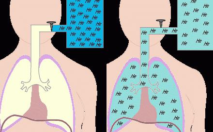
The lungs are literally riddled with hundreds of branches of bronchi, with alveoli the size of a pinhead located at their ends. There are up to 300 million of them in the body of a healthy person. The alveoli play an important role: they supply blood vessels with oxygen and, having a branched system, are able to provide a large area for gas exchange. Just imagine: they can cover the entire surface of a tennis court!
In appearance, the lungs resemble semi-cones, the bases of which are adjacent to the diaphragm, and the tops with rounded ends protrude 2-3 cm above the collarbone. The human lungs are a rather unique organ. The anatomy of the right and left lobes is different. So, the first one is slightly larger in volume than the second one, while it is somewhat shorter and wider. Each half of the organ is covered with pleura, consisting of two layers: one is fused with the chest, the other with the surface of the lung. The outer pleura contains glandular cells that produce fluid into the pleural cavity.
The inner surface of each lung has a depression called the hilum. They include the bronchi, the base of which looks like a branching tree, and the pulmonary artery, and a pair of pulmonary veins emerge.
Human lungs. Their functions
Of course, there are no secondary organs in the human body. The lungs are also important in ensuring human life. What kind of work do they do?
- The main functions of the lungs are to carry out the respiratory process. A person lives while he breathes. If the supply of oxygen to the body is cut off, death will occur.
- The job of the human lungs is to remove carbon dioxide, thereby maintaining the acid-base balance in the body. Through these organs, a person gets rid of volatile substances: alcohol, ammonia, acetone, chloroform, ether.
- The functions of the human lungs do not end there. The paired organ is still involved in which comes into contact with air. As a result, an interesting chemical reaction occurs. Oxygen molecules in the air and carbon dioxide molecules in dirty blood change places, i.e. oxygen replaces carbon dioxide.
- The various functions of the lungs allow them to participate in the water exchange occurring in the body. Up to 20% of the liquid is removed through them.
- The lungs are active participants in the process of thermoregulation. They release 10% of their heat into the atmosphere when they exhale.
- Regulation is not complete without the participation of the lungs in this process.
How do the lungs work?
The functions of the human lungs are to transport the oxygen contained in the air into the blood, use it, and remove carbon dioxide from the body. The lungs are fairly large soft organs with spongy tissue. The inhaled air enters the air sacs. They are separated from each other by thin walls with capillaries.
There are only small cells between the blood and the air. Therefore, thin walls do not create obstacles for inhaled gases, which facilitates good passage through them. In this case, the functions of the human lungs are to use necessary and remove unnecessary gases. Lung tissue is very elastic. When you inhale, the chest expands and the lungs increase in volume.
The windpipe, represented by the nose, pharynx, larynx, trachea, looks like a tube 10-15 cm long, divided into two parts called bronchi. Air passing through them enters the air sacs. And when you exhale, the volume of the lungs decreases, the chest decreases in size, and the pulmonary valve partially closes, which allows air to escape again. This is how human lungs work.

Their structure and functions are such that the capacity of this organ is measured by the amount of inhaled and exhaled air. So, for men it is equal to seven pints, for women - five. The lungs are never empty. The air remaining after exhalation is called residual air. When you inhale, it mixes with fresh air. Therefore, breathing is a conscious and at the same time unconscious process that occurs constantly. A person breathes when he sleeps, but he does not think about it. In this case, if you wish, you can interrupt your breathing for a short time. For example, while underwater.
Interesting facts about lung function
They are capable of pumping 10 thousand liters of inhaled air per day. But it is not always crystal clear. Along with oxygen, dust, many microbes and foreign particles enter our body. Therefore, the lungs perform the function of protection against all unwanted impurities in the air.
The walls of the bronchi have many tiny villi. They are needed to trap germs and dust. And the mucus, which is produced by the cells of the walls of the respiratory tract, lubricates these villi, and is then expelled when coughing.
It consists of organs and tissues that fully provide ventilation and respiration. The functions of the respiratory system lie in the implementation of gas exchange - the main link in metabolism. The latter is responsible only for pulmonary (external) respiration. It includes:
1. consisting of the nose and its cavity, larynx, trachea, bronchi.
The nose and its cavity heat, humidify and filter the inhaled air. Its cleansing is achieved through numerous hard hairs and goblet cells with cilia.
The larynx is located between the root of the tongue and the trachea. Its cavity is divided by the mucous membrane in the form of two folds. They are not completely fused in the middle. The gap between them is called the glottis.
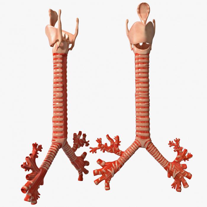
The trachea originates from the larynx. In the chest it is divided into bronchi: right and left.
2. Lungs with densely branched vessels, bronchioles and alveolar sacs. They begin the gradual division of the main bronchi into small tubes called bronchioles. They make up the smallest structural elements of the lung - lobules.
The pulmonary artery carries blood from the right ventricle of the heart. It is divided into left and right. The branching of the arteries follows the bronchi, entwining the alveoli and forming small capillaries.
3. The musculoskeletal system, thanks to which a person is not limited in breathing movements.
These are the ribs, muscles, diaphragm. They monitor the integrity of the airways and maintain them during various postures and body movements. Muscles, contracting and relaxing, contribute to changes. The diaphragm is designed to separate the thoracic cavity from the abdominal cavity. It is the main muscle involved in normal inhalation.
A man breathes through his nose. Next, the air passes through the airways and enters the human lungs, the structure and functions of which ensure the further functioning of the respiratory system. This is a purely physiological factor. This type of breathing is called nasal breathing. In the cavity of this organ, heating, humidification and purification of the air occurs. If the nasal mucosa is irritated, the person sneezes and protective mucus begins to be released. Nasal breathing may be difficult. Then the air enters the throat through the mouth. Such breathing is said to be oral and, in fact, pathological. In this case, the functions of the nasal cavity are disrupted, which causes various respiratory diseases.

From the pharynx, air is directed to the larynx, which performs other functions besides conducting oxygen further into the respiratory tract, in particular, reflexogenic. If this organ is irritated, a cough or spasm appears. In addition, the larynx is involved in sound production. This is important for any person, since his communication with other people occurs through speech. They continue to heat and humidify the air, but this is not their main function. By performing certain work, they regulate the volume of inhaled air.
Respiratory system. Functions
The air around us contains oxygen, which can penetrate into our body through the skin. But its quantity is not enough to support life. This is why the respiratory system exists. The circulatory system transports necessary substances and gases. The structure of the respiratory system is such that it is able to supply the body with oxygen and remove carbon dioxide from it. It performs the following functions:
- Regulates, conducts, humidifies and degreases the air, removes dust particles.
- Protects the respiratory tract from food particles.
- Carries air into the trachea from the larynx.
- Improves gas exchange between the lungs and blood.
- Transports venous blood to the lungs.
- Saturates blood with oxygen and removes carbon dioxide.
- Performs a protective function.
- Detains and resolves blood clots, particles of foreign origin, emboli.
- Performs the metabolism of necessary substances.
An interesting fact is that with age, the functionality of the respiratory system becomes limited. The level of ventilation of the lungs and the work of breathing decreases. The causes of such disorders can be various changes in the bones and muscles of a person. As a result, the shape of the chest changes and its mobility decreases. This leads to a decrease in the capabilities of the respiratory system.
Breathing phases
When you inhale, oxygen from the alveoli of the lungs enters the blood, namely the red blood cells. From here, on the contrary, carbon dioxide passes into the air, which contained oxygen. From the moment air enters until air leaves the lungs, its pressure in the organ increases, which stimulates the diffusion of gases.
When you exhale, a pressure greater than atmospheric pressure is created in the alveoli of the lungs. The diffusion of gases: carbon dioxide and oxygen begins to take place more actively.
Every time after exhalation there is a pause. This happens because there is no diffusion of gases, since the pressure of the air remaining in the lungs is insignificant, much lower than atmospheric pressure.
As long as I breathe, I live. Breathing process
- The baby in the womb receives oxygen through her blood, so the baby's lungs do not take part in the process; they are filled with fluid. When a baby is born and takes its first breath, the lungs begin to work. The structure and functions are such that they are able to provide the human body with oxygen and remove carbon dioxide.
- Signals about the amount of oxygen required in a specific period of time are given by the respiratory center, which is located in the brain. Thus, during sleep, much less oxygen is required than during working hours.
- The volume of air entering the lungs is regulated by messages sent by the brain.

- When this signal arrives, the diaphragm expands, which leads to stretching of the chest. This maximizes the volume that the lungs occupy when they expand during inhalation.
- During exhalation, the diaphragm and intercostal muscles relax, and the volume of the chest decreases. This causes air to be pushed out of the lungs.
Types of breathing
- Clavicular. When a person hunches, his shoulders are raised and his stomach is compressed. This indicates insufficient oxygen supply to the body.
- Chest breathing. It is characterized by expansion of the chest due to the intercostal muscles. Such functions help saturate the body with oxygen. This method, purely physiologically, is more suitable for pregnant women.
- Deep breathing fills the lower organs with air. Most often, athletes and men breathe this way. This method is convenient during physical activity.
It is not without reason that they say that breathing is a mirror of mental health. Thus, the psychiatrist Lowen noticed an amazing relationship between the nature and type of a person’s emotional disorder. In people prone to schizophrenia, breathing involves the upper chest. And a person with a neurotic type of character breathes more with his stomach. Typically, people use mixed breathing, which involves both the chest and the diaphragm.
Lungs of people who smoke
Smoking causes severe damage to the organs. Tobacco smoke contains tar, nicotine and hydrogen cyanide. These harmful substances have the ability to settle on the lung tissue, resulting in the death of the organ epithelium. The lungs of a healthy person are not subject to such processes.
People who smoke have dirty gray or black lungs due to the accumulation of a huge number of dead cells. But these are not all negative aspects. Lung functions are significantly reduced. Negative processes begin, leading to inflammation. As a result, a person suffers from chronic obstructive pulmonary diseases, which contribute to the development of respiratory failure. It, in turn, causes numerous disorders that occur due to a lack of oxygen in the body tissues.

Social advertising constantly shows clips and pictures with the difference between the lungs of a healthy person and a smoker. And many people who have never picked up a cigarette breathe a sigh of relief. But you shouldn’t get your hopes up too much, thinking that the terrible sight that is the lungs of a smoker has nothing to do with you. The interesting thing is that at first glance there is no particular external difference. Neither an x-ray nor conventional fluorography will show whether the person being examined smokes or not. Moreover, not a single pathologist can determine with one hundred percent certainty whether a person had an addiction to smoking during life until he detects typical signs: the condition of the bronchi, yellowing of the fingers, and so on. Why? It turns out that harmful substances floating in the polluted air of cities, entering our body, just like tobacco smoke, enter the lungs...
The structure and functions of this organ are designed to protect the body. It is known that toxins destroy lung tissue, which subsequently, due to the accumulation of dead cells, acquires a dark color.
Interesting things about breathing and the respiratory system
- The lungs are the size of a human palm.
- The volume of the paired organ is 5 liters. But it is not fully used. To ensure normal breathing, 0.5 liters is enough. The volume of residual air is one and a half liters. If you count, then exactly three liters of air volume are always in reserve.
- The older a person is, the less frequent his breathing. In one minute, a newborn inhales and exhales thirty-five times, a teenager twenty, an adult fifteen times.
- In one hour a person takes a thousand breaths, in a day - twenty-six thousand, in a year - nine million. Moreover, men and women do not breathe the same way. In one year, the former take 670 million inhalations and exhalations, and the latter - 746.
- In one minute, it is vital for a person to receive eight and a half liters of air volume.
Based on all of the above, we conclude: you need to take care of your lungs. If you have any doubts about the health of your respiratory system, consult your doctor.
The lungs are the organs that provide human breathing. These paired organs are located in the chest cavity, adjacent to the heart on the left and right. The lungs have the shape of semi-cones, the base adjacent to the diaphragm, the apex protruding 2-3 cm above the collarbone. The right lung has three lobes, the left - two. The skeleton of the lungs consists of tree-like branching bronchi. Each lung is covered on the outside by a serous membrane - the pulmonary pleura. The lungs lie in the pleural sac, formed by the pulmonary pleura (visceral) and the parietal pleura (parietal) lining the inside of the chest cavity. Each pleura contains glandular cells on the outside that produce fluid into the cavity between the layers of the pleura (pleural cavity). On the inner (cardial) surface of each lung there is a depression - the hilum of the lungs. The pulmonary artery and bronchi enter the gates of the lungs, and two pulmonary veins exit. The pulmonary arteries branch parallel to the bronchi.
Lung tissue consists of pyramidal lobules, with their bases facing the surface. The apex of each lobule includes a bronchus, which sequentially divides to form terminal bronchioles (18-20). Each bronchiole ends with an acinus, a structural and functional element of the lungs. The acini consist of alveolar bronchioles, which are divided into alveolar ducts. Each alveolar duct ends in two alveolar sacs.
Alveoli are hemispherical protrusions consisting of connective tissue fibers. They are lined with a layer of epithelial cells and abundantly intertwined with blood capillaries. It is in the alveoli that the main function of the lungs is carried out - the processes of gas exchange between atmospheric air and blood. In this case, as a result of diffusion, oxygen and carbon dioxide, overcoming the diffusion barrier (alveolar epithelium, basement membrane, blood capillary wall), penetrate from the erythrocyte to the alveoli and vice versa.
Lung functions
The most important function of the lungs is gas exchange - supplying hemoglobin with oxygen and removing carbon dioxide. The intake of oxygen-enriched air and the removal of carbon dioxide-saturated air is carried out thanks to the active movements of the chest and diaphragm, as well as the contractility of the lungs themselves. But there are other functions of the lungs. The lungs take an active part in maintaining the required concentration of ions in the body (acid-base balance), and are capable of removing many substances (aromatic substances, esters, and others). The lungs also regulate the body’s water balance: approximately 0.5 liters of water per day evaporates through the lungs. In extreme situations (for example, hyperthermia), this figure can reach up to 10 liters per day.
Ventilation of the lungs is carried out due to the pressure difference. During inhalation, pulmonary pressure is much lower than atmospheric pressure, allowing air to enter the lungs. When you exhale, the pressure in the lungs is higher than atmospheric pressure.
There are two types of breathing: costal (chest) and diaphragmatic (abdominal).
- Costal breathing
At the points where the ribs are attached to the spinal column, there are pairs of muscles that are attached at one end to the vertebra and at the other to the rib. There are external and internal intercostal muscles. The external intercostal muscles provide the process of inhalation. Exhalation is normally passive, but in case of pathology, the act of exhalation is assisted by the internal intercostal muscles.
- Diaphragmatic breathing
Diaphragmatic breathing is carried out with the participation of the diaphragm. When relaxed, the diaphragm has a dome shape. When its muscles contract, the dome flattens, the volume of the chest cavity increases, the pressure in the lungs decreases compared to atmospheric pressure, and inhalation occurs. When the diaphragmatic muscles relax as a result of the pressure difference, the diaphragm returns to its original position.
Regulation of the breathing process
Breathing is regulated by the centers of inhalation and exhalation. The respiratory center is located in the medulla oblongata. Receptors that regulate breathing are located in the walls of blood vessels (chemoreceptors sensitive to the concentration of carbon dioxide and oxygen) and on the walls of the bronchi (receptors sensitive to changes in pressure in the bronchi - baroreceptors). There are also receptive fields in the carotid sinus (the divergence of the internal and external carotid arteries).
Lungs of a smoker
In the process of smoking, the lungs are subjected to severe shock. Tobacco smoke that penetrates the lungs of a smoker contains tobacco tar (tar), hydrogen cyanide, and nicotine. All these substances settle in the lung tissue, as a result the epithelium of the lungs simply begins to die. The lungs of a smoker are a dirty gray or even just a black mass of dying cells. Naturally, the functionality of such lungs is significantly reduced. In the lungs of a smoker, ciliary dyskinesia develops, spasm of the bronchi occurs, as a result of which bronchial secretions accumulate, chronic pneumonia develops, and bronchiectasis is formed. All this leads to the development of COPD - chronic obstructive pulmonary disease.
Pneumonia
One of the most common severe pulmonary diseases is pneumonia. The term “pneumonia” includes a group of diseases with different etiologies, pathogenesis, and clinical features. Classic bacterial pneumonia is characterized by hyperthermia, cough with purulent sputum, and in some cases (when the visceral pleura is involved in the process) – pleural pain. With the development of pneumonia, the lumen of the alveoli expands, exudative fluid accumulates in them, red blood cells penetrate into them, and the alveoli are filled with fibrin and leukocytes. To diagnose bacterial pneumonia, X-ray methods, microbiological examination of sputum, laboratory tests, and study of blood gas composition are used. The basis of treatment is antibacterial therapy.
From the first days of life, a person is inextricably linked with biology. Acquaintance with this science begins at school, but we have to deal with biological processes or phenomena every day. Later in the article we will look at what biology is. The definition of this term will help to better understand what is included in the range of interests of this science.
What does biology study?
The first thing considered when studying any science is the theoretical explanation of its meaning. So, there are several formulated definitions of what biology is. We'll look at a few of them. For example:
- Biology is the science of all living organisms living on Earth, their interactions with each other and with the environment. This explanation is most common in school educational literature.
- Biology is a set of teachings that deals with the consideration and knowledge of living objects of nature. Humans, animals, plants, microorganisms are all representatives of living organisms.
- And the shortest definition is: biology is the science of life.
The origin of the term has ancient Greek roots. If translated literally, then we will have another definition of what biology is. The word consists of two parts: “bio” - “life”, and “logos” - “teaching”. That is, everything that is related to life in one way or another falls within the scope of the study of biology.

Subsections of biology
The definition of biology will become more complete when listing the sections included in this science:
- Zoology. She studies the animal world, classifies animals, their internal and external morphology, life activity, relationship with the world, and influence on human life. In addition, zoology examines rare and extinct species of animals.
- Botany. This is a branch of biology related to the plant world. She studies plant species, their structure and physiological processes. In addition to the basic issues related to plant morphology, this category of biology studies the use of plants in industry and human life.
- Anatomy examines the internal and external structure of the human and animal body, organ systems, and the interaction of systems.
Each biological section has a number of its own subcategories, each of which deals with the study of narrower topics of the section. In this case, there will be several definitions of biology.
What does biology study?
Since the definitions of biology state that it is the science of living things, therefore, the objects of its study are living organisms. These include:
- Human;
- plants;
- animals;
- microorganisms.
Biology deals with the study of more precise structures of the body. These include:
- Cellular, molecular - this is the consideration of organisms at the level of cells and smaller components.
- Tissue - a complex of cells of one direction develops into tissue structures.
- Organ - cells and tissues that perform one function form organs.
- Organismal - a system of cells, tissues and organs and their interaction with each other, forms a full-fledged living organism.
- Population - the structure is aimed at studying the life of individuals of one species in a single territory, as well as their interaction within the system and with other species.
- Biosphere.
Biology is closely related to medicine, so its teachings are also medical topics. The study of microorganisms, as well as the molecular structures of living substances, helps to obtain new medications to combat various diseases.

What sciences does biology overlap with?
Biology is a science that has close interaction with various sciences in other areas. These include:
- Chemistry. Biology and chemistry have a close intertwining of topics and are inextricably linked with each other. After all, various biochemical processes continuously occur in biological objects. A simple example is the respiration of organisms, photosynthesis of plants, and metabolism.
- Physics. Even in biology there is a subsection called biophysics, which studies the physical processes associated with the life of organisms.
As you can see, biology is a multifaceted science. The definition of what biology is can be paraphrased in different ways, but the meaning remains the same - it is the study of living organisms.
The lungs are a paired respiratory organ. They are located in the pleural cavities and carry out gas exchange between the air surrounding the body and the blood.
The right and left lungs are located in the chest. Each lung is surrounded by a membrane - the pleura - from neighboring anatomical structures. Between the pleura surrounding the lungs and the chest there is another layer of pleura - the parietal layer, which lines the inner surface of the chest.
Between the pulmonary pleura and the parietal pleura there is a slit-like closed space - the pleural cavity. In the pleural cavity there is a small amount of liquid that moistens the adjacent smooth layers of the parietal and pulmonary pleura, eliminating friction between them. When breathing, the volume of the lungs increases or decreases. In this case, the pulmonary pleura (VISCERAL) slides freely along the inner surface of the parietal pleura. In places where the parietal pleura transitions from the costal surface to the diaphragm and mediastinum, depressions are formed - pleural sinuses.
The lungs, located in the pleural sacs, are separated by the MEDIA, which includes the heart, aorta, inferior vena cava, esophagus and other organs. The organs of the mediastinum are also covered by pleura, which is called the mediastinal pleura. In the upper part of the chest, on the right and left sides, the parietal pleura connects with the mediastinal pleura and forms the DOME OF THE PLEURA (right and left). Below, the lungs lie on the diaphragm. The right lung is shorter and wider than the left lung because the right dome of the diaphragm is higher than the left dome of the diaphragm. The left lung is narrower and longer than the right lung, because part of the left half of the chest is occupied by the heart. In front, from the sides, from behind and above, the lungs are in contact with the chest.
The shape of the lung resembles a truncated cone. The average height of the right lung is 27.1 cm in men and 21.6 cm in women. The average height of the left lung is 29.8 cm in men and 23 cm in women. The average width of the base of the right lung in men is 13.5 cm in men and 12.2 cm in women. The average width of the base of the left lung in men is 12.9 cm and in women - 10.8 cm. The average length of the right lung in living people, measured on x-rays, is 24.46 +-2.39 cm, the weight of one lung is 374.+-14 g.
In each lung there is an apex, a base and three surfaces - costal, medial (facing the mediastinum) and diaphragmatic. The surfaces of the lung are separated by edges. The anterior margin separates the costal surface from the medial surface. The lower edge separates the costal and medial surfaces from the diaphragmatic surface.
Each lung is divided into lobes by slits protruding deeply into the lung tissue. The lobes are also lined with visceral pleura. The right lung has three lobes - upper, middle and lower, while the left lung has only two lobes - upper and lower. On the medial surface of each lung, approximately in the center, there is a funnel-shaped depression - the PORTAL OF THE LUNG. The root of the lung enters the gate of each lung.
The root of the lung consists of the main bronchus, pulmonary artery, pulmonary veins (two), lymphatic vessels, nerve plexuses, bronchial arteries and veins. The hilum of the lung also contains lymph nodes. The location of vascular formations in the root (hilum) of the lung is usually such that the upper part of the hilum is occupied by the main bronchus, nerve plexuses, pulmonary artery, lymph nodes, and the lower part of the hilum is occupied by the pulmonary veins. At the gate of the right lung at the top lies the main bronchus, below it is the pulmonary artery and below it are two pulmonary veins. At the hilum of the left lung at the top there is the pulmonary artery, below it is the main bronchus and even lower are two pulmonary veins. At the hilum of the lungs, the main bronchi divide into lobar bronchi.
The lobes of the lungs are divided into bronchopulmonary SEGMENTS - pulmonary areas, more or less separated from the same neighboring areas by layers of connective tissue. The right lung has three segments in the upper lobe, two segments in the middle lobe, and five segments in the lower lobe. The left lung has five segments in the upper lobe and five segments in the lower lobe. The segmental structure of the lungs is associated with the order of branching of the bronchi in the lungs: at the hilum of the lungs, the main bronchi are divided into lobar bronchi; The lobar bronchi, in turn, enter the gates of the lung lobes and are divided into segmental bronchi - according to the number of pulmonary segments.
Segmental bronchi enter the bronchopulmonary segment and are divided into branches, numbering 9 - 10 orders of branching. The bronchopulmonary segment itself consists of pulmonary lobules. The segmental bronchus and segmental artery pass through the center of the segment. Along the border of adjacent segments, in the connective tissue septum, a segmental vein runs, draining blood from the segments. The segment with its base faces the surface of the lung, and its apex faces the root.
Articles and publications:
Research in the field of embryology and its significance for the progress of biology
Embryonic development has attracted attention since ancient times. However, until the 18th century. embryology was in its infancy. Directions and achievements of embryology in the 18th century. had not only a theoretical, but also a fundamental methodological...
Molecular basis of evolution, developmental differentiation and aging
It is known that some DNA fragments can move from one place to another within one chromosome or be integrated into another chromosome. The existence of jumping genes was first shown by B. McClintock while studying the genetics of chickens...