A benign neoplasm of dense consistency, localized in the joint capsule, is usually called hygroma. The shape of the tumor can be round or oblong, the diameter reaches 0.5-3 cm. It appears on the child’s hand in the wrist area or on the back of the hand, as well as on the leg. The formation has a capsule, inside of which there is a serous fluid with mucus impurities. It can be easily felt when pressed.
In most diagnosed cases, the cyst does not pose a serious danger, since it is not capable of degenerating into a malignant form. But as the child grows up, he experiences aesthetic discomfort, which is the reason for its removal.
The main cause of the tumor has not yet been established. Hygroma in a child appears against the background of predisposing factors.
These include:
hereditary tendency;
untreated hand injuries;
inflammatory processes in joint tissue;
insufficient or excessive physical activity.
An independent reduction in the size of a hygroma, as well as its complete resorption, has not been recorded even once, so there is no point in entertaining yourself with illusions.
Treatment methods
What to do if a cyst has formed in a joint on your arm? The most effective treatment method is surgical removal. The probability of relapse is 8-20%.
Main indications for the operation:
constant growth of the cyst;
sensation of pain when moving the arm or at rest;
limited motor activity in the joint;
aesthetic discomfort.
Treatment of hygroma by excision involves the use of local anesthesia. The average duration of the operation is 30 minutes. Sutures are removed after 7 days. If the cyst is large, the operation is performed under general anesthesia.
Hygroma is subject to mandatory treatment, and the smaller its size, the easier it is to remove.
Contents [Show]
Symptoms of hygroma in a child
Usually, hygroma in a child does not cause any anxiety or severe pain for a long time. Parents simply notice a small lump on a certain area of the child's skin. The development of a tumor can last several months or even years; it grows and gradually increases in size. After some time, this leads to increased discomfort and pain, especially during physical activity. First of all, these signs are associated with a decrease in the range of motion of the joints where the hygroma was found, as well as the “neighborhood” of the tumor with the nerves.
What are the main symptoms of hygroma in a child? The tumor can be palpated; it resembles an elastic, inactive ball with a smooth surface, the base of which is securely attached to the skeletal bones or nearby tissues. Most often, such a tumor is single, but sometimes a hygroma in a child manifests itself in the form of “rice bodies” with pronounced fluctuation and high mobility. When palpated, such a tumor does not initially cause pain. Also, the child does not have an increase in temperature. However, as the hygroma develops, some important changes can be identified:
- the growth of round formations reaching 6 centimeters in diameter;
- soft elasticity and smoothness of the surface of new growths (in most cases);
- with strong compression of the tumor or movements in the joint, a nagging pain is formed; sometimes children may experience radiating or dull pain, especially after active physical activity (for example, outdoor games, physical education class, etc.);
- thickening and roughness of the skin over the hygroma;
- redness of the skin during the inflammatory process (hyperemia).
It should be noted that hygromas appear both in the form of tumors that are soft and elastic to the touch, and hard tumor-like formations. However, in both cases there is a clear limitation by hygroma. In this case, the skin over the tumor almost always moves freely. As a result of the child’s active movements, the hygroma can increase in size, and then, at rest, again acquire its original appearance.
Unfortunately, independent reduction and complete resorption of hygroma is impossible. Mostly, their treatment requires surgery. However, in this case, an important positive factor should be noted: such tumors never develop into malignant tumors.
Hygroma on a child's hand
Hygroma in a child can occur on different parts of the body, most often on the arm or leg. In many cases, the hygroma is located on the back of the hand. It is a compacted formation resulting from the filling of some tissues with fluid. This tumor most often develops from the joint capsule, less often from the tendons. Pediatrics does not have clear explanations for the reasons for the appearance of such neoplasms in children. The tumor can be the result of an untreated hand injury, inflammation of the joint, systematic physical activity, as well as a hereditary predisposition.
Hygroma on a child’s hand is mainly localized on the palm or back of the wrist. This is essentially a cyst with a cavity containing a gelatinous mass. Over time, this mass accumulates, forming a compaction that can be easily felt when pressed. Sometimes a hygroma appears on the flexor muscles of the child’s fingers (finger hygroma).
By its nature, hygroma in a child differs from other tumor-like formations - atheroma, lipoma, fibroma, and never develops into a malignant form. Quite often, cyst-like lumps appear in the area of the child’s wrist joint. As a rule, this process occurs due to a fracture, frequent impacts or dislocation of the radius, as well as as a result of improper treatment of injuries of this kind.
In any case, if a hygroma is detected on a child’s hand, it is necessary to urgently consult a doctor in order to promptly begin treatment of the disease.
Hygroma of the wrist in a child
A sudden hygroma of the wrist in a child initially does not have any pronounced pain, but it interferes with the full functioning of the joints and in the future, with strong physical exertion or systematic flexion of the wrist, can cause severe pain. This is explained by a violation of flexion and extension movements, as a result of which the child cannot lead his usual lifestyle. That is why such a tumor, which resembles a cystic formation, most often requires immediate surgical intervention.
The cause of hygroma of the wrist joint in a child can be monotonous movements or overstrain of the muscle groups of the hand. Children who play the violin or piano, spend a lot of time at the computer, etc. often suffer from such hygroma.
A hygroma in a child that appears on the wrist looks like a tumor that reaches several centimeters in diameter. Those cases are dangerous when the hygroma is located in the area of the radial artery - in the carpal joint under the palm. This complicates the surgical process due to the radial artery, which cannot be damaged. If the operation is carried out carelessly, the child faces injury to the artery, which subsequently leads to disruption of the blood supply to the hand.
Hygroma on a child's leg
Hygroma on a child’s leg can occur both in the knee area, most often below the knee, and in other areas. In medicine, it is not uncommon for a tumor to be located in the ankle joint. It should be noted that such lump-shaped seals are very painful, and this, in turn, affects the activity and mobility of children. As the hygroma on the leg develops, the child often complains of pain when moving, and this should immediately alert his parents. In this case, you cannot do without medical help.
Hygroma in a child, localized on the leg, mainly manifests itself due to heavy loads, as well as systematic injuries to the tendons or joints of the leg. For example, a tumor of the knee progresses quite quickly and can lead to a number of complications in the future. It is caused by the accumulation of excess fluid in the cavities of the synovial bursa as a result of injury to the knee joint or overstrain. Popliteal hygroma in a child is caused by muscle stagnation and interferes with the flexion movements of the leg. As a result, it becomes increasingly difficult for the child to walk, so such a tumor requires immediate surgical intervention, i.e. removal.
As for hygroma of a child’s foot, its localization is associated with the ankle joint. The tumor may also develop on the back of the metatarsophalangeal bones. At the very beginning, a protruding compaction of small size appears on the leg. It does not cause pain in the child, but in the absence of timely treatment it can reach very impressive sizes. Naturally, such progression of hygroma provokes compression of nearby vessels and nerves located in the foot, and leads to severe pain in the child. The pain increases significantly with various physical activities, wearing uncomfortable shoes, and additional leg injuries. If the hygroma is damaged, it can lead to severe inflammation of the muscle tissue. It is for this reason that the hygroma should be removed even before its pathological course begins.
Hygroma of the foot in a child
Often, active games cause various injuries in children, in particular, severe bruises, dislocations of the foot or fingers. As a result of such injuries, hygroma of the child’s foot may occur. Mostly this tumor develops on the back of the phalangeal bones or in the ankle area. It develops very quickly, while causing discomfort to the child while walking.
Acute pain syndrome is associated with hygroma of the foot, since the tumor is localized near the nerve endings. The child complains of pain and refuses to wear shoes. In addition, in this case, there is a risk of injury when wearing tight shoes: the tumor increases and provokes compression of blood vessels and nerve endings. Injury to foot hygroma leads to the development of an inflammatory process, so the tumor must be removed as quickly as possible, otherwise a pathological exacerbation of the disease is possible.
Hygroma in a child that occurs in the foot area is treated conservatively and through surgery. The first method of treatment is to crush the hygroma or pump out its puncture. It is marked by recurrent manifestations of the disease due to the complete preservation of the capsule producing synovial fluid. Surgical treatment of foot hygroma includes excision or laser removal of the tumor. A successful operation aimed at complete excision of the hygroma capsule significantly reduces the number of recurrent manifestations.
Pediatricians do not recommend treating hygroma in a child at home. Indications for complete tumor removal are factors such as cosmetic defects, rapid tumor growth, discomfort and severe pain, and the development of complications in the form of suppuration, swelling and inflammation.
Hygroma of the knee joint in children
Hygroma of the knee joint in children in most cases develops due to the accumulation of tumor fluid in the synovial bursa of the joint. Among the reasons for the development of a tumor are injuries in the knee joint, constant tension and stress on the joint due to excessively active movements of the child.
Symptoms of hygroma of the knee joint in a child, first of all, may be visual changes. Almost always, in the area of the child’s kneecap there is a spherical seal of different diameters, which is an accumulation of fluid. The child does not experience any pain, and sometimes does not even notice the development of the tumor. However, with strong physical exertion on the knees, as well as excessive mobility, the child may experience pain.
Modern medicine offers several methods for treating hygroma of the knee joint in children. At the initial stage of the disease, you can use various massage practices using medicinal herbs, as well as UHF therapy. In general, these treatment methods are quite effective, but there is a risk of relapse. Therefore, surgery is a more reliable method for getting rid of a disease such as hygroma in a child.
Hygroma under the knee in a child
Hygroma in a child is a kind of cystic formation that suddenly appears in different parts of the body, including on the leg, namely? under the knee. In modern medicine, such a tumor is called a “Becker cyst.”
Visually, hygroma under the knee in a child appears in the form of a dense subcutaneous lump-shaped tumor localized in the upper part of the popliteal fossa. Such a tumor is characterized by a slight displacement of the convexity to the inside of the knee. As a rule, the occurrence of a Becker cyst in a child is not associated with any specific disease of the knee joint. Most likely, the development of such a pathology is provoked by physical activity, as well as excessive mobility of the child, or a knee injury. However, medicine currently does not know the exact causes of this disease.
Localization of the tumor under the knee causes a number of negative symptoms in the child, primarily? compression of the neurovascular bundle, resulting in trophic disorders, pain, and paresthesia. In addition, cosmetic defects occur, and if the disease is neglected, complications such as inflammation of the joints are possible. Therefore, at the first detection of a hygroma under the knee in a child, it is necessary to consult a doctor in order to decide on further treatment.
Hygroma of the popliteal fossa in a child
Hygroma in a child often occurs in the popliteal fossa. Such a tumor is called a “Becker cyst” in medicine. This is a dense tumor-like neoplasm, which is located in the upper part of the popliteal fossa, with a slight inward displacement. A Becker cyst is directly connected to the cavity of the knee joint and contains joint fluid. In children, as a rule, there is no correlation between the occurrence of hygroma of the popliteal fossa and the presence of any disease of the knee joint. This feature is mainly noticed in adults.
In the effective treatment of Becker's cyst in children, the determining factor is dynamic observation. This is especially true for young children, because their rate of spontaneous tumor disappearance is much higher. In order to have a successful outcome of treatment, parents should take care to avoid all kinds of stress on the child’s injured limb, including sports.
Hygroma of the popliteal fossa in a child is treated by surgical intervention in cases where the cyst increases in size or retains its parameters after 2-3 years. Parents of the child should remember the risk of possible recurrent manifestations, and therefore? the need for repeated surgery.
Surgical treatment of popliteal fossa hygroma involves separating the cyst and completely emptying it of its contents. When the tumor connects to the joint cavity, plastic duplication of the cyst gate is performed.
Treatment and prevention of hygroma in a child
“Hygroma, or synovial cyst, is a benign tumor that forms in the joint capsule and can be round or irregular in shape. Hygroma in children can be hard to the touch, but more often it is soft because it consists of a viscous liquid. Often this cyst does not cause pain in the child and does not pose a serious danger to his health, but in some cases the tumor can become malignant, so it cannot be ignored.
Causes of hygroma
According to statistics, hygroma most often occurs in children aged 6-10 years. Provoking factors for this disease may be:
- too high and prolonged physical activity;
- physiological disorders - for example, pathologies in the development of joints;
- injuries;
- increased mobility, or, conversely, lack of physical activity.
In other words, if a child spends too much time on the computer or TV, he is much more likely to develop hygroma than those children who prefer to play in the yard. On the other hand, regular heavy physical training, typical for children who attend sports clubs, can also contribute to the development of this tumor. Children who engage in traumatic sports – football, basketball, boxing, etc. – are especially at risk. »
Symptoms of the disease
The first sign of hygroma is the appearance of a small tumor on the child’s body. It may appear on the arm, shoulders, ankle or knee joint. In most cases, hygroma in a child does not cause pain symptoms, but over time it can increase in size and cause a lot of inconvenience to the baby. If the tumor becomes very large, it will cause pain to the child when there is stress on the area of the body where it is located. The movement of the joints where the tumor is located becomes difficult, and it becomes difficult for the child to do normal daily activities. Sometimes the tumor can become inflamed, red, and cause severe pain.
Diagnosis and treatment
To identify hygroma, doctors take fluid from the cyst. This sample is studied in the laboratory, after which an accurate diagnosis is made. In addition, a computed tomography and ultrasound may be prescribed. In rare cases, hygroma in children is diagnosed using radiography. In almost half of the cases, hygromas go away on their own, without medical intervention. If the cyst is located in a place where it does not cause discomfort to the child, the doctor can only prescribe regular examinations to determine the growth of the tumor. If the tumor is small, gentle treatment such as electrophoresis, ultraviolet irradiation, mud therapy, paraffin baths, etc. is most often used. If the hygroma is large, puncture or surgical removal may be indicated. Puncture of a hygroma is the removal of fluid from it using a syringe, followed by the introduction of an anti-inflammatory hormonal drug into the resulting cavity. Two thirds of children are completely cured of this disease after one such procedure. A third of patients experience relapses. A more effective way to treat hygroma is surgery, which involves removing the tumor. The probability of relapse in this case is much lower - only 5% of children. If the child is under 10 years old, the operation is performed under general anesthesia. For older children, local anesthesia is more often used.
Folk remedies
Some folk recipes may be no less effective than conservative therapy methods. They can be used for small hygromas, having previously received the doctor’s approval.
- Cabbage juice. You need to take a fresh forkful of white cabbage, grind it through a meat grinder, and squeeze the resulting mass through cheesecloth. This juice should be drunk before each meal, dividing the glass of juice into one day. The duration of treatment is a month.
- Tea mushroom. A compress made from pieces of kombucha, which is applied to the sore area, helps to resolve the hygroma.
- Honey and cabbage. A cabbage leaf smeared with honey and applied to the tumor overnight also has a good effect.
- Honey and aloe. You will need aloe juice and honey, mixed in equal proportions. Rye flour is added to this mixture in such an amount to form a dough-like mass. A cake is made from it, which is applied to the tumor overnight, wrapped in cellophane on top and tied with warm woolen cloth.
- Copper plate. A copper plate with a diameter a couple of centimeters larger than the hygroma is taken, calcined over a fire, then washed in a saline solution. This plate must be applied to the hygroma, wrapped with a bandage on top. After 3 days, remove the plate, burn it again on fire, rinse it and apply it to the tumor again.
Prevention of hygroma
To prevent the occurrence of hygroma in a child, you must follow the following recommendations:
- Do not overload the child physically. Loads should be regular, but moderate.
- Your child’s shoes should be comfortable and made from high-quality materials.
- Periodically take a course of taking chondroprotectors.
- Avoid injuries, and follow safety precautions when engaging in traumatic sports.
- Visit your doctor regularly.
- The diet should be balanced, containing calcium and protein.
By following these simple tips, you can reduce the risk of hygroma to a minimum, and if you already had a cyst, you can significantly reduce the risk of its reoccurrence.
Hygroma(ganglion) is a cyst, always located in the joint area and formed by a dense closed capsule filled with liquid contents. In other words, a hygroma is a bag of fluid, which in medical language is called an accumulation of fluid of a serous-mucosal or serous-fibrinous nature inside a dense bag. Cysts are also called tumor-like or tumor-like formations, because they look similar to benign tumors, but in fact have completely different properties and anatomical structure.
The hygroma bursa may consist of an area of bulging synovial membrane of the joint or of connective tissue formed from the sheath of the tendon that strengthens the joint. This means that the hygroma always forms in the immediate vicinity of the joint and is an organ-specific cyst that is not found in any other organs or tissues. Liquid containing proteins, mucus, fibrin and some other components accumulates inside the bag. Depending on which components predominate in the liquid filling the hygroma bag, it can have a different consistency - from liquid to jelly-like.
In the development of hygroma, an important role is played by inflammation of the joint capsule or its parts (bursitis, synovitis, etc.), as well as trauma and stretching of the tendons that fix and hold various muscles in the joint area. It is the inflammation or stretching of the anatomical structures related to the joint that causes a local disruption of their properties with the formation of a protrusion, which forms a hygroma capsule. Gradually, this capsule fills with fluid, which is soaked from the surrounding tissues or produced by the cells of the internal part of the capsule, and a hygroma is formed.
Hygroma - general characteristics and types
Hygroma has the appearance of a rounded dense ball, which can be slightly shifted to the side under the skin. To the touch the cyst has an elastic structure. The skin over the hygroma has a constant pattern, but, as a rule, thickened and flaky. If the hygroma is small, then the skin over it is often completely normal.
According to the anatomical structure, a hygroma is a cyst formed from the synovial bursa of a joint or from the tendon sheath, through which the muscles are attached to the bones in the joint area. That is, hygroma is formed from tissues located either in the structure of the joint or in the immediate vicinity of it. This explains the fact that these cysts are always localized in the joint area.
Hygroma can form in two main ways. The first possible mechanism for the formation of hygroma is as follows: a crack or small tear forms in the dense fibrous capsule of the joint, isolating it from surrounding tissues. Through the resulting hole, the synovial membrane begins to protrude, covering the dense fibrous capsule from the inside. When a sufficiently large part of the synovial membrane protrudes through a crack in the fibrous capsule of the joint, a free cavity is formed, which is gradually filled with fluid. Typically, the fluid comes from the joint. When the entire protrusion is filled with fluid, the hygroma will be fully formed and will begin to bulge under the skin in the form of a round, dense ball in the joint area. Such hygromas are called synovial cysts and form near large joints, such as the knee, elbow, etc.
The second mechanism for the formation of hygromas is associated with the formation of a capsule of connective tissue present on the bones in the immediate vicinity of the joints. The fact is that muscles are attached to bones using tendons. Moreover, each tendon in the area of direct connection with the bone has a sheath formed by connective tissue. It is these connective tissue tendon sheaths that are the substrate for the formation of the cystic cavity of the hygroma.
Tendon sheaths can become injured, inflamed and destroyed, resulting in loose pieces of connective tissue. These pieces form a cavity into which fluid from blood and lymphatic vessels penetrates. Fluid is also produced by some cells lining the inner surface of the cystic cavity. When the cavity is completely filled with liquid, a formed hygroma appears. These types of hygromas are called myxoid cysts and are formed in the area of small joints, such as the wrist, interphalangeal, etc.
Thus, there are two types of hygromas - myxoid and synovial. However, they differ from each other only in the mechanism of formation and localization, and the principles of treatment and clinical symptoms for cysts of both types are the same. And since the synovial membrane and tendon sheaths are present in the area of each joint, hygromas can be localized near any joint. However, most often cysts form on the dorsum of the wrist joint.
Inside the cystic cavity of the hygroma there is a fluid containing proteins, fibrin and mucus. In some cases, the hygroma fluid contains an admixture of blood. As the cyst continues to exist, its contents become increasingly dense, since the volume of water remains the same, and the amount of protein, fibrin and mucus increases. Therefore, small hygromas, as a rule, contain a thick jelly-like mass inside, and relatively large ones contain a yellowish liquid mixed with blood, fibrin threads, cholesterol crystals and so-called rice bodies.
Hygromas can form in people of any age, including children and the elderly. However, most often these cysts form in people 20–30 years old. Moreover, women have a greater tendency to develop hygromas compared to men.
Hygromas are not dangerous because they never become malignant and do not turn into a cancerous tumor. If someone encounters a malignant hygroma, this means that he was misdiagnosed and in fact there was a completely different tumor.
Since the hygroma is not dangerous, it can be left alone provided that it does not cause concern. However, the cyst often provokes pain due to compression of surrounding tissues, and also reduces freedom of movement in the joint. In these cases, it is recommended to remove the hygroma.
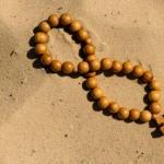
Hygroma - photo
Hygroma on the wrist.
Hygroma in the area of the interphalangeal joint of the thumb.
Hygroma in children
Hygroma in children is no different from that in adults, since it has identical properties and is localized in the same areas. In childhood, hygromas are formed, as a rule, against the background of joint injuries and excessive physical exertion associated with training, competitions or heavy physical labor. The principles of therapy and rehabilitation in children are the same as in adults, so it is not advisable to separately consider hygromas of childhood.
The exact reasons for the appearance of hygromas have not been established, so there are several theories, each of which explains only one aspect and does not cover other nuances associated with the process of cyst formation. These theories are of interest to doctors and researchers, but they are almost never used in practical medicine.
For practicing physicians, knowledge of a number of factors that can contribute to the formation of hygroma is of greater importance. Such factors include chronic inflammatory diseases of the tissues of the joint capsule of the tendon sheaths of the muscles, such as:
- Bursitis;
- Tenosynovitis;
- Tendinitis.
With long-term, sluggish listed inflammatory diseases, the formation of a cyst shell occurs, which is gradually filled with fluid leaking from numerous small blood vessels. As a result, the capsule fills and a hygroma is formed.
In addition, a predisposing factor for hygroma is frequent and prolonged injury, compression and overstrain of any joint or tissues around it. This factor is the leading factor in the formation of hygroma in people whose work involves frequent injuries, compression or overstrain of the joint (for example, typists, pianists, cooks, laundresses, etc.).
Hygroma of the wrist joint very often occurs in women after childbirth, as they begin to lift the child by inserting their palms into his armpits, which leads to severe strain on the wrist. In addition, hygromas on the joints of the feet often form in men and women when wearing tight and constrictive shoes.
Separately, it is worth noting any previous joint surgery as a predisposing factor for the formation of hygroma.
Symptoms of hygroma
Regardless of location, all hygromas are characterized by a spectrum of similar clinical manifestations, which may have different nuances when the cyst is localized in the area of a particular joint.
Clinical manifestations are mainly determined by the size of the hygroma. Moreover, the following pattern is characteristic of hygromas: the larger the cyst, the more pronounced the symptoms and the more varied the person’s complaints.
Small hygromas do not cause any inconvenience to a person and do not manifest clinical symptoms. The main complaint of people with small cysts is their unaesthetic appearance. However, as the hygroma increases, it begins to compress the surrounding tissues, nerves and blood vessels, which is manifested by a constantly present dull pain of a nagging nature. The pain intensifies when the joint in the area of which the hygroma is located is tense. For example, if the cyst is located in the area of the wrist joint, then the pain will intensify when stirring something in a container (for example, sugar in tea, cake cream in a bowl, etc.), lifting heavy objects, etc. If the hygroma is located in the area of the knee joint, then the pain will intensify when walking, standing for long periods of time, running, etc.
If the hygroma strongly compresses the blood vessels and nerves, then the person will experience impaired sensitivity and mobility in areas of the body located further than the affected joint. For example, if the hygroma is localized on the wrist, then sensitivity and mobility will be impaired in the entire hand, etc. Sensory disorders can be of two types:
Hyperesthesia(increased sensitivity of the skin, in which even light touches seem unpleasant, painful, etc.).
Paresthesia(feeling of goosebumps, numbness of the skin, etc.).
In addition to sensory disturbances, a large hygroma can cause constant neuralgic pain due to compression of the nerve, as well as venous stagnation and deterioration of blood microcirculation in areas located further than the affected joint. Impaired microcirculation and venous stagnation lead to constant paleness and coldness of the skin.
Externally, hygroma of any localization looks like a rounded bulge covered with skin. If you shine a flashlight on the cyst in complete darkness, you can see that it is a translucent bubble filled with some kind of liquid.
The skin over the hygromas usually has an unchanged pattern, but becomes thinner and colored in relatively dark shades. If a person’s joint area is subjected to compression and trauma, the skin over the hygroma may be thickened and rough, or even flaky. When palpated, the skin over the hygroma is mobile and soft enough, so it can easily be moved away from the cyst to the side. If the cyst becomes inflamed, the skin over it becomes red and swollen, and even light pressure on the formation causes pain.
The hygroma itself is painless and quite mobile when palpated, since it can be slightly moved in any direction. The surface of the formation is smooth, and the consistency is soft or densely elastic. By lightly tapping one side of the hygroma, fluctuations can be detected. To do this, a finger is placed on the surface of the hygroma on one side, and light blows are applied to the wall of the cyst on the other. In this case, the fluid present in the cyst hits the opposite wall, and a finger placed on its surface feels this movement.
Brief description of hygromas of various localizations
Let us consider the features of the development and manifestations of hygromas localized in the area of various joints.
Hygroma of the wrist (wrist joint)
Hygroma of the wrist (wrist joint) can be localized on the dorsal and palmar sides. Hygroma most often forms on the back of the wrist. A cyst is a sac filled with fluid, which is almost invisible at first, but gradually bulges more and more. The dimensions of the wrist hygroma are 3–6 cm in diameter.
It is formed from prolonged and constant physical stress on the joint during monotonous work, for example, seamstresses, embroiderers, typists, etc. Also, wrist hygroma can form as a result of an untreated injury.
At first, the cyst does not manifest itself clinically, but after some time, due to compression of the nerves and blood vessels, pain may appear, especially severe in the thumb, and difficulties in the functioning of the hand, for example, poor bending of the fingers, inability to perform precise movements, etc.
Hygroma of the hand
Hygroma of the hand is a bulging nodule on the back of the hand. As a rule, it develops after injuries (bruises or sprains) or against the background of prolonged physical stress on the hand, which may occur in musicians and some athletes (javelin throwing, shot throwing, archery, etc.).
The hygroma of this localization has a small size (no more than 2 cm in diameter), very high density and tension of the walls, and is also practically motionless. Hygroma of the hand does not manifest itself clinically in any way, since it very rarely compresses blood vessels and nerves.
Hygroma on the finger
Hygroma on the finger can be localized on the lateral, palmar or dorsal surfaces. Moreover, on the back of the finger, hygromas are much smaller than those on the palmar surface. The formation on the back side is dense, small, and has a regular round shape. As a rule, it does not manifest itself with any symptoms, and only with bruises it can hurt.
Hygroma of the palmar surface of the fingers is large and can spread to two phalanges. Due to its large size, the formation often compresses the nerves, which provokes severe pain similar to neuralgia.
Very rarely, a hygroma forms in the area where the finger joins the palm. In this case, the formation is very small (maximum 3 - 4 mm in diameter) and painful even with light pressure.
Hygroma on the hand
Hygroma on the hand can be located in the wrist or elbow joint, as well as on the back of the hand, on the palm and on the fingers. Characteristics of hygromas of the wrist, fingers and hand are presented in the sections above, so we will consider only the formation localized in the area of the elbow joint.
Hygroma of the elbow joint usually occurs due to trauma and is small in size. However, due to the fact that there is little soft tissue in the elbow area, even a small hygroma can compress the nerves and blood vessels, which causes prolonged aching dull pain, as well as impaired sensitivity and movement in the entire arm below the elbow joint.
Hygroma of the knee joint (popliteal)
Hygroma of the knee joint (popliteal) is also called
Baker's cyst, and usually develops against the background of rheumatoid arthritis, arthrosis and hematomas in the joint cavity. Most often, the cyst bulges in the area under the knee, since it is in this part that there is enough free space to place the formation between the skin and the structures of the joint. In very rare cases, the cyst bulges on the side of the knee, and almost never occurs on the front.
The size of the knee joint hygroma is quite large - up to 8 - 10 cm in diameter. When you press on the surface of the cyst, it softens as the fluid goes into the cavity of the knee joint. However, after some time, the hygroma becomes tense and dense again as the fluid returns.
Knee hygroma interferes with normal movement, flexion and extension of the leg. In addition, the formation compresses the nerves, which causes weakness and pain in the muscles of the lower leg, as well as paleness of the skin below the knee and a crawling sensation.
Hygroma of the ankle joint
Hygroma of the ankle joint is rarely formed, as a rule, only as a result of severe traumatic damage to the periarticular tissues (rupture, tendon sprain, dislocation, etc.). The cyst is usually small, but due to the small amount of soft tissue in this area, it often compresses the nerves and blood vessels, which is manifested by pain, impaired sensitivity and mobility of both the entire foot and its toes.
Hygroma of the foot
Hygroma of the foot is formed from prolonged and heavy physical activity associated with both sports and intense work. In addition, a cyst of this localization is formed quite often due to compression and trauma to the tissues by uncomfortable, constricting and tight shoes. Hygroma on the foot often hurts due to the need to wear shoes.
Hygroma on the leg
Hygroma on the leg can be localized in the area of the ankle or knee joints, as well as the back or plantar side of the foot. The characteristics of these formations are described in the relevant sections.
Hygroma of the neck
Hygroma of the neck is a congenital anomaly of the development of lymphatic vessels in a child. As a rule, hygromas on the neck are combined with congenital malformations of other organs in children. Therefore, if cysts of this localization are detected, you should contact a specialized genetic clinic for consultation and development of optimal treatment tactics. As a rule, hygromas are removed immediately after detection, since these “bumps” can cause suffocation, difficulty swallowing, etc. in the child.
DiagnosticsDiagnostics
Hygroma is quite simple, since in most cases a simple examination, palpation of the formation and detailed questioning about the circumstances of its appearance are sufficient. If in doubt, the doctor may prescribe a biopsy, computed tomography, x-ray or ultrasound of the formation to confirm or refute the diagnosis of hygroma.
Treatment of hygromaGeneral principles of therapy
Hygroma can be treated using conservative and surgical methods. Surgical methods include an operation during which the capsule is removed and pathologically altered tissues surrounding the hygroma are excised.
Conservative methods of treating hygroma include the following:
- Hygroma puncture with fluid suction;
- Hygroma crushing;
- Laser evaporation of hygroma;
- Physiotherapeutic treatment;
- Treatment of hygroma with propolis ointment;
- Traditional methods of treatment.
It should be noted that the only methods of therapy that guarantee complete cure of hygroma with no relapses in the future are laser evaporation and surgery, during which the tumor is removed along with the capsule, and the damaged surrounding tissue is excised. Such radical removal of the hygroma with the capsule, combined with excision of the affected surrounding tissue, ensures that it does not form again in this area for a very long period of time.
All other conservative methods of treating hygroma provide a temporary cure, since after a short period of absence the hygroma appears again. However, conservative treatment methods can reduce pain and ensure normal motor activity and sensitivity of the affected joint, so they can be used as symptomatic therapy.
Currently, doctors believe that it is necessary to surgically remove a hygroma if it grows rapidly, causes pain, or puts pressure on nerves and blood vessels, interfering with normal movements and disrupting sensitivity and blood circulation in the tissues. If the cyst does not hurt, does not increase in size, does not limit movements and does not impair sensitivity, then its surgical removal is carried out only at the request of the person, mainly to eliminate a cosmetic defect. In such situations, the hygroma can be left alone, simply observing the formation and using various conservative methods of therapy aimed at temporarily alleviating the condition.
Removal of hygroma (operation)
Surgical removal of hygroma is usually performed under local anesthesia, which provides excellent pain relief, but at the same time does not eliminate tactile sensitivity, due to which a person feels the doctor’s touch. Sometimes, in addition to pain-relieving injections, the anesthesiologist gives a mask with nitrous oxide, which a person can put on his face himself when he deems it necessary to enhance the effect of anesthesia. In rare cases, when a person cannot tolerate drugs for local anesthesia or when the location of the hygroma is complex, the operation is performed under general anesthesia.
An operation to remove hygroma is mandatory if a person has the following conditions, which are considered absolute indications:
- Pain at rest or with normal movements;
- Sharp limitation of range of motion in the joint;
- Rapid growth of hygroma;
- Unaesthetic appearance.
The operation is performed using conventional or arthroscopic techniques. The usual surgical technique involves making a skin incision over the hygroma, followed by spreading the edges of the wound to the sides and holding them in this position. After this, the upper part of the hygroma capsule is grabbed with forceps and held while the rest of the cyst is cut off from the surrounding tissue with the jaws of scissors. When the cyst is completely cut off from the surrounding tissues, it is pulled out, the edges of the wound are aligned and sutures are applied. Sutures are removed 7–10 days after surgery.
The arthroscopic technique of performing the operation involves introducing special manipulators in the form of long and thin tubes through a small puncture. With one manipulator, the doctor holds the instruments and removes the cyst, cutting it off from the surrounding tissue in the same way as during a normal operation, and the other is attached to a camera and a light source, which ensures that the image is transmitted to the screen. It is on this screen that the doctor sees everything he does.
Arthroscopy is a gentle and less traumatic operation compared to conventional surgery. Therefore, if possible, it is best to remove the hygroma arthroscopically.
Laser removal
Laser removal of hygroma is a modern, low-traumatic method of radical treatment that provides the same effect as surgery. Laser removal of hygroma is performed using local anesthesia to completely eliminate any discomfort during the procedure.
The essence of laser removal of hygroma is to dissect the skin over the cyst with a laser beam and provide access directly to the tumor capsule. After this, the surgeon grabs the capsule with forceps and pulls it up a little. Then a laser beam cuts off the cyst capsule from the tissue, after which it tightens the edges of the wound and applies sutures. The laser cuts through the skin and soft tissues bloodlessly, thereby minimizing trauma, resulting in healing occurring much faster than after conventional surgery.
After laser removal of the hygroma, a sterile bandage must be applied to the joint. In addition, for 2–3 days the joint is fixed with a brace or plaster cast, which provides the most favorable conditions for tissue healing and restoration of their structure, which reduces the risk of relapses and complications to a minimum.
Laser removal of hygroma is cosmetic, since it leaves an almost invisible scar on the skin, which is much more aesthetically pleasing than that after conventional surgery.
Treatment of hygroma without surgery
Treatment of hygroma without surgery involves the use of a variety of conservative methods aimed at eliminating unpleasant symptoms. The most effective conservative method is puncture of the hygroma with suction of fluid. This method allows you to remove the cyst for a while, but in 80% of people it appears again, since the formation shell remains intact.
The method of so-called crushing of the hygroma is not recommended because, firstly, it is very painful, and secondly, it leads to the re-formation of a much larger cyst. The essence of crushing is strong pressure exerted on the cyst, as a result of which its shell bursts and the liquid spreads throughout the tissues. However, after some time, a new full-fledged capsule is again formed from the pieces of the shell, which is filled with liquid and, accordingly, the hygroma appears again.
Physiotherapeutic methods are used to reduce the severity of inflammatory processes in the hygroma, to relieve pain and to neutralize the effects of compression of nearby tissues. The following physiotherapeutic techniques are most effective:
- UHF– improves blood microcirculation and tissue regeneration processes, and also relieves inflammation. It is recommended to do 1 procedure per day lasting 10–12 minutes for 8–10 days.
- Ultrasound– relaxes muscles, improves microcirculation, saturates tissues with oxygen and reduces the severity of inflammation. It is recommended to do 1 procedure per day lasting 10 minutes for 8 – 10 days.
- Magnetic therapy – reduces the severity of inflammation. It is recommended to do 1 procedure per day lasting 10 – 15 minutes for 10 days.
- Paraffin wraps– reduce the severity of inflammation, relieve pain, relieve swelling. It is recommended to do 1 procedure per day lasting 20 minutes for 10 days.
During the entire course of physical therapy, a tight bandage should be applied to the hygroma, and movements and physical activity on the affected joint should be limited. If you follow these recommendations in a gentle manner, the hygroma will stop hurting for a while, and the manifestations of compression of the nerves and blood vessels will disappear.
Another fairly effective method of conservative treatment of hygroma is the regular use of propolis ointment. This method allows you to completely remove the hygroma, but it takes quite a lot of time. For treatment, you should prepare an ointment by mixing two tablespoons of crushed propolis with 100 g of melted butter, and heating this composition over low heat for 3 hours. The finished ointment is filtered, cooled and applied to the hygroma 2 times a day until the cyst is completely resolved.
Puncture of wrist hygroma - video Hygroma of the knee joint (Baker's cyst): description, symptoms and diagnosis, treatment (puncture, removal) - video Puncture of Baker's cyst (popliteal hygroma) under ultrasound control - video After removal of the hygroma
After removing the hygroma, it is necessary to immobilize the joint in the area of which surgery was performed for several days. To do this, you can apply a plaster splint or brace to the joint. After 2 - 3 days (maximum 5), the fixing bandage should be removed and simple gymnastics should begin, aimed at developing the joint and preventing the formation of adhesions in its cavity, which in the future can make it inactive.
It is very important to start moving the joint 2–3 days after surgery, since during this period the adhesions are still thin and easily torn. And if you leave the joint without movement for 2 - 3 weeks, until the skin has completely fused, then the adhesions inside the joint will harden and become dense, and it will be very difficult and painful to break them. As a result, if a person does not endure the pain associated with rupture of the adhesions, he will have to forever come to terms with the fact that the joint will not move fully.
As gymnastic exercises, you can perform any movements in the joints, trying to achieve maximum amplitude. During movements in the joints, you should not load the muscles by holding dumbbells, heavy objects, etc. in your hands or feet. It will be possible to use the joints to their full potential no earlier than 2–3 months after the operation.
Folk remedies
The range of folk remedies used in the treatment of hygroma is very wide and very diverse. However, unfortunately, not a single folk method guarantees getting rid of hygroma and, in fact, its effects are equivalent to physiotherapy. However, traditional methods can be used to reduce the severity of pain, relieve inflammation, improve blood circulation and joint mobility.
The most effective and safest are the following folk remedies for treating hygroma:
- Compress with ficus tincture. Pour half a glass of crushed fresh ficus leaves with alcohol or vodka and leave for 24 hours. Then moisten gauze in the infusion, apply it to the hygroma, cover it with film and insulate it with a woolen bandage. The compress is changed every two hours. Duration of therapy is 2 weeks.
- Ointment made from clay and sea salt. To prepare the ointment, mix one tablespoon of crushed red clay and sea salt. It is necessary to add water drop by drop to the mixture so that the result is a thick paste. This paste is applied to the hygroma and secured with a bandage, leaving for 10 - 12 hours. After this, the bandage is changed. Treatment is carried out for 3 – 4 weeks.
- Chestnut compress. Grind fresh chestnuts in a meat grinder and apply the pulp to the hygroma, securing it with a bandage. Change the compress every 3 - 4 hours, and carry out the course of treatment for 1 - 2 weeks.
Hygroma is a benign capsule-shaped tumor. Hygroma in children at the initial stage does not cause discomfort, so it is noticed by chance, after increasing in size. This pathology never becomes malignant. Most often it occurs in 6-10 year old children, but sometimes it can form during fetal development. Most often, a child develops hygroma of the wrist or knee joint.
Hygroma in children is a periarticular neoplasm of a non-malignant nature, which can cause pain when moving.
What it is?
Hygroma (synovial cyst, tendon ganglion) is a dense neoplasm filled with a transparent liquid of a viscous structure. Formed under the skin, it is formed from the synovial membrane that extends between nearby tendons or joints. Appearance of the neoplasm:
- diameter from 0.5 to 3 cm;
- irregular or rounded shape;
- dense or soft consistency;
- does not hurt;
- does not cause discomfort.
Localization
The main locations of the ganglion are:
- hand (palm, back of wrist);
- finger (flexor muscles);
- leg (knee, area under the knee, popliteal fossa);
- foot;
- metatarsophalangeal bones;
- joint (elbow, ankle, wrist, shoulder, knee);
- back of the head;
- brain.
 The reasons for the development of hygroma can be excessive loads, injuries, and hereditary factors.
 Why does it happen?
The exact causes of hygroma have not been established, but there are a number of factors that presumably contribute to its appearance:
- hereditary predisposition;
- inflammatory process in the joint;
- joint pathology;
- injury;
- stretching;
- muscle strain;
- improper treatment of bruises, fractures, dislocations.
Symptoms
Characteristic symptoms of beginning hygroma:
- inactive compaction under the skin;
- not connected to the skin, moves under it;
- gradual growth of the tumor;
- doesn't bother;
- single (more often);
- several small moving lumps (very rare).
Signs of the disease during its long-term development (several months or years):
- significant increase in size (up to 6 cm in diameter);
- smoothness and soft consistency;
- nagging or dull pain (after compression or physical activity);
- roughness, thickening or peeling of the skin over the tumor;
- inflammation and hyperemia;
- numbness, hyperesthesia, paresthesia;
- inflammation of the joints (in advanced cases).
 Children's hygroma can cause groaning pain, numbness, and inflammation.
 Hygroma on the back of the head is the most dangerous type of this disease, which can cause the death of a child.
The types of tumors, depending on the characteristic symptoms and consequences for the child’s health, are presented in the table:
Hygroma Kinds Symptoms Consequences Hygroma of the wrist Severe pain when bending and straightening Interferes with the functioning of joints, disrupts blood circulation in the hand Hygroma of the ankle joint A dense lump forms, which hurts very much Limits the child's mobility Popliteal hygroma Progresses quickly, very painful Muscle stagnation, problems with leg flexion and extension Hygroma of the foot Severe pain syndrome Compression of nearby nerves and blood vessels, inflammation of muscle tissue Hygroma of the knee joint Visual changes - displacement of the seal to the inside of the knee Joint inflammation If a child develops a lump, it is necessary to urgently seek advice from professionals in order to make a correct diagnosis and exclude more dangerous options (including malignant neoplasms). This may help:
The study of children's hygroma includes an initial examination and hardware examination as prescribed by a doctor.
- pediatrician;
- surgeon;
- orthopedist;
- traumatologist;
- oncologist.
To diagnose a ganglion, different methods are used:
- taking anamnesis;
- visual inspection;
- palpation;
- X-ray examination;
- ultrasound diagnostics of compaction and nearby soft tissues;
- CT scan;
- aspiration;
- histology.
Hygroma treatment
If the lump is small in size, does not hurt, does not affect nerve endings and blood vessels, it can simply be observed by a doctor once every six months. Ganglion therapy in children must be carried out to eliminate discomfort in order to avoid complications and relapses. For this, various methods are used, the choice of which depends on the size of the tumor, its location and the age of the child:
- electrophoresis;
- ultraviolet irradiation;
- mud therapy;
- paraffin applications and baths;
- massage;
- laser therapy;
- puncture;
- surgical excision.
Hygroma in a child in 40−60% of cases regresses on its own; conservative treatment is effective for small tumors, and surgical treatment for large ones.
Ganglion removal
Conservative therapeutic methods cannot be applied to all types of hygromas; they are not always effective enough, and relapses are often observed after them. There are 3 types of surgery to remove a tumor:
- puncture;
- excision;
- endoscopic removal.
Radical removal of hygroma in children may be recommended for large tumors, frequent pain, and active growth.
- with a large size;
- intensive growth;
- absence of regression (within 2-3 years);
- cosmetic defect;
- associated pain and discomfort;
- limitation of motor functions;
- inflammatory processes;
- relapses of hygroma.
During puncture, fluid is removed from the ganglion with a needle, while the capsule itself remains untouched. Sclerosing, physiotherapeutic, anti-inflammatory steroid agents and therapeutic mud are introduced into the resulting cavity. This method is considered quite outdated and not as effective as excision. Endoscopic removal is a modern method that allows you to remove the tumor more gently - during the operation a small incision is made, which allows you to avoid damaging the surrounding tissues.
During surgical removal, the capsule is completely excised along with all its contents. At the moment, this method is the most effective and does not cause relapses (if there is no degenerative tissue left). Children are operated on under general anesthesia, and children over 10 years old - under local anesthesia. The operation to remove a synovial cyst is simple and lasts 20-30 minutes; the child spends several hours under the supervision of doctors, and then he is sent home. After about a week, the stitches are removed. The complete recovery process lasts a maximum of 2 months.
You can try to cure a small childhood hygroma with folk remedies, but you should not get carried away with self-medication and hesitate to visit a specialist.
Sometimes a small lump forms on the ankle, wrist, under the knee, on the finger or hand, which gradually thickens and increases in size. This benign formation is caused by the accumulation of fluid in the joints and is called a ganglion or hygroma in children. It is not capable of degenerating into a malignant form, but it causes some discomfort and therefore requires measures. As soon as a child has a hygroma on his leg or arm, he will need to consult an orthopedist. He will examine the knee joint, assess the condition of the wrist joint and determine the etiology of the disease.
Causes and consequences
Hygroma in children is congenital or acquired. Its appearance is facilitated by:
- frequent injuries,
- genetic predisposition,
- monotonous activity
- excessive physical activity,
- passive lifestyle.
An anomaly in the popliteal fossa most often occurs as a result of active play by children, when the knee suffers from an excess of movements: children fall and get bruised. So, over time, a small growth forms under the knee, making it difficult to bend the knee. If the affected area in the popliteal fossa is left untreated, there is a risk of rheumatoid arthritis or arthrosis.
With flat feet, a hygroma of the foot can form, which is a capsular cyst on the sole. Failure to contact the clinic in a timely manner may result in compression of the nerves, which will have a detrimental effect on health. If you ignore damage to the ankle, motor activity and sensitivity of the limb will also significantly decrease due to compression of the nerve fibers during foot hygroma. This can later lead to dangerous knee stiffness, changes in gait, and lameness.
The peculiarity of tumor-like formations on the wrist, finger or hand is that they grow when ignored. If the ganglion has affected the wrist joint, immediate surgical intervention is required, since the advanced pathological process will lead to limited mobility.
Symptoms
Hygroma in children, depending on the location, has different symptoms. Its characteristic feature is the formation of compactions of various sizes. Its appearance may be accompanied by:
- painful sensations,
- swelling,
- redness,
- temperature rise,
- roughness of the skin.
What to do if typical signs are detected? Get examined at the clinic.
Diagnostics
If an abnormal growth appears on the skin, you should consult an orthopedist. To identify hygroma in children, he performs a puncture, histological analysis, radiography and ultrasound examination. After collecting anamnesis, the doctor tells how and how to treat the disease.
Treatment
Treatment should only be carried out by a certified orthopedist, since the most preferable method is the removal of hygroma, which is carried out in medical centers under the supervision of competent doctors. Self-medication at home is dangerous and useless. There are many tips on how and how to treat the pathology, but effective therapy can only be carried out in a medical center. If the ganglion is independently crushed, the cystic fluid will pass into the joint cavity, and then the hygroma on the child’s arm will reappear. Hygroma on a child’s leg also has a recurrent nature if not treated correctly. Only a qualified doctor knows what to do to eliminate the tumor.
How and with what to treat
Therapeutic measures are developed strictly individually. These include: taking medications and undergoing physical therapy. In case of a complicated form, an operation is performed to remove the hygroma, excision of the lesion and treatment of the affected area with a laser.
Prevention
Hygroma in children is associated with injury and high physical activity. To prevent its occurrence, it is necessary to adjust the daily routine and correctly distribute sports activities. During training, you should use elastic bandages, avoid overloading, and if injuries occur, immediately visit an orthopedist.
You can find a specialist on our portal yourself or using the help desk (the call is free).
Hygroma (ganglion, synovial cyst) is a benign tumor that is localized in the places where the tendons are closest to the bone or connective tissue. The formation is a capsule filled with serous fluid mixed with mucus and fibrin.
In children, hygroma most often occurs in the periarticular area of the arms and legs. The neoplasm does not have the ability to undergo malignancy - a malignant change in cells.
The tumor has an oval or irregular shape and a dense structure. Average tumor size? 0.5-3 cm. Hygroma looks like a lump or small nodule with a hard or soft consistency. The skin on the surface of the formation does not change. There is usually no peeling or redness on the bump.
Locations of hygroma:
- Wrist;
- Brushes;
- Knee-joint;
- Foot.
In isolated cases, the tumor forms in the neck or brain. Then it poses a threat to the health and life of the child.
Hygroma may consist of one or several parts. Single-chamber tumors have a dense consistency. These bumps resemble cartilage or bone in structure. When the tumor consists of several parts, it is elastic to the touch and has the ability to increase in size. In addition, multi-chamber hygromas can grow deep into the tissue, which complicates their removal.
On palpation, the lump is mobile and not fused to the skin. It is always associated with a joint or tendon sheath.
The shell and contents of the neoplasm are formed from connective tissue, the cells of which change properties as a result of continuous pathological division.
Causes
Synovial cysts often appear in children of high school age. Factors that provoke the degeneration of connective tissue cells:
- Genetic predisposition;
- Joint injuries;
- Sprained tendons, ligaments;
- Excessive physical activity;
- Lack of physical activity.
At risk are children who play sports that involve systematic injuries to the limbs: boxing, football, basketball. The development of a tumor is often associated with monotonous activities: playing musical instruments, embroidering, working on a computer.
High and regularly repeated loads lead to thinning of the joint. In this state, the ability of the movable joint to hold liquid inside the shell is lost. Because of this, a lump with jelly-like contents appears in the joint capsule. It gradually hardens and grows.
There is another theory. Its founders associate the formation of hygroma with changes in cell structure. Some of the degenerated elements are transformed into a capsule surrounding the serous fluid. Cells of another type fill the membrane with thick contents. Together they form a rolling bump.
When a child attends sports clubs, the elastic fluid that fills the joint cavity begins to be produced in increased volume. Part of the capsule protrudes due to increased pressure, forming a subcutaneous tubercle. The same process occurs during monotonous activities associated with regular joint tension. 
In some cases, a tumor may appear without apparent cause or prerequisites.
How to recognize a hygroma
The tumor is always located next to the joint. Hygroma is a subcutaneous lump with elastic or soft contents. Usually the formation is single, but sometimes several pieces grow.
The child may feel discomfort in the affected area during increased stress on the joint. When pressing on the tumor, acute pain occurs. This symptom is typical for seals located near nerves and tendons. Children often complain of dull pain in the area of the lump that occurs after physical activity.
If the lump is small and localized under a ligament, it can go unnoticed for a long time. The tumor in this case manifests itself as pain when bending an arm or leg. 
In 1/3 of situations, hygroma does not cause stiffness or discomfort in the child. It may go unnoticed if it doesn't start to grow.
Features of a tumor on the hand
Most often, hygroma in a child is localized on the wrist. This feature is associated with the complex structure of the nearby joint. Moreover, in 70% the tumor is found on the outside of the arm. A tumor on the wrist can cause inconvenience if it presses on a nerve or vessel. In this case, the child complains of tingling in the hand, dull pain when moving or grasping an object. Treatment is required when the hygroma causes discomfort and increases rapidly.

Localization of the tumor in the foot area usually requires surgical intervention. The lump is often damaged by shoes and causes discomfort when walking. Regular damage can lead to muscle tissue inflammation. For this reason, treatment is indicated even for small tumors.
Hygroma under the knee is removed if it interferes with bending the leg. Small lesions usually go away without treatment. However, in such tumors it is necessary to observe the dynamics of development.
Diagnostics
If you find a suspicious lump in a child, which is most often localized on the wrist, back of the hand or under the knee, you need to contact a surgeon. Additionally, the doctor may refer you to a traumatologist or orthopedist.
To distinguish a hygroma from a tumor of another type, studies are prescribed:
- Radiography;
- Magnetic resonance imaging.
Diagnostic results help to exclude osteoarticular pathologies and study the structure of the tumor. Material is rarely taken for histological examination, because the tumor does not have the ability to become malignant. A study of the contents is carried out to distinguish the ganglion from pathologies of the sebaceous glands and adipose tissue.
In most cases, a doctor's examination and an ultrasound examination are sufficient. Other procedures are performed for the purpose of differential diagnosis with similar neoplasms and when planning surgical intervention. 
Conservative therapy
Hygroma resolves spontaneously in 50% of cases, without any measures. If the lump does not hurt or cause discomfort when moving, after diagnosis you need to periodically monitor the dynamics of its development.
The doctor chooses the method of treatment based on the medical history, the age of the child, and the location of the tumor.
If the growth of a tumor is provoked by excessive physical activity, it may be enough to relieve the pressure on the affected joint for self-healing. Conservative therapy is rarely used in practice due to the high rate of relapses.
If the tumor does not exceed 3 cm, the child’s parents may be offered the following methods:

Such methods of therapy rarely bring the desired effect. The percentage of tumor reappearance after conservative treatment is 80-90%.
In certain cases, the doctor may prescribe drug therapy and puncture of the tumor. The growth dynamics of the compaction are influenced by hormonal and sclerosing agents. Puncture is a procedure in which the contents of the tumor are pumped out using a special needle and endoscope. After complete removal of the fluid, antiseptics and antibacterial drugs are injected into the shell of the synovial cyst.
Warming, electrophoresis and exposure to ultraviolet rays are prescribed as additional methods. These procedures are not used as independent treatment.
The most effective method of treating hygroma in a child is surgical excision.

Tumor removal is carried out in the following cases:
- Dynamic growth;
- Limitation of joint mobility and loss of sensitivity;
- Pain syndrome;
- Threat of injury, infection entering the cavity of the capsule;
- Cosmetic discomfort.
Large tumors are always removed surgically. Excision is necessary if the lump does not resolve within 2-3 years or begins to grow rapidly.
Hygroma removal methods:
- Surgery;
- Laser exposure.
Surgical method
In young children, the operation is performed under general anesthesia. If the child is over 10 years old, the hygroma is removed after local anesthesia is administered. An incision is made in the affected area. A tourniquet is applied above the excision area to bleed the operated area.
After removing the contents and the capsule itself, the cavity is washed with an antiseptic solution and sutured. A rigid bandage is used to immobilize the joint. This measure is necessary to prevent relapse. A drainage may be placed in the wound for 2-3 days if indicated. The duration of the operation is about 30 minutes.
The risk of relapse after removal of hygroma is 5-20%. Re-growth of the tumor occurs if some of the abnormal cells remain near the joint. The changed elements begin to actively divide. This process leads to the formation of a new compaction of the same nature.
Laser beam heating
The feasibility of laser removal of hygroma is determined using X-rays or magnetic resonance imaging. The advantage of the technique compared to surgical excision:
- The procedure takes less time;
- Neat seam;
- Evaporation of cyst fluid occurs without involvement of surrounding tissues;
- There is no scar left after healing;
- Fast rehabilitation.
The operation is performed under local anesthesia. An incision is made over the tumor. Hygroma is removed by exposure to a high-energy beam. The manipulation is carried out with a carbon dioxide device, the action of which ensures coagulation of pathological cells and disinfection of the cavity. After laser exposure, the child is given a suture and an immobilizing bandage. The operation takes no more than 15 minutes.
Laser removal of hygroma is the most gentle procedure compared to surgical excision. But beam heating has disadvantages. The mouth of the capsule is not sutured, which often leads to relapse.
Traditional methods of treatment
Unconventional methods of eliminating hygroma are used as additional measures of conservative therapy.
Folk recipes will help you get rid of the bump:

Since the ganglion is difficult to recognize without the help of a doctor, home remedies are used after consultation with a surgeon. Compresses and heating can be dangerous for tumors that look like hygroma.
Complications
The most common complication of hygroma is re-filling of the cavity with serous fluid. Relapse occurs due to poor-quality treatment after surgery, lack of rest for the joint for a specified period, or failure to comply with the dressing regimen.
Possible consequences of surgical intervention include purulent tenosynovitis. This complication rarely occurs in children. Inflammation occurs when an infection gets into the wound.
Forecast
Children have a high probability of spontaneous disappearance of hygroma. It often resolves as the load on the joint decreases. Tumors located near large vessels pose a danger. The prognosis is favorable if the operation is performed on time.
The tumor does not threaten the child's life. If the lump grows rapidly, the operation should not be postponed. Timely removal of hygroma will save the child from joint problems in the future.