The liver is an unpaired organ, which is the largest gland in the human body. This body belongs to digestive system. Its importance for the functioning of the entire organism is determined by its topographic and anatomical location. To begin with, it should be noted that the topography of the liver is the structure of the gland, that is, the study of its layers and location in the body. The topography of the liver also includes the study of its blood supply and innervation.
The topography of the liver is very important in operative surgery, as it will become a guide for doctors. After all, each person is individual, and the structure of the organ and vascular system not identical.
The liver is very important for the whole body; its dysfunction leads to pathologies of all systems. Its main function is to cleanse the body of toxins and various harmful substances that are in the blood. In addition, it removes excess other substances from the body, such as hormones, vitamins and other metabolic products.
- Produces bile.
- Produces protein.
- It stores glycogen, vitamins and various microelements; it is a so-called storage facility, and when they are deficient in the body, it releases the required amount.
- Produces cholesterol, and regulates fat and carbohydrate metabolic processes.
Organ segments
Previously, the liver was divided only into lobes. Today, the idea of the structure of the liver has changed and expanded greatly. There is a whole science about the segmental structure of the liver. It has 5 tubular systems:
- Arterial vessels.
- Portal system - branches of the portal vein.
- Caval system – veins of hepatic localization.
- Bile ducts.
- Vessels of the lymphatic type.
The portal system and the caval system do not touch in any way. Unlike other segments, which are always parallel to each other. As a result of such proximity, bundles of individual structures with innervation are formed.
Location
The liver is a gland that is located in the peritoneum. In the area of the right side there is most of organ, but it is also localized in the epigastric part, and a small part of it is still in the left hypochondrium. Since the liver is shaped like a triangle, and its edges are sloping. The base of the liver is the right lobe, and the acute angle is left lobe, which is much smaller. This arrangement of the organ is due to a complex ligamentous system.
 The top edge of the gland is adjacent to the diaphragm. On the upper right, the organ is located at the same level with the V-shaped costal cartilage. On the left side, in the upper edge, the left lobe of the organ is located with the VI costal cartilage at the same level.
WITH right side the bottom edge corresponds to the location of the costal arch, then rushing towards left side, the organ emerges from behind the costal arch, at the place where the VII and X costal cartilages connect. On the left side, the left lobe extends beyond the ribs, in the area where the VII and VIII costal cartilages are connected.
The right border of the organ is located along the midline of the axillary region. The upper point on the right side is located on the same line with the VII rib, and the lower right is on the line with the XI rib. If we consider the posterior projection of the liver, then the upper border is at the same level with the IX thoracic vertebra. And the lowest point from the back is located on the line of the XI thoracic vertebra.
The liver changes its location during breathing, that is, when inhaling and exhaling, it rises and falls by 3 cm. From below, the gland is adjacent to other organs, while there are squeezes on the gland. Namely, from the colon, kidney, the stomach is adjacent to the left side of the organ, the back part borders on the esophagus.
The duodenum is adjacent to the posterior plane of the organ. There is also a depression from the gallbladder, which is located between the two lobes of the liver. And the duodenal indentation is located near the hepatic gate. From above, the left lobe of the liver is adjacent to the heart, and an indentation is also formed.
The liver is a parenchymal organ with a soft consistency. Weighs from 1.5 to 2 kg in an adult. It has 2 surfaces:

- The longitudinal one on the right is the depression from the gallbladder. The inferior vena cava passes through the posterior part of this recess. To the right of this depression is the right lobe of the organ.
- Longitudinal on the left - the round ligament and umbilical vein are localized in it. At the back, a fibrous cord is located in this recess. To the left of this depression is the left lobe of the organ.
- The transverse groove is the portal of the liver. It is in this place that the main bile duct, blood vessels and nerves are located.
Between these structures is the quadrate segment of the liver. There is also another segment called the caudate lobe. It is localized between the hepatic hilum and the recesses with the inferior vena cava and venous ligament.
Liver structure
The liver consists of 2 structures - the right and left lobes. These are the structural and functional units of the organ. There is connective tissue between them.
The right and left lobules look like hexagonal prisms with a flat base but a convex top. The hepatic lobules consist of beams and lobulated sinusoidal hemocapillaries.
 These structural elements are located radially, starting from the periphery of the organ and heading towards the center, to the place where the portal vein passes. The beams are made up of 2 rows of hepatocytes. And lobular sinusoidal hemocapillaries consist of flat endothelial cells.
Hepatocytes are arranged in rows, and between them there are also bile capillaries. Their diameter does not exceed 1 micron. It is characteristic that they do not have a membrane, but are limited to the plasmalemma of hepatocytes, which are located nearby. Bile capillaries pass along the hepatic beam and smoothly pass into other structures - cholangioles. These are tubes that flow into the bile ducts located between the lobes of the gland.
The connective tissue that is localized between the two lobes does not develop. Its development and growth can only occur as a result of various pathologies. For example, cirrhosis.
Ligaments of the liver
The ligamentous apparatus is formed by the peritoneum. These ligaments pass to the liver from the diaphragm, namely from its lower part. The ligament passing from the diaphragm is the coronary ligament of the liver. At the edges, the coronary ligament has triangular plates.
The visceral surface of the liver has its own ligamentous apparatus. Ligaments extend from it to organs that are located nearby.
There is also a falciform ligament, which is located between the convex part of the liver and the diaphragm. The round ligament of the liver arises from this ligament. It runs from the umbilicus to the left branch of the portal vein.
There are 2 more triangular ligaments - right and left. On the right side, it goes from the diaphragm to the right lobe of the organ. But in some people it may be completely absent, and in most people the right triangular ligament is poorly developed. As for the triangular ligament on the left side, it runs from the bottom of the diaphragm to the convex surface of the left lobe.
Blood supply
The liver is an organ that cleanses the blood of toxins. Accordingly, blood enters it from both arteries and veins. Namely, the vessels that supply the liver with blood are the hepatic artery and the portal vein. These are the 2 largest vessels. In this case, only 25% of the blood comes from the artery, and the remaining 75% is venous blood, that is, it is blood from the portal vein.
The blood supply to the liver is divided into 2 parts. And between these parts there is a border that passes through the top of the gallbladder, as well as the inferior vena cava. Such a boundary is a hypothetical plane. It is slightly tilted to the left. That is, each half of the organ is autonomously supplied with blood and also has an outflow of bile and blood autonomous from each other. And these 2 halves are divided into 4 segments, the blood supply pattern of which is the same. Small deviations from the general scheme are allowed.
The hepatic artery supplies the organ with blood and oxygen. This vessel is a branch of the aorta. Further, these vessels branch throughout the organ: lobular, segmental, interlobular and veins.
Additional vessels, which only 30% of people have, can also supply the liver with blood. These are vessels that branch from the following arteries:
- celiac;
- left gastric;
- superior mesenteric;
- gastroduodenal.
Sometimes these vessels branch from the aorta or phrenic artery. But this happens in rare cases.
The portal vein carries blood from other organs. This vein has 2 to 4 roots, including the superior mesenteric vein and the splenic vein. In addition, there may also be superior mesenteric and left gastric. But these branches are rarely observed as the roots of the portal vein.
Large vessels enter the thickness of the organ in its lower part in the center. This place is called Glisson's Gate. In most people, this is where the hepatic artery and portal vein divide.
The outflow of blood occurs through 3 veins. They come together at the back of the liver and gradually enter the inferior vena cava, which runs in the inferior vena cava. There are also small thin-walled veins, which range in size from 2 to 8 mm, and they pass outside the gland.
The main duct for the outflow of bile runs through the porta hepatis. The right and left ducts flow into it. They drain bile from the right and left parts of the liver. About 75% of all bile passes through them. Bile plays a very important role in the human body. It is involved in the process of thermoregulation, sweating, and also helps cleanse the body. Bile also promotes pigmentation of the skin.
 During operations, surgeons pay special attention to the vascular-ductal structures, which are located close to the surface of the organ. Such ducts are located 1.5 cm deep in the organ. In the depths of the organ there are only small ducts (3rd and 4th order), they are located in the area of the diaphragmatic surface.
Lymphatic drainage occurs through nodes that are located near the hepatic gate, as well as lymph nodes located in the hepatoduodenal ligament and nodes in the retroperitoneal space. There are superficial and deep lymph nodes.
Superficial lymphatic vessels are localized near the veins of the liver, they run parallel and go to the back of the organ, and then go to the lymph nodes located in chest cavity. At the same time, they pass through the diaphragm or through the slits that are in it.
Deep hepatic lymph vessels can be:
- Ascending.
- Descending. They are localized near the portal vein and its branches, the hepatic artery and ducts. They are directed to the lymph nodes, which are located near the hepatic artery, and near the aorta and inferior vena cava.
Innervation
The process of innervation of the liver occurs due to the action mainly of the vagus nerves, as well as those localized in the celiac plexus and the right phrenic nerve.
The splanchnic nerves are responsible for the innervation of the sympathetic type, and the vagus nerves in in this case for the parasympathetic system.
The vagus nerves, the plexuses of which are localized at the hepatic gate, are divided into anterior and posterior hepatic plexuses. The branches that arise from the phrenic nerve are localized along the inferior vena cava. The fibers of this nerve are in the structure of the hepatic plexuses and therefore these fibers become effective sources in the process of innervation of both the liver and the gallbladder.
Who said that it is impossible to cure severe liver diseases?
- Many methods have been tried, but nothing helps...
- And now you are ready to take advantage of any opportunity that will give you the long-awaited well-being!
An effective treatment for the liver does exist. Follow the link and find out what doctors recommend!
The liver (hepar) is located in the right hypochondrium, pancreas and partially in the left hypochondrium (Fig. 137). The position of the falciform ligament divides the upper surface of the liver into right and left parts. On the lower surface of the organ there are right and left longitudinal grooves and a transverse groove - the hilum of the liver. Along these grooves, located in the shape of the letter H, the liver is divided into four lobes: the right and left lobes, between which there is a square lobe (lobus quadratus) in front and a caudate, or Spigelian, lobe (lobus caudatus Spigelii) in the back.
The upper border of the liver rises along the right midclavicular line to the cartilage of the 5th rib, along the midline of the body to the base of the xiphoid process of the sternum and along the left sternal line to the cartilage of the 6th rib. The lower border of the organ, corresponding to its anterior edge, is located on the right along the edge of the costal arch, along the midline of the body within middle third the distance between the xiphoid process and the navel and, going to the left, goes to the junction of the cartilages of the VII and VIII ribs. To the left, the liver ends within the space between the left sternal and left parasternal lines. From the back, the posterior surface of the liver is projected within the boundaries of the lower edge of the IX and middle of the XI thoracic vertebrae. The upper surface of the liver is adjacent to the diaphragm, the anterior surface is adjacent to the diaphragm and the anterior abdominal wall, posterior - to the spine, legs of the diaphragm, aorta, esophagus and inferior vena cava, lower - to the right bend of the colon, the upper pole of the right kidney with the adrenal gland, the initial segment duodenum and stomach (pylorus, lesser curvature and cardia). The liver is located mesoperitoneally. It is surrounded by peritoneum above and below and lacks it on the back side. The serous membrane covering the organ passes to adjacent anatomical elements and forms ligaments.
The arrow shows the entrance to the omental bag.
1 - square lobe of the liver; 2 - round ligament of the liver; 3 - caudate lobe of the liver; 4 - left lobe of the liver; 5 - spleen; 6 - diaphragm; 7 - stomach; 8 - colon: 9 - duodenum; 10 - right lobe of the liver; 11 - elements of the hepatoduodenal ligament; 12 - gallbladder. A - posterior surface of the liver; 13 - abdominal area; 14 - inferior vena cava.
The crescent-shaped, or hanging, ligament of the liver (lig. falciforme, s. suspensorium hepatis) is a double layer of the peritoneum, following along the sagittal plane from the upper surface of the liver to the diaphragm. This ligament is thickened at the free edge and appears as the round ligament of the liver (lig. teres hepatis). This ligament runs from the liver to the navel and contains the empty v. umbilicalis. The coronary ligament of the liver (lig. coronarium) is the leaves of the peritoneum of the suspensory ligament that diverge to the right and left. It ends on the sides like lig. triangulare dextra et sinistra and follows from the posterosuperior edge of the organ to the diaphragm. The existing hepatogastric and hepatoduodenal ligaments have been described previously. The hepatorenal ligament (lig. hepatorenale) is a not always pronounced fold of the peritoneum, following from the porta hepatis to the right kidney.
The liver receives arterial blood through its own hepatic artery (a. hepatica propria), which at the gate of the organ is divided into right and left branches that go to the corresponding lobes of the organ. Blood also flows to the liver through the portal vein, formed from the confluence of the venous branches of the stomach, spleen, intestines, and pancreas. At the porta hepatis, the vein divides into branches for the right and left lobes of the organ. The outflow of blood from the liver occurs through 2-4 hepatic veins, flowing into the inferior vena cava in the area of its contact with the posterior surface of the organ.
The lymphatic vessels of the liver are divided into superficial and deep. The outflow of lymph is carried out: 1) along the paths that follow through the gates of the liver; into the hepatoduodenal ligament at nod. lymph, hepatici proprii, nod. lymph., hepatici communis and further to nod. lymph, coeliaci; 2) through vessels heading through the veins of the liver, into The lymph nodes nod. lymph, subdiaphragmatici, nod. lymph, supradiaphramatici, nod. lymph, retrosternalis, further into the ductus lymphaticus dexter.
The liver is innervated by branches coming from the solar plexus, vagus and right phrenic nerves. These branches, passing through the hepatoduodenal ligament, form the anterior and posterior hepatic plexuses.
The liver occupies the right hypochondrium, the epigastric region and partly the left hypochondrium. The left border is projected along the left midclavicular line in the 5th intercostal space, along the right parasternal line on the 5th costal cartilage, along the right midclavicular line in the 4th intercostal space, along the right midaxillary line on the 8th rib and at the spine on the 11th rib. The lower edge is along the midaxillary line at the 10th intercostal space, then comes out from under the costal arch, goes obliquely upward, projecting along the midline of the body in the middle of the distance between the navel and the base of the xiphoid process. The lower edge intersects the left part of the costal arch at the level of the VI costal cartilage.
The liver has two surfaces: upper (diaphragmatic) and lower (visceral), as well as two edges. The lower edge is sharp with two notches - the impression from the gallbladder and the notch of the round ligament of the liver. The posterior edge is rounded and faces the posterior abdominal wall. The upper surface is convex and smooth. The lower one is uneven, has two longitudinal and one transverse grooves (indentations from adjacent organs). The transverse groove corresponds to the porta hepatis. The right longitudinal groove is the fossa of the gallbladder in the anterior part and the groove of the inferior vena cava in the posterior part. The left longitudinal groove is a deep gap separating the left lobe of the liver from the right. It contains the round ligament of the liver. The liver consists of right and left lobes. On the diaphragmatic surface the border is the falciform ligament, on the lower surface there is a longitudinal groove. In addition, there are quadrate and caudate lobes. Square - between the anterior sections of the longitudinal grooves, caudate - between their posterior sections. The lobes are separated from each other by a transverse groove.
Gate of the liver
Anterior border- posterior edge of the quadrate lobe; right - right lobe; posterior - caudate lobe and partially right; left - left lobe. The liver is covered by peritoneum on all sides except for the hilum and the surface adjacent to the diaphragm. The peritoneal covering, when passing from the liver to the surrounding organs, forms a ligamentous apparatus.
Round ligament of the liver- from the navel in the groove of the same name to the gate. The anterior part of the falciform ligament merges with it.
Falciform ligament- between the diaphragm and the upper convex surface. From the back to the right and left it passes into the coronary ligament.
Coronary ligament- transition of the parietal peritoneum from the lower surface of the posterior part of the diaphragm to the visceral one.
With the help of the hepatogastric and hepatoduodenal ligaments, the liver is connected to the organs of the same name.
Between the leaves of the hepatoduodenal ligament there passes the hepatic artery, the common bile duct with the common hepatic and cystic duct, the portal vein, etc. A lobe, sector and segment are called a section of the liver that has a separate blood supply, bile outflow and lymph drainage. In addition to the two lobes, there are 5 sectors and 8 most constant segments. The segments, grouped around the gates, form sectors. Venous circulation in the liver is represented by the portal vein system, which brings blood to the organ, and the hepatic vein system, which drains blood into the inferior vena cava. The arterial blood supply begins from the celiac trunk and is represented by the common, then the proper hepatic artery, which is divided into the left and right lobes.
Holotopia: located mostly in the right hypochondrium, occupies the epigastric region and partially the left hypochondrium.
Skeletotopia:
1. upper limit: along the left midclavicular line – V intercostal space; along the right parasternal – V costal cartilage; along the right midclavicular line – IV intercostal space; along the right mid-axillary – VIII rib; at the spine - XI rib.
2. lower limit: along the right mid-axillary line – X intercostal space; along the midline - the middle of the distance between the navel and the base of the xiphoid process; The left costal arch crosses at the level of the VI costal cartilage. Relation to the peritoneum: mesoperitoneal organ (the hilum and dorsal surface are not covered).
Syntopy: top – diaphragm; in front – the anterior abdominal wall and diaphragm; behind – X and IX thoracic vertebrae, crura of the diaphragm, esophagus, aorta, right adrenal gland, inferior vena cava; below - the stomach, bulb, superior flexure and upper quarter of the descending duodenum, right flexure of the colon, upper pole of the right kidney, gall bladder.
Structure
The liver has two surfaces: upper (diaphragmatic) and lower (visceral), as well as two edges. The lower edge is sharp with two notches - the impression from the gallbladder and the notch of the round ligament of the liver. The posterior edge is rounded and faces the posterior abdominal wall. The upper surface is convex and smooth. The lower one is uneven, has two longitudinal and one transverse grooves (indentations from adjacent organs). The transverse groove corresponds to the porta hepatis. The right longitudinal groove is the fossa of the gallbladder in the anterior part and the groove of the inferior vena cava in the posterior part. The left longitudinal groove is a deep gap that separates the left lobe of the liver from the right. It contains the round ligament of the liver. The liver consists of the right and left lobes, the border between which is the falciform ligament on the diaphragmatic surface, and the longitudinal groove on the lower one. In addition, there are quadrate and caudate lobes. Square - between the anterior sections of the longitudinal grooves, caudate - between their posterior sections. These lobes are separated by a transverse groove. In addition to the two lobes, there are 5 sectors and 8 most constant segments. The segments, grouped around the gates, form sectors. A lobe, sector and segment are called areas of the liver that have a separate blood supply, bile outflow and lymph drainage.
Ligamentous apparatus
Coronary ligament fixes the liver to the lower surface of the diaphragm in the frontal plane. At the right and left edges of the liver, it passes into the right and left triangular ligaments.
Falciform ligament located in the sagittal plane between the diaphragm and the convex diaphragmatic surface of the liver at the border of its right and left lobes.
Round ligament of the liver located between the umbilicus and the porta hepatis in the free edge of the falciform ligament and is a partially obliterated umbilical vein.
From the visceral surface of the liver, the hepatogastric, hepatoduodenal and hepatorenal ligaments are directed to the corresponding organs.
Blood supply
Proper hepatic artery is a branch of the common hepatic artery, and the latter is a branch of the celiac trunk. It passes to the left of the common bile duct between the layers of the hepatoduodenal ligament to the portal of the liver and is divided into right and left branches. The right branch supplies the right lobe of the liver, and, as a rule, gives off the cystic branch to the gallbladder, the left branch supplies the left lobe of the liver.
Portal vein drains venous blood from all unpaired organs to the liver abdominal cavity. Its trunk is formed behind the head of the pancreas from the splenic and superior mesenteric veins.
Umbilical vein is located in the round ligament of the liver and flows into the left trunk of the portal vein; obliterated near the umbilical ring.
Periumbilical veins are located in the round ligament of the liver, flow into the portal vein; carry blood from the anterior abdominal wall.
Venous drainage from the liver carried out by a system of 3–4 hepatic veins, which flow into the inferior vena cava at the place where it is closely adjacent to the posterior surface of the liver.
Innervation
Nerve branches coming from the celiac plexus, the vagus and the right phrenic nerves take part in the innervation of the liver. At the gate of the liver, the anterior and posterior hepatic plexuses are formed from them, the nerve conductors of which spread through the connective tissue layers throughout the entire organ.
Lymphatic drainage
suturing the wall of the sigmoid colon with interrupted sutures around the entire circumference of the surgical wound, connecting the serous layer with the parietal peritoneum;
opening of the intestinal lumen after the formation of adhesions between the visceral and parietal peritoneum (after 3-4 days);
suturing the edges of the mucous membrane to the skin.
Imposition of the unnatural anus –
creating an opening in the colon through which all intestinal contents are expelled without entering the underlying parts of the intestine.
Indications: tumors, wounds, cicatricial narrowing of the rectum, rectal amputation.
Classification: temporary and permanent, single-barrel (Hartmann operation) and double-barrel (Maidl operation).
Technique for applying a single-barreled unnatural anus:
layer-by-layer opening of the abdominal cavity with an oblique variable incision in the left groin area;
piercing the intestinal mesentery in the avascular zone and passing a rubber tube through the window;
suturing the afferent and efferent loops together under the tube with 3-4 interrupted seromuscular sutures (formation of a “spur”);
suturing the parietal peritoneum to the edges of the skin incision;
suturing the “double-barreled shotgun” removed from the abdominal cavity with serous-muscular sutures along the entire circumference to the parietal peritoneum;
transverse section of the anterior wall of a thick sewn-in
intestines (the resulting “spur” protrudes upward and eliminates the possibility of feces getting into the outlet loop).
Features of the jejunum and ileum in newborns and children
Primary department small intestine, as well as its terminal section, are located much higher in children than in adults: the initial one lies at the level of the XII thoracic vertebra, and the terminal one - the IV lumbar vertebra. With age, these sections gradually descend, and by the age of 12-14 the duodenum
The jejunal flexure is located at the level of the second lumbar vertebra, and the ileocecal angle is located in the right iliac region.
Loops of the small intestine in children of the first year of life in upper section covered by the liver, and throughout the rest of the length they are directly adjacent to the anterior abdominal wall. With the development of the greater omentum, the area of contact of the small intestine with the anterior abdominal wall gradually decreases. By the age of 6-7 years, the omentum completely covers the intestinal loops in front. The relative length of the small intestine in children under 3 years of age is greater than in adults.
Malformations of the jejunum and ileum
Atresia - can be single or multiple, combined with various anomalies in the development of the mesentery (mesenteric defects) and blood vessels, and have different localizations.
Stenoses are associated with the formation of membranes from the mucous membrane, and sometimes from other layers of the intestinal wall, with more or less holes.
Duplication of the small intestine - in the form of thick-walled cystic formations or elongated additional segments of the intestine in the shape of a horn or double-barreled shotgun (located on the mesenteric edge or side wall).
Congenital volvulus is caused by incomplete rotation of the midgut.
Features of the colon in newborns and children
The cecum in newborns is located at the level of the iliac crest and only by the age of 14 reaches the iliac fossa. In some cases, the process is delayed, and then in older children one can encounter a high position of the cecum and appendix. With an excessively long mesentery, the cecum becomes mobile and can be located in various parts of the abdominal cavity. The cecum in children in the first months of life has a funnel-shaped or conical shape and acquires its normal appearance only by the age of 7 years. The muscular sphincter of the ileocecal region is not developed at the time of birth and the contents of the intestines can pass freely in both directions.
The base of the appendix in children is funnel-shaped, and the border between it and the cecum is smoothed. The hole leading to the appendix gapes, and only by the end of the first year of life its sphincter is formed.
The transverse colon in newborns has additional bends, its mesentery is mobile, the length is 1.5-2 cm. Then the mesentery gradually thickens, lengthens, and by 1.5 years it reaches 5-8 cm.
Colon malformations
Megacolon (Hirschsprung's disease) is a sharp expansion of the entire colon or its individual sections. The muscle fibers, as well as the mucous layer of the expanded part of the intestine, are sharply thickened. It is currently believed that the main cause of megacolon is underdevelopment of the nodes of the Auerbach plexus. As a result, the tone of the sympathetic nerve plexus predominates, which leads to a state of constant spasm of this part of the intestine. These changes are most pronounced in the distal sigmoid and rectum. Dilatation of the proximal bowel is secondary to continuous overcoming of resistance. There are four types of megacolon: gigantism, megadolichocolon, mechanical megacolon, Fawali Hirschsprung's disease itself with the presence of a spastic zone and an expansion of the diameter of the proximal part.
Operations for Hirschsprung's disease are performed at 2-3 years of age using the abdominal-perineal method. The intervention includes resection of the entire aganglionic zone and the adjacent section of the dilated intestine for 6-12 cm with the formation of an anastomosis between the proximal part of the resected intestine and the final part of the rectum. The large intestine is brought down to the perineum through distal section straight or through a tunnel formed in the retrorectal tissue.
Atresia of the colon - manifests itself in two forms: membranous (there is a membrane of varying thickness that covers the entire lumen of the intestine) and saccular (one of the segments ends in a blind pouch, and the rest retains its normal shape).
Colon stenosis is a narrowing of the intestinal lumen as a result of the presence of a thin membrane or local thickening of the intestinal wall.
Duplication of the colon - cystic, diverticular and tubular (tubular) forms.
TOPOGRAPHIC ANATOMY AND OPERATIONS ON PARENCYMATOUS ORGANS
TOPOGRAPHIC ANATOMY OF PARENCYMATOUS ORGANS
Topographic anatomy of the liver Holotopia: located mostly in the right sub-
rib, occupies the epigastric region and partially the left hypochondrium.
upper border: along the left midclavicular line – V intercostal space; along the right parasternal – V costal cartilage; along the right midclavicular line – IV intercostal space; along the right mid-axillary – VIII rib; at the spine - XI rib.
Lower border: along the right mid-axillary line – X intercostal space; along the midline - the middle of the distance between the navel and the base of the xiphoid process; left
The costal arch crosses at the level of the VI costal cartilage. Relation to the peritoneum: mesoperitoneal organ (not
the hilum and dorsal surface are covered).
Syntopy: top – diaphragm; in front – the anterior abdominal wall and diaphragm; behind – X and IX thoracic vertebrae, crura of the diaphragm, esophagus, aorta, right adrenal gland, inferior vena cava; below - the stomach, bulb, superior flexure and upper quarter of the descending duodenum, right flexure of the colon, upper pole of the right kidney, gall bladder.
The liver has two surfaces: upper (diaphragmatic) and lower (visceral), as well as two edges. The lower edge is sharp with two notches - the impression from the gallbladder and the notch of the round ligament of the liver. The posterior edge is rounded and faces the posterior abdominal wall. The upper surface is convex and smooth. The lower one is uneven, has two longitudinal and one transverse grooves (indentations from adjacent organs). Transverse
The naya groove corresponds to the porta hepatis. Right longitudinal
groove - the fossa of the gallbladder in the anterior part and the groove of the inferior vena cava in the posterior part. The left longitudinal groove is a deep gap that separates the left lobe of the liver from the right. It contains the round ligament of the liver. The liver consists of the right and left lobes, the border between which is the falciform ligament on the diaphragmatic surface, and the longitudinal groove on the lower one. In addition, there are quadrate and caudate lobes. Square - between the anterior sections of the longitudinal grooves, caudate - between their posterior sections. These lobes are separated by a transverse groove. In addition to the two lobes, there are 5 sectors and 8 most constant segments. The segments, grouped around the gates, form sectors. A lobe, sector and segment are called areas of the liver that have a separate blood supply, bile outflow and lymph drainage.
The coronary ligament fixes the liver to the lower surface of the diaphragm in the frontal plane. At the right and left edges of the liver, it passes into the right and left triangular ligaments.
The falciform ligament is located in the sagittal plane between the diaphragm and the convex diaphragmatic surface of the liver at the border of its right and left lobes.
The round ligament of the liver is located between the umbilicus and the porta hepatis in the free edge of the falciform ligament and is a partially obliterated umbilical vein.
From the visceral surface of the liver, the hepatogastric, hepatoduodenal and hepatorenal ligaments are directed to the corresponding organs.
Feature circulatory system The liver is that blood is delivered to it by two vessels: the proper hepatic artery and the portal vein.
The proper hepatic artery is a branch of the common hepatic artery, and the latter is a branch of the celiac trunk. It passes to the left of the common bile duct between the layers of the hepatoduodenal ligament to the portal of the liver and is divided into right and left branches. The right branch supplies the right lobe of the liver, and, as a rule, gives off the cystic branch to the gallbladder, the left branch supplies the left lobe of the liver.
The portal vein drains venous blood from all unpaired abdominal organs to the liver. Its trunk is formed behind the head of the pancreas from the splenic and superior mesenteric veins.
The umbilical vein is located in the round ligament of the liver and flows into the left trunk of the portal vein; obliterated near the umbilical ring.
The peri-umbilical veins are located in the round ligament of the liver and empty into the portal vein; carry blood from the anterior abdominal wall.
Venous drainage from the liver is carried out by a system of 3-4 hepatic veins, which flow into the inferior vena cava in the place where it is closely adjacent to the posterior surface of the liver.
Nerve branches coming from the celiac plexus, the vagus and the right phrenic nerves take part in the innervation of the liver. At the gate of the liver, the anterior and posterior hepatic plexuses are formed from them, the nerve conductors of which spread through the connective tissue layers throughout the entire organ.
Lymphatic drainage from the liver occurs in the lymph nodes located at the gates of the liver, in the right or left gastric, celiac, preaortic, lower diaphragmatic and lumbar nodes.
Topographic anatomy of the gallbladder
Gallbladder It is a pear-shaped reservoir for bile, located between the right and quadrate lobes of the liver. It distinguishes between the bottom, body and neck. The neck of the gallbladder continues into the cystic duct, is directed towards the portal of the liver and lies together with the cystic duct in the hepatoduodenal ligament.
Skeletotopy: the bottom of the gallbladder is determined anteriorly, at the point of intersection of the outer edge of the right rectus abdominis muscle with the costal arch, posteriorly - at the level of the upper edge of the L2 vertebra.
The attitude towards the peritoneum of the gallbladder is subject to large individual fluctuations. Usually located mesoperitoneally in relation to the peritoneum. However, there is an intrahepatic position, when almost the entire gallbladder, with the exception of its bottom, is surrounded by liver parenchyma. At
intraperitoneal position, when the gallbladder has a pronounced mesentery, it may twist with subsequent necrosis of the gallbladder.
Syntopy: in front and above - the liver, on the right and below - the right flexure of the colon, on the left - the pylorus.
Blood supply from the cystic artery. Venous drainage occurs through the cystic vein, which flows into the right branch of the portal vein.
Lymphatic drainage occurs from the lymphatic vessels of the bladder to the first-order lymph nodes located at the gate of the liver.
Innervation from the hepatic nerve plexus.
Topography of extrahepatic biliary tract
The extrahepatic bile ducts include the right and left hepatic ducts, the common hepatic duct, the cystic duct and the common bile duct. General hepatic duct is formed at the porta hepatis from the confluence of the right and left hepatic ducts.
The cystic duct in the hepatoduodenal ligament merges at an acute angle with the common hepatic duct, forming the common bile duct. Depending on the location, the common bile duct is conventionally divided into four parts: supraduodenal, retroduodenal, pancreatic and intramural.
The first part of the duct passes through the thickness of the hepatoduodenal ligament to the upper level of the duodenum, the second part of the duct is located behind the upper part of the duodenum. Both of these parts are most susceptible to injury during operations on the stomach and duodenum.
The third part of the common bile duct passes either deep into the head of the pancreas or behind it. It can be compressed by a tumor of the head of the pancreas, resulting in obstructive jaundice. The fourth part in an oblique direction pierces the posterior wall of the duodenum and opens on its major papilla. In 80% of cases, the final sections of the common bile duct and the pancreatic duct merge, forming a
cheno-pancreatic ampulla, in the circumference of which the ring-shaped sphincter of the ampulla (sphincter of Oddi) is formed.
In the final section of the common bile and pancreatic ducts there is a powerful accumulation of sympathetic, parasympathetic and sensory nerve conductors and intramural microganglia, which provide complex regulation of the activity of the sphincter of Oddi.
Topographic anatomy of the pancreas
The pancreas is an organ that has excretory and incretory functions. The gland is divided into a head, body and tail. A hook-shaped process sometimes extends from the lower edge of the head.
The head is surrounded above, to the right and below, respectively, by the upper, descending and lower horizontal parts of the duodenum. She has:
the anterior surface, to which the antral part of the stomach is adjacent above the mesentery of the transverse colon, and below - the loops of the small intestine;
the posterior surface to which the right renal artery and vein, the common bile duct and the inferior vena cava adjoin;
top and bottom edges.
the front surface to which it is adjacent back wall stomach;
posterior surface to which the aorta, splenic and superior mesenteric veins are adjacent;
the lower surface, to which the duodenojejunal flexure adjoins from below;
upper, lower and anterior edges.
the anterior surface to which the fundus of the stomach is adjacent;
posterior surface adjacent to the left kidney, its vessels and adrenal gland.
The pancreatic duct passes through the entire gland from the tail to the head, which, connecting with the bile duct or separately from it, opens into the descending part of the duodenum at the large duodenal papilla.
Sometimes an accessory pancreatic duct opens on the small duodenal papilla, located approximately 2 cm above the large one.
gastropancreatic - the transition of the peritoneum from the upper edge of the gland to the posterior surface of the body, cardia and fundus of the stomach (the left gastric artery runs along its edge);
pylorogastric - the transition of the peritoneum from the upper edge of the body of the gland to the antrum of the stomach.
Holotopy: In the epigastric region proper and left hypochondrium. It is projected along a horizontal line through the middle of the distance between the xiphoid process and the navel.
Skeletotopy: head – L1, body – Th12, tail – Th11. The organ is in an oblique position, and its longitudinal axis is directed from right to left and from bottom to top. Sometimes the gland occupies a transverse position, in which all its sections are located at the same level, as well as a downward position, when the tail is bent downwards.
Relation to the peritoneum: retroperitoneal organ. The blood supply is carried out from the pools of the general
coronary, splenic and superior mesenteric arteries. The head is supplied with blood by the upper and lower pancreas
doctoduodenal arteries (from the gastroduodenal and superior mesenteric arteries, respectively).
The body and tail of the pancreas receive blood from the splenic artery, which gives off 2 to 9 pancreatic branches, among which the largest is a. pancreatica magna.
Venous outflow is carried out into the portal vein system through the pancreatic-duodenal and splenic veins.
The pancreas is innervated by the celiac, superior mesenteric, splenic, hepatic and left renal nerve plexuses.
Lymphatic drainage occurs in the regional nodes of the first order (upper and lower pancreatic-duodenal, upper and lower pancreatic, splenic, retropyloric), as well as in the nodes of the second order, which are the celiac nodes.
In contact with
The liver (hepar) is located in the right hypochondrium, pancreas and partially in the left hypochondrium (Fig. 137). The position of the falciform ligament divides the upper surface of the liver into right and left parts. On the lower surface of the organ there are right and left longitudinal grooves and a transverse groove - the hilum of the liver. Along these grooves, located in the shape of the letter H, the liver is divided into four lobes: the right and left lobes, between which there is a square lobe (lobus quadratus) in front and a caudate, or Spigelian, lobe (lobus caudatus Spigelii) in the back.
The upper border of the liver rises along the right midclavicular line to the cartilage of the 5th rib, along the midline of the body to the base of the xiphoid process of the sternum and along the left sternal line to the cartilage of the 6th rib. The lower border of the organ, corresponding to its anterior edge, is located on the right along the edge of the costal arch, along the midline of the body within the middle third of the distance between the xiphoid process and the navel and, going to the left, goes to the junction of the cartilages of the VII and VIII ribs. To the left, the liver ends within the space between the left sternal and left parasternal lines. From the back, the posterior surface of the liver is projected within the boundaries of the lower edge of the IX and middle of the XI thoracic vertebrae. The upper surface of the liver is adjacent to the diaphragm, the anterior surface is adjacent to the diaphragm and the anterior abdominal wall, the posterior surface is adjacent to the spine, legs of the diaphragm, aorta, esophagus and inferior vena cava, the lower surface is adjacent to the right bend of the colon, the upper pole of the right kidney with the adrenal gland, the initial segment of the duodenum intestines and stomach (pylorus, lesser curvature and cardia). The liver is located mesoperitoneally. It is surrounded by peritoneum above and below and lacks it on the back side. The serous membrane covering the organ passes to adjacent anatomical elements and forms ligaments.
Rice. 137. Topography of the liver. The small seal has been removed.
The arrow shows the entrance to the omental bag.
1 - square lobe of the liver; 2 - round ligament of the liver; 3 - caudate lobe of the liver; 4 - left lobe of the liver; 5 - spleen; 6 - diaphragm; 7 - stomach; 8 - colon: 9 - duodenum; 10 - right lobe of the liver; 11 - elements of the hepatoduodenal ligament; 12 - gallbladder. A - posterior surface of the liver; 13 - abdominal area; 14 - inferior vena cava.
The crescent-shaped, or hanging, ligament of the liver (lig. falciforme, s. suspensorium hepatis) is a double layer of the peritoneum, following along the sagittal plane from the upper surface of the liver to the diaphragm. This ligament is thickened at the free edge and appears as the round ligament of the liver (lig. teres hepatis). This ligament runs from the liver to the navel and contains the empty v. umbilicalis. The coronary ligament of the liver (lig. coronarium) is the leaves of the peritoneum of the suspensory ligament that diverge to the right and left. It ends on the sides like lig. triangulare dextra et sinistra and follows from the posterosuperior edge of the organ to the diaphragm. The existing hepatogastric and hepatoduodenal ligaments have been described previously. The hepatorenal ligament (lig. hepatorenale) is a not always pronounced fold of the peritoneum, following from the porta hepatis to the right kidney.
The liver receives arterial blood through its own hepatic artery (a. hepatica propria), which at the gate of the organ is divided into right and left branches that go to the corresponding lobes of the organ. Blood also flows to the liver through the portal vein, formed from the confluence of the venous branches of the stomach, spleen, intestines, and pancreas. At the porta hepatis, the vein divides into branches for the right and left lobes of the organ. The outflow of blood from the liver occurs through 2-4 hepatic veins, flowing into the inferior vena cava in the area of its contact with the posterior surface of the organ.
The lymphatic vessels of the liver are divided into superficial and deep. The outflow of lymph is carried out: 1) along the paths that follow through the gates of the liver; into the hepatoduodenal ligament at nod. lymph, hepatici proprii, nod. lymph., hepatici communis and further to nod. lymph, coeliaci; 2) through vessels heading through the veins of the liver to the lymph nodes nod. lymph, subdiaphragmatici, nod. lymph, supradiaphramatici, nod. lymph, retrosternalis, further into the ductus lymphaticus dexter.
The liver is innervated by branches coming from the solar plexus, vagus and right phrenic nerves. These branches, passing through the hepatoduodenal ligament, form the anterior and posterior hepatic plexuses.
LIVER (inflammation - hepatitis) - a large digestive gland that produces bile, has a mass of 1500 g. It is involved in all types of metabolism, including carbohydrate (glycogen depot); the liver performs hormonal and barrier functions (neutralizes toxic substances absorbed from the intestines, as well as products of protein metabolism); in the embryonic period it produces red blood cells; the liver is also a blood depot. The liver is located under the diaphragm and has two surfaces- top diaphragmatic surface and lower visceral surface and two edges - bottom edge and the rear edge is rounded. It goes from the diaphragm to the liver frontally coronoid ligament, along the edges of which are formed right and left triangular ligaments. It goes sagittally from the diaphragm to the liver falciform ligament, which divides the liver into right and left lobes. The free edge of the falciform ligament has round ligament of the liver(overgrown umbilical vein of the embryo, running from the umbilicus to the gate of the liver), which bends over the lower edge of the liver, forming a notch round ligament, and lies on the visceral surface of the liver in the groove of the same name.
On the visceral surface of the liver there are two longitudinal grooves and one transverse. Left longitudinal sulcus – represented front – groove of the round ligament of the liver, A behind - groove of the venous ligament(overgrown venous duct (Arantsiev), connecting the embryo's umbilical vein with the inferior vena cava, bypassing the liver). The right longitudinal groove runs parallel to the left and front presented gallbladder fossa, A behind– groove of the inferior vena cava. Longitudinal grooves are connected by a transverse groove - gate of the liver. The gates of the liver include the hepatic artery, portal vein, and nerves, and the common hepatic duct and lymphatic vessels exit. On the visceral surface of the liver there are square fraction- located in front of the portal of the liver; caudate lobe-located behind the porta hepatis. The liver has depressions from organs in contact with it. The liver is covered by peritoneum on three sides ( mesoperitial), with the exception of its rear surface. Structure liver. Under the serous membrane of the liver is fibrous membrane (Glisson's capsule). It enters the substance of the liver in the hilum area and continues in the connective tissue septa between the lobules of the liver. Liver lobules - have prismatic shape and are built from interconnected liver plates (“beams”)- in the form of radially located double rows of liver cells. In the center of each lobe is central vein, v. centralis. Inside each liver plate, between two rows of liver cells, there is bile duct, ductulus bilifer, which goes from the center of the lobule (closed end) to the periphery, where it flows into the bile interlobular duct. The hepatic artery and portal vein enter through the porta hepatis. Proper hepatic artery, a. hepatica propria- carries arterial blood, and portal vein, v. portae- carries venous blood from unpaired abdominal organs. Inside the liver, the artery and portal vein branch to interlobular arteries and veins and, together with the bile interlobular duct (carrying bile), form hepatic triad. From interlobular veins they go inside the lobules sinusoidal capillaries, located radially between the hepatic plates and carrying blood from the periphery of the lobule to its center - to the central vein. From interlobular arteries capillaries also depart, which flow into primary departments sinusoidal capillaries. Central veins emerging from the lobules they merge and ultimately form hepatic veins (2-3), flowing into the inferior vena cava. The bile interlobular ducts merge with each other and ultimately form right and left hepatic ducts, ductus hepaticus dexter et sinister(from the right and left lobes of the liver) and at the porta hepatis they merge, forming. Thus, in the liver, in addition to the usual channel (artery - capillaries - veins), there is wonderful venous network, rete mirabile venosum(portal vein - capillaries - hepatic veins) in connection with the neutralizing function of the liver (all blood that is absorbed in the stomach and intestines must be cleared in the liver before entering the general venous bed). At the level of the liver, a porto-caval anastomosis occurs (v. portae – v. cava inferior). Borders of the liver . The liver is projected onto the anterior abdominal wall in the epigastric region. The upper and lower borders of the liver converge with each other on the right and left. Upper limit The liver begins in the X intercostal space on the right in the midaxillary line. From here it rises steeply upward and reaches the IV intercostal space along the right midclavicular line, from here the border descends to the left, crossing the base of the xiphoid process of the sternum and ends in the V intercostal space in the middle of the distance between the left midclavicular and parasternal lines. Bottom line begins in the same place in the X intercostal space, runs along the lower edge of the right costal arch and crosses the costal arch at the level of attachment of the VIII costal cartilage to the VII left costal cartilage and in the V intercostal space connects with upper limit. Segmental structure of the liver . Segment The liver is a pyramidal section of its parenchyma adjacent to the hepatic triad: branches of the portal vein, the proper hepatic artery and the third-order bile duct. The following segments are distinguished in the liver (Quinaud classification): C I– caudate segment left lobe, C II - posterior segment left lobe, C III - anterior segment left lobe, C IV - square segment left lobe, C V - middle upper anterior segment right lobe, From VI- lateral inferoanterior segment right lobe, C VII - lateral inferoposterior segment right lobe, C VIII- middle superoposterior segment right lobe. Segments are grouped into sectors: 1. left lateral sector corresponds to the C II segment (monosegmental sector); 2. left paramedian sector formed by C III and C IV segments; 3. right paramedian sector make up C V and C VIII segments; 4. right lateral sector includes C VI and C VII segments; 5. left dorsal sector corresponds to the C I segment (monosegmental sector).
GALL BLADDER, vesica fellea s. biliaris – is a reservoir for the accumulation of bile. The volume is 30-50 cm 3, length 8-12 cm, and width 4-5 cm. It lies in the liver fossa of the same name and is pear-shaped. The gallbladder has bottom, fundus, which is projected at the level of intersection of the rectus muscle with the right costal arch. The gallbladder has body, corpus And neck, collum, which continues into cystic duct, ductus cysticus. The mucous membrane of the bladder forms folds, and in the neck and cystic duct there is spiral fold, plica spiralis. Muscularis consists of smooth muscle cells. Serosa covers the bubble from the bottom surface. Cystic duct, ductus cysticus And common hepatic duct, ductus hepaticus communis connect and form common bile duct, ductus choledochus. The common bile duct lies between the two layers hepatic duodenal ligament, lig. hepatoduodenal, (between two leaves are located from right to left - ductus choledochus, v. portae, a. hepatica propria). Then it goes down, goes behind the upper part of the duodenum, pierces the medial wall of the descending part of the duodenum and opens along with the pancreatic duct on the major papilla (Papilla of Vater).
Age characteristics – the newborn has a liver large sizes and occupies more than half the volume of the abdominal cavity. The weight of the liver of a newborn is 135 g (4-4.5% of body weight), in an adult (2-3% of body weight). The left lobe of the liver is equal to or larger than the right. In a newborn, the lower edge of the liver along the right midclavicular line protrudes from under the costal arch by 2.5-4 cm, and along the anterior midline by 3.5-4 cm below the xiphoid process. In children 3-7 years old, the lower edge of the liver is 1.5 cm-2 cm below the costal arch. After 7 years, the lower edge of the liver does not come out from under the costal arch. In children, the liver is very mobile and its position easily changes with changes in body position.
Digestive system tests:
1. In the stomach of a newborn, the following are weakly expressed:
a) cardiac part+
c) pyloric region.+
e) front wall
2. Projection of the bottom of the gallbladder:
a) intersection of the outer edge of the right rectus abdominis muscle with the costal arch +
b) intersection of the outer edge of the left rectus abdominis muscle with the costal arch
c) umbilical region
d) left side area
e) at the level of the xiphoid process
3. Functions of the pancreas:
a) digestive +
b) immune
c) hematopoietic
d) endocrine +
e) detoxification
4. The stomach is projected:
a) in the epigastric region +
d) in the umbilical region
e) in the left side area
5. Mesenteric parts of the small intestine:
a) ascending part of the duodenum
b) jejunum +
c) ileum +
d) descending part of the duodenum
e) horizontal part of the duodenum
6. The right quarter of the stomach is projected:
a) in the epigastric region +
b) in the right hypochondrium
c) in the left hypochondrium
d) in the umbilical region
e) in the right lateral area
7. Features of the colon in a newborn:
a) there are no haustra +
b) there are no tapes
c) there are no omental processes +
d) there are no semilunar folds
e) there is no muscular layer
8. The ileum is projected:
a) in the left side area
b) in the right hypochondrium
c) in the umbilical region +
d) in the right side area +
d) in the epigastric region
9. The gates of the liver include:
a) common hepatic duct
b) portal vein +
c) hepatic artery proper +
d) lymphatic vessels
e) common hepatic artery
10. Organs with which the head of the pancreas comes into contact:
a) transverse colon +
b) stomach +
V) right kidney
d) duodenum +
e) right adrenal gland
11. The stomach in children takes on the shape of an adult during the period:
a) from 3 to 5 years
b) from 5 to 7 years
c) from 7 to 11 years +
d) 12 to 15 years
12. The jejunum is projected:
a) in the left side area +
b) in the right hypochondrium
c) in the umbilical region +
d) in the right lateral area
d) in the left hypochondrium
13. The duodenum in a newborn has the shape:
a) horseshoe-shaped
b) a vertical loop
c) horizontal loop
d) ring-shaped +
d) spiral
14. Liver lobes:
a) right lobe +
b) posterior lobe
c) square fraction +
d) caudate lobe +
e) anterior lobe
15. Parts of the pancreas:
a) tail +
b) head +
d) isthmus
16. The greater curvature of the stomach when it is filled is projected:
a) in the epigastric region
b) in the right hypochondrium
c) in the left hypochondrium
d) in the umbilical region +
d) in the hypogastric region
17. The descending part of the duodenum is located at the level of:
a) I-II lumbar vertebrae
b) II-III lumbar vertebrae
c) III-IV lumbar vertebrae
d) I-III lumbar vertebrae +
e) X thoracic – I lumbar vertebra
18.Vv. hepaticae fall into:
b) v. cava superior
c) v. cava inferior +
d) vv. centrales
19.Indicate the place where the accessory duct of the pancreas opens:
a) major duodenal papilla
b) minor duodenal papilla +
c) hepatic-pancreatic ampulla
d) longitudinal fold of the duodenum
e) into the lumen of the horizontal part of the duodenum
20. The stomach in front is in contact with:
a) with diaphragm +
b) with the pancreas
c) with the left kidney
d) with the left lobe of the liver +
d) with the spleen
21. Parts of the stomach:
b) cardiac part +
d) pyloric part +
22. The posterior surface of the stomach is in contact with:
a) with stuffing box +
b) with transverse colon +
c) with the left kidney +
d) with the pancreas +
d) with jejunum
23.The length of the small intestine of children is equal to the length of the small intestine of an adult:
a) 1-3 years (early childhood)
b) 4-7 years (first childhood)
c) 8-12 years (second childhood) +
d) 13-16 years (adolescence)
d) up to 1 year
24.Position of the pancreas in relation to the peritoneum:
a) intraperitoneal position
b) mesoperitoneal position
c) extraperitoneal position +
d) intraperitoneal position, in the presence of mesentery
e) intra-mesoperitoneal position
25. Ducts forming the common hepatic duct:
a) cystic duct
b) right hepatic duct +
c) left hepatic duct +
d) common bile duct
e) lobular duct
26.Stomach lining:
a) adventitia
b) muscular layer +
c) submucosa+
d) mucous membrane+
e) serous membrane+
27. The sigmoid colon is projected:
b) in the left hypochondrium
c) in the right groin area
d) in the left groin area +
d) in the epigastrium
28. The ribbons, haustra and omental processes in children are finally formed:
a) by 2-3 years
b) by 4-5 years
c) by 6-7 years +
d) by 13-14 years old
d) by the age of 15-16
29. From the porta hepatis emerge:
a) common hepatic duct +
b) portal vein
c) the actual hepatic artery
d) celiac artery
e) common hepatic artery
30. Parts of the gallbladder:
c) top
d) neck +
31.Layers of the muscular lining of the stomach:
a) outer circular layer
b) internal oblique fibers +
c) middle circular layer +
d) outer longitudinal layer +
e) middle oblique longitudinal layer
32.Loops of the mesenteric part of the small intestine are projected into the areas:
a) wombs (mesogastrium) +
b) epigastrium (epigastrium)
c) hypogastrium (hypogastrium) +
d) left hypochondrium
e) right hypochondrium
33. Sections of the intestine, in the walls of which there are intestinal villi:
a) transverse colon
b) jejunum +
c) ileum +
d) rectum
34. Number of liver segments:
35. Projection of the pancreas onto the anterior abdominal wall:
a) the epigastric region itself +
b) right hypochondrium
c) left hypochondrium +
d) umbilical region
e) right lateral area
36. At the junction of the stomach and duodenum there are:
a) gatekeeper valve +
b) corner notch
c) pyloric sphincter +
d) sphincter of the stomach wall
e) the mouth of the common bile duct
37. The main forms of the adult stomach:
a) hook shape +
b) horn shape +
c) stocking shape +
d) spindle shape
d) ellipse shape
38. The cecum is projected:
a) in the epigastrium
b) in the womb
c) in the right lateral area
d) in the right groin area +
e) in the right hypochondrium
39. Ductus pancreaticus connects:
a) with ductus pancreaticus accessorius
b) with ductus hepaticus communis
c) with ductus choledochus +
d) with ductus cysticus
e) with ductus hepaticus dexter
40. Liver lobes:
a) right lobe +
b) posterior lobe
c) square fraction +
d) caudate lobe +
e) anterior lobe
41. The ascending colon is projected:
a) in the epigastrium
b) in the womb
c) in the right side area +
d) in the right groin area
d) in the umbilical region
42. The largest folds in the area of the lesser curvature of the stomach:
a) oblique sweets
b) transverse folds
c) longitudinal folds +
d) ring-shaped folds
e) semilunar folds
43. The transverse colon is projected:
a) in the epigastrium +
b) umbilical region +
c) in the right lateral area
d) in the right groin area
d) in the substomach
44. Projection of the liver onto the surface of the body:
a) right hypochondrium +
b) the epigastric region itself +
c) left hypochondrium +
d) umbilical region
e) right groin area
45. Position of the stomach in relation to the peritoneum:
a) intraperitoneal+
b) mesoperitoneal
c) extraperitoneal
d) meso-, intraperitoneal
e) retroperitoneal
46. The descending colon is projected:
a) in the right hypochondrium
b) in the left hypochondrium +
c) in the left side area +
d) in the right lateral area
d) in the umbilical region
47. Parts secreted from the duodenum:
a) ascending part +
b) horizontal part +
c) descending part +
d) upper part +
d) back
48.Anatomical structures of the mucous membrane of the small intestine:
a) single lymphoid nodules +
b) semilunar folds
c) intestinal glands +
d) group lymphoid nodules +
e) intestinal villi +
49. Adjacent to the pancreas at the back are:
a) jejunum
b) transverse colon
c) abdominal aorta +
d) stomach
e) superior vena cava
50. Peculiarities of the liver structure in a newborn:
a) relatively larger in size than an adult +
b) relative mass is greater than that of an adult +
c) the left share is equal to the right +
d) the left lobe is smaller than the right
d) relatively smaller in size than an adult
15278 0
Occupying the zone of the right subdiaphragmatic space, the liver is in contact with a number of important formations in this area - the diaphragm, stomach, duodenum, head of the pancreas, transverse colon, right adrenal gland and kidney.
During development pathological processes they may involve neighboring organs ( purulent diseases, tumors, alveococcosis), and when these organs are damaged, the liver can be secondarily involved in the pathological process.
The liver is almost completely covered by the peritoneal layer. The latter is absent along the ligaments that attach the organ to the diaphragm, in the zone of entry of vessels into it and the exit of the bile ducts (Glissonian gate) and in a small trapezoidal area of the posterior sections, where it is in direct contact with the retroperitoneal tissue. In this zone, through the bed of the resected XII rib or through the subcostal incision on the right, it is possible to achieve extraperitoneal access to the posterior parts of the organ, in particular for opening and draining posterior abscesses.
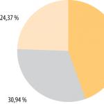
On the lower surface of the organ, approximately in the center, there is a place where large vessels enter the liver and exit the bile ducts - Glissonian gate. Arterial blood enters the liver through its own hepatic artery (a. hepaticapropria) and through the portal vein (v. portae). The quantitative ratio of blood entering the liver through these vessels is approximately such that about 25% flows through the hepatic artery and 75% through the portal vein.
According to most researchers, only in 2/3 of cases the hepatic artery and portal vein are divided at the gate into right and left branches. Sometimes there are other options, the frequency of which fluctuates. According to the B.C. Shapkin, bifurcation of the portal vein occurs in 86%, trifurcation in 6.55%, and quadrifurcation in 1.58% of cases. Bifurcation of the hepatic artery occurs in 66%.


The outflow of blood from the liver occurs through a system of three hepatic veins, which, approaching each other on the posterior surface of the liver, flow into the inferior vena cava, passing in the groove of the same name on the border of the right and left halves of the liver. The sections of thin-walled hepatic veins passing outside the liver are very short (2-8 mm), which greatly complicates their isolated ligation and often leads to damage to the walls of blood vessels when attempting to isolate them. It is more convenient to ligate them through the liver tissue [Krylova, 1963].

The right and left bile ducts, draining the corresponding halves of the organ, in 75% merge into the hepatic duct at the hilum.
From a practical point of view, one should take into account the fact that large vascular-ductal structures of the liver are located closer to its lower surface. These formations lie at a depth of 1.5-2 cm from the surface, which should be taken into account during surgical interventions. On the diaphragmatic surface of the liver, at a considerable depth, only vessels and ducts of the 3rd-4th order are found.

Lymphatic drainage from the liver occurs through the lymph nodes of the portal, hepatoduodenal ligament and retroperitoneal space.

Innervation of the liver occurs due to the hepatic plexuses of the abdominal cavity with the participation of the Latarget nerve from the anterior trunk of the vagus nerve.
The duplications of the peritoneum, passing to the liver from the diaphragm and abdominal organs, form a ligamentous apparatus that fixes the organ to the diaphragm, the posterior abdominal wall and connects the liver with the stomach and duodenum. The liver is attached to the diaphragm using the falciform, semilunar and triangular ligaments, which do not correspond to the structural elements of the organ and perform only the functions of its fixation.
From the gate to the area of the duodenum there passes the hepatoduodenal ligament (lig. hepato-duodenale), which is of great practical importance, the free edge of which is limited in front by the hole leading into the cavity of the lesser omentum (foramen Winzlomi), and in its thickness there are vital formations of the Glissonian gate (hepatic artery, portal vein and common bile duct). To the left, the hepatoduodenal ligament passes into the hepatogastric ligament, which makes up a significant part of the lesser omentum.
From the inner surface of the navel to the area of the Glissonian gate stretches a cord-like ligament - the round ligament of the liver (lig. teres hepatis). In its thickness lies the umbilical vein (v. umbilicalis), which flows into the main trunk or left branch of the portal vein. Due to the fact that after birth this vessel is obliterated only 1-2 cm from the navel, it is possible to bougienage it and directly penetrate the portal system for the purpose of contrast studies, manometry or infusion medicinal substances[Doviner D.G., 1954; Ostroverkhov G.E., Nikolsky AD., 1964; Gonsales-Carbalhaes O., 1959].
Of particular importance in surgical anatomy liver have studies on its segmental structure. Domestic and foreign researchers have clearly proven that the liver consists of separate segments, each of which has its own autonomous blood supply from the hepatic artery and portal vein systems. Outflow from these segments is also carried out in isolation venous blood and bile. The vessels of individual segments, according to most authors, do not have wide anastomoses, but, as shown by the anatomical studies of A.S. Yalynsky (1975), such messages exist, although perhaps not powerful enough.
Based on blood supply, the liver can be divided into two large parts. The boundary between them is a hypothetical plane passing through the apex of the gallbladder and the inferior vena cava. This plane is slightly inclined to the left and passes through the same hypothetical Rex-Cantle line. Each half of the organ has an autonomous blood supply and outflow of blood and bile. In turn, the right and left halves of the liver are divided into four segments each. With small details, these schemes repeat each other.
The topography of the vascular-ductal structures of each segment (the vessels and ducts included in it) is practically important. This topography has been described by several authors [Umbrumyants O.A., 1968; Patel J., Leger L., 1975].


It is necessary to emphasize the promise of such studies for the purposes of practical surgery. At the same time, excellent knowledge of the intraorgan formations of the liver is absolutely necessary for carrying out modern interventions on this organ using any method. It is also important to know the projection of the vascular-ductal structures of the liver onto its surface, which is especially important for the implementation of interventions for the removal of bile.