Hearts must adhere to the following rules.
Positionpatient. It is necessary to listen to the patient in various positions - vertical, horizontal and lying on the left side. This must be done, since sound phenomena that occur in the heart with various valvular heart defects can be heard in one or another position of the patient. For example, a murmur with mitral stenosis is better heard when auscultating the patient lying on his left side; a pericardial friction murmur is better heard at the base of the heart when the patient's torso is tilted forward.
Positionparamedic The paramedic is positioned to the right of the patient and so that it is convenient and free to use the phonendoscope over all points of listening to the heart.
Heart listening points
- dotmitralvalve- apex of the heart (1-2 cm inside from the midclavicular line to the left in the 5th intercostal space);
- dotaorticvalve- II intercostal space at the sternum;
- dotpulmonary clipanaarteries- II intercostal space on the left at the edge of the sternum;
- pointstricuspidvalve- at the base xiphoid process sternum;
- dotBotkin(V point) - at the bridge of attachment of the III-IV ribs to the sternum on the left, sounds (murmurs) from the mitral and aortic valves are transmitted here.
Normally, heart sounds are clear (loud), rhythmic, and pure.
Toneshearts can weaken (with mitral valve insufficiency - I sound) or intensify (I sound at the apex of the heart with mitral stenosis).
Weakening of the second tone over the aorta - with hypotension.
Of practical importance is gainIItonesneither<) aorthat- for hypertension and atherosclerosis; gainabovepulmonaryartery- with mitral heart defects.
If the second tone is more sonorous over the aorta, then accentIItonesaboveaorta; if above the trunk of the pulmonary artery - accentIItonesabovepulmonaryartery.
In case of severe myocardial pathology, bifurcation of the second tone is possible - a 3-member heart rhythm appears (rhythm halopa, rhythm reminiscent of the sound of a galloping horse).
Auscultation video
Heart rate
Normally, the number of heart contractions (HR) is 60-80 per minute. Heart rate may increase (tahicardia) or decrease (bradycardia). Tachycardia can occur in healthy people during running, physical work, increased emotional state (disappears after 2-3 minutes) and various pathological conditions (fever, myocarditis, thyrotoxicosis, anemia, blood loss, etc.). Heart rate in these cases can increase to 100-120 or more per minute.
Bradycardia can be observed in healthy people (athletes, vagotonics) and in pathological cases (narrowing of the aortic mouth, typhoid fever, meningitis, overdose of digitalis drugs, etc.), a decrease in pulse (less than 40 beats per minute) is observed in case of rhythm disturbance (complete atrioventricular block ).
Normally, the heart rate completely coincides with the pulse rate. With atrial fibrillation (atrial fibrillation), weak contractions of the heart do not drive blood to the periphery, therefore the pulse rate is calculated only by the pulse waves that have arrived, the number of which is less than the heart rate. The difference between heart rate and pulse rate is called deficitpulse
Heart sounds may be muffled or muffled (poor, sometimes barely audible) when the contractile function of the heart is reduced in heart failure.
Heart sounds may be arrhythmic (arrhythmic) - evidence of disturbances in the conduction system of the heart or in the myocardium.
Heart sounds may be unclear, and murmurs are heard instead of sounds.
Organicnoises arise due to organic lesions of the heart and heart valves. They can be rough and worsen after physical activity.
Functionalnoises occur without organic damage to the heart: with anemia, thyrotoxicosis, nervous excitement.
Distinctivesignsfunctionalnoise functional murmurs in most cases are systolic and are better heard at the apex of the heart and above the pulmonary artery. They are always soft, unstable (can disappear after physical activity), are not carried out anywhere and are not detected by palpation of the heart, and are not accompanied by an increase in the size of the heart.
There are noises systolic(occur after a long pause between the first and second heart sounds) and diastolic(during a long pause between the II and I heart sounds). Combinations of both noises are possible.
Having determined the place of the best listening to noise, knowing the values of the listening points, you can determine the place! pathological process. For example, if the murmur is best heard at the apex of the heart, mitral valve pathology is suspected.
Having determined the nature of the noise, we assume what it is! this is a pathology. Systolic murmur - mitral valve insufficiency During the systole phase, the valves do not close tightly, and the noise of blood flowing back from the ventricle through the gap formed between the valve flaps into the atrium is heard. Diastolic murmur - stenosis of the left venous opening (through the narrowed opening in the diastole phase, blood from the atrium enters the left ventricle with noise).
Noise conductivity
Murmurs arising from valvular heart defects can be heard not only at the listening points, but also at some distance from them. They are usually carried out along the blood flow from the place of their origin or through the dense heart muscle during its contraction. Thus, a sharp systolic murmur with stenosis of the aortic mouth extends far along the blood flow to the periphery and is well audible on the carotid subclavian arteries, on the clavicles, and on the thoracic spine. In case of mitral valve insufficiency, the noise is conducted to the left axillary region. The diastolic murmur of aortic valve insufficiency is conducted down to the left ventricle and is often best heard at Botkin's point. The position of the patient during auscultation is important: murmurs with mitral defects are better heard in the patient's position on the back and left side, with aortic defects - in an upright position.
Extracardiac murmurs include noisefrictionpericard. It occurs in diseases of the pericardium, the leaves of which become rough, are not associated with heart sounds, resemble the sound of rustling paper, and are best heard at the left edge of the sternum (ask the patient to hold his breath - a pericardial friction noise is heard). The pericardial friction noise is inconsistent, disappears at times, and intensifies when the stethoscope is pressed on the chest.
Diagnostic methods such as percussion, palpation, and auscultation of the heart, which are outdated at first glance, do not lose their relevance today. Listening to the patient's cardiac activity using a phonendoscope is actively used for initial diagnosis both at the prehospital stage in acute conditions and in hospitals and clinics. Today, experts recommend performing auscultation sequentially at five main points, which allow the tones of a working organ to be distinguished in as much detail as possible.
Auscultation at certain points of the chest makes it possible to determine such indicators of cardiac activity as the frequency and rhythm of contractions, the presence of noises that are normally absent, the purity and melody of the sound of the heart, and heart tones. In this case, inaccurate application of the phonendoscope can significantly distort the sounds heard, which can lead to an error in diagnosis.
It is important! The search for the necessary points is carried out by palpation in the corresponding anatomical areas. However, specialists with extensive experience are able to detect with maximum accuracy the necessary places for listening visually, without resorting to palpation.
When performing auscultation, the degree of hairiness of the patient's chest is important. A significant layer of hair can make auscultation impossible even with proper placement of the necessary points.
Location and search for heart auscultation points
The examination points correspond to the projection of the heart valves on the chest, as well as to the places where studying their work is most convenient and productive. The conduction of noise is also influenced by the blood flow in the vessels, which leads to the formation of a certain sound-conducting effect.
Listening to the patient’s cardiac activity is carried out in a strict sequence corresponding to the list below:
- Apex impulse. This point is the first auscultated area, and allows you to evaluate the work of both the mitral valve and the atrioventricular orifice on the left. Searching for it is carried out visually or with the help of hands. In cases where there is no visible apex beat, the doctor resorts to the percussion method. In this case, the boundaries of the heart are determined by tapping, and the phonendoscope olive is set to the boundary of the relative dullness of the heart. While listening, the patient is asked to hold his breath after inhaling and exhaling.
- The second listening point is determined in the area of the second intercostal space to the right of the sternum. As in the first case, the study is carried out after the patient holds his breath while exhaling. Here the work of the aortic valves and the aortic valve is determined.
- The third point is the area of listening to the operation of the valve apparatus of the pulmonary artery. It is determined in the area of the second intercostal space to the left of the sternum. It is important that after examination at the third point, it is necessary to repeat the first and second stages of the procedure. All three points of cardiac auscultation should have the same volume of heart sounds.
- The fourth research point is located in the area of the xiphoid process of the sternum and the place of attachment of the fifth rib to it. The examination performed here reveals pathology of the tricuspid valve and atrioventricular orifice on the right.
- The fifth point is an additional area for listening to the functioning of the aortic valves. It is located in the area of the third intercostal space to the left of the sternum. The examination is also carried out while the patient exhales while breathing is held.
It is important! The research on the above points is standard, aimed at the initial diagnosis. If there is a suspicion of the presence of a pathological process, auscultation is also carried out with the patient in the lateral position. In this case, the examination begins from the first point, after which the phonendoscope is shifted to the anterior-axillary and mid-axillary line. This measure makes it possible to achieve a clearer sound of noise, which contributes to a more accurate diagnosis.
In some cases, the technique of auscultation of the heart after physical activity is also used. In this case, listening is performed first at standard points, and then along the entire projection of the heart, determining the ability of noise to be carried beyond the contour of the organ, as well as the direction of their conduction. Thus, the murmur of the tricuspid valve, as a rule, is diverted to the right, the mitral valve is diverted to the left, and the murmur of the aorta radiates to the carotid arteries.
Assessment of noise at auscultation points
Pathological noises, the presence of which was determined during listening, are subject to a certain assessment by a specialist. Thus, the time of their occurrence (during systole or diastole), the localization and conduction of noises, their volume, timbre, and duration are determined. The position of the patient’s body in which the murmur is best heard is also of diagnostic importance, as well as its dynamic characteristics (increase, decrease). 
When assessing detected murmurs, it is important to take into account the age of the patient being examined. Thus, young children are characterized by good hearing of three or four heart sounds, which is not a pathology. At the same time, a similar phenomenon in adults in most cases indicates the presence of severe heart disease. In addition, there are other age-related features of the cardiovascular system, the failure to take them into account can lead to an erroneous diagnosis with subsequent incorrect treatment.
Are you completely unfamiliar with auscultation as an examination method? But you are wrong. You encountered this method already in deep childhood and continue to encounter it to this day. And there is nothing surprising about this. It’s just that the name came to us from eighteenth-century France, when the doctor Rene Laennec in 1816 proposed a new technique for listening to patients.
The new technique was based on the use of a special instrument, which was called a stethoscope, and which, in one form or another, you saw doctors hanging around their necks. Of course, that ancient stethoscope has evolved over two hundred years into a modern and very common instrument. The first action of any therapist when meeting a patient is to touch and listen.
Before the proposal of Rene Laennec, listening to the work of the heart took place by putting the ear to the patient's chest. From a diagnostic point of view, such an application was not very informative, but there were no other options. Laennec himself describes in his works how he accidentally managed to find a more effective option.
In one “narrow” situation, he remembered the acoustic effect when, putting his ear to the end of a log, one could hear the touch of a needle to the other end. The effect of sound wave transmission was used for the proposed stethoscope.
Without going into physical processes, we note that sound effects accompany vibrations of the heart valves, contractions of the walls of blood vessels, as well as the movement of blood through the cardiovascular system. As an example, you sometimes hear water flowing through the pipes in your apartment. Blood flowing through the vessels will also be heard.
The stethoscope allowed René Laennec to hear the heartbeats more clearly than could have been done if he had again put his ear directly to his chest. The design of the stethoscope that Laennec proposed was a wooden tube with a bell.
In this form, the structure existed until the beginning of the 20th century (almost a hundred years). An improvement in the form of a membrane glued to the bell was made by N.S. Korotkov (Russian surgeon). As a result, a virtually new instrument appeared - a phonendoscope.
A century of experience using the stethoscope has led to the experimental understanding that the human internal organs produce sound vibrations of different frequencies.
For reference. The heart and intestines produce low-frequency vibrations, while the lungs and blood vessels produce high-frequency vibrations. It turned out that when using a stethoscope, low-frequency vibrations drowned out high-frequency ones.
 The membrane used by N.S. Korotkov made it possible to muffle low frequencies, which made it possible to hear high frequencies well. This is the difference between a stethoscope and a phonendoscope.
The modern instrument is already a combined device - a stethophonendoscope. The head is combined from a membrane on one side and a “bell” on the other (see positions 5 and 6 in the figure). If the doctor wants to listen to the heart, he puts the head with a “bell” to the body; if he wants the lungs, he puts the head with a membrane to the body.
Everything together (head, sound-conducting tube, tee, headbands with olives) affects the quality of sound transmission, depending on the manufacturer and material of manufacture.
What is auscultation
Auscultation is almost always used during initial examinations by general practitioners. When coming with any complaint, the doctor will ask the patient to undress to the waist. First, he will conduct a visual examination, and then remove the stethoscope from the neck and begin listening.First of all, auscultation of the heart is performed to understand its condition. This simple procedure, which takes very little time, is one of the most important diagnostic methods that allows for a comprehensive assessment of the functioning of the cardiovascular system. It allows you to listen and evaluate the tones, rhythm and tempo of heartbeats.
Using only a stethophonendoscope and accumulated experience allows you to accurately assess the patient’s current condition. For this reason, the auscultation method is used in all medical institutions, both in the city and in regions where there is no expensive diagnostic equipment.
Auscultation can provide information in the presence of diseases such as:
- heart disease. This disease is characterized by the appearance of noise, as well as additional tones that appear due to gross disturbances of hemodynamics (blood movement) when moving in the heart chambers.
- pericarditis. This disease is characterized by inflammation of the pericardial sac, which is reflected in the sound of the pericardium - friction noise (dry pericarditis) or muffled heart sounds (effusive pericarditis).
- (infective endocarditis), in which noises and tones characteristic of heart defects occur.
1.What is auscultation?
2.Types of auscultation, their advantages and disadvantages.
3.Physical basis of auscultation.
4. General rules of auscultation. Rules and techniques for auscultation of the lungs.
5. Classification of respiratory sounds.
6. The mechanism of occurrence of vesicular respiration and its characteristics are normal.
7. Quantitative and qualitative changes in vesicular respiration, their diagnostic significance.
8.What is bronchial breathing? Where is it heard normally?
9.Pathological bronchial breathing. Causes, types, diagnostic value.
10.Mixed breathing: causes, diagnostic significance.
11.What are additional breath sounds?
12.What is wheezing, how are they classified?
13. Causes and mechanism of dry wheezing.
14. Causes and mechanism of occurrence of moist rales.
15.Diagnostic value of detecting wheezing.
16.What is crepitation? The mechanism of its occurrence and differences from moist rales.
17. Pleural friction noise and the mechanism of its occurrence, distinctive features.
18. Specific auscultatory phenomena detected in hydropneumothorax.
19.What is bronchophony? Methodology for its detection and diagnostic value.
1. What is auscultation?
Auscultation is a research method that consists of listening to sound phenomena that arise in the body as a result of vibrations of certain elements of it, and in judging the physical state of the body based on the nature of the sound.
2. Types of auscultation, their advantages and disadvantages.
Direct auscultation (performed by applying the ear to the patient).
Mediocre auscultation (performed using a stethoscope or phonendoscope).
The advantages of direct auscultation are:
allows you to listen to weaker and higher sounds;
allows you to listen to a large area of the body at once;
natural sounds are heard;
the doctor receives tactile sensations (this is important when listening to inconsistent heart sounds - III, IV).
The disadvantages of direct auscultation are:
difficulty localizing sounds, especially when listening to the heart;
inability to listen to a number of areas of the chest (supraclavicular and axillary areas), neck;
unhygienic.
The advantages of mediocre auscultation are:
hygiene;
convenience for the patient and the doctor;
the ability to localize the location of sounds;
the ability to listen to areas inaccessible to the direct method;
a flexible stethoscope allows you to listen to the patient in any position of his body;
convenience when examining young children, severe and immobile patients;
sound amplification.
The disadvantages of mediocre auscultation are:
sound distortion (you need to use one stethoscope, the doctor is tied to it);
a large number of additional noises caused by the use of a stethoscope.
3. Physical basis of auscultation.
The emergence of sound is the result of pendulum-like movements (oscillations) of a body brought out of a state of rest. If a body is homogeneous in its composition, then it undergoes periodic oscillations; if it is heterogeneous, it undergoes non-periodic oscillations. Vibrations of the body cause in the surrounding air a series of alternating condensations and rarefactions, which, spreading in all directions in the form of a sound wave, reach our ear and cause irritation of the hearing aid with the same sequence and frequency with which the body, removed from a state of equilibrium, vibrates. In the case of periodic oscillations, the sound sensation has a tonality characteristic of musical sounds and is designated as tone; in the case of non-periodic oscillations, a sound sensation without tonality arises - noise.
The strength or volume of the sound depends on the amplitude of the oscillating body (direct dependence). The volume of sound is also affected by the distance from the sound source. The greater the depth of the sound source (for example, in the lung), the lower the sound volume. The pitch of the tone depends on the number of vibrations per second performed by the body; the more vibrations, the higher the pitch and vice versa.
Sound phenomena arising in the lung are transmitted through the air columns in the bronchi to the chest wall, causing vibrations in it, and then through the surrounding air to the ear of the examiner. The conditions for sound conduction in the lungs are not entirely favorable due to the elasticity of the bronchial walls and their high ability to vibrate, and, consequently, to propagate the sound wave in all directions. As a result, the amplitude of vibrations by the time the sound wave reaches the chest is significantly reduced, and the sound reaches each individual part of the chest significantly weakened.
With inflammatory compaction of the lung tissue, the tissue between the bronchi is also saturated with inflammatory exudate. It becomes dense and forms, as it were, a continuation of the bronchial wall. It is less capable of vibrations than the walls of the bronchus and inhibits them. Therefore, in the air column itself, enclosed in the bronchus, less energy spreads to the sides. Thus, through the compacted lung, sounds arising in the respiratory system are better transmitted to the chest wall through the air in the bronchus, and, therefore, reach the ear less attenuated than through a normal lung.
If the conducting medium has the same oscillation frequency as the frequency of conducted sound, then the air column enclosed in the bronchus (in the cavity) plays the role of a resonator and the sound is amplified. This is observed in a condensed lung (sound to the ear is carried out without weakening). Thus, compaction of the lung tissue prevents the sound from weakening and contributes to the manifestation of the amplifying effect of the resonator.
Since the conductor of sound is mainly air in the bronchus, bronchial patency is a necessary condition for listening to the lung.
If in any place of the bronchus there is a narrowing of it, so that air from the wide part, having passed through the narrowing, enters the wide part, then air turbulences arise at the place of narrowing. As a result, vibrations of the bronchial wall occur, and a noise called stenotic occurs in the area of narrowing. There is a direct dependence of the noise volume on the degree of narrowing and air flow speed. However, with a significant narrowing, a lower speed is sufficient; with a small narrowing, a higher current speed is needed to generate noise.
4. General rules of auscultation. Rules and techniques for auscultation of the lungs.
General rules of auscultation.
The room where the listening is performed should be as quiet as possible.
The room where the auscultation is performed must be warm, since cold-induced trembling and muscle tension distort the nature of breathing and other auscultatory sounds.
The parts of the body to be auscultated must be exposed.
If necessary, the hair in the auscultated part of the body should be shaved and moistened with oil, soap solution or water.
The bell of the stethoscope should be warm, it should be warmed up.
The bell of the stethoscope should be attached to the patient's body with its entire edge, hermetically.
The stethoscope should be fixed to the patient’s body without touching the tubes, since touching them creates additional noise.
The stethoscope should not be pressed tightly against the patient’s body, since pressing it tightly dampens vibrations of the body surface, which leads to a weakening of the sound. The exception is listening to high-pitched sounds, which are better heard with significant pressure on the chest with a stethoscope.
The stems of the stethoscope should fit snugly against the walls of the external auditory canal, which ensures tightness and closure of the acoustic system, but they should not cause pain.
It is advisable to use the same stethoscope.
The position of the patient and the doctor should be comfortable.
You should guide the patient, his breathing, and other actions that are appropriate in terms of listening.
You should devote as much time as possible to listening to patients, as this is one of the most difficult research methods to master.
While listening to the patient, you should learn to distract yourself from other extraneous noises coming from outside. You should learn to concentrate your attention and hearing on the sounds that come from the stethoscope.
Rules for auscultation of the lungs.
It is best to listen to the lungs with the patient sitting or standing.
It is necessary to follow the sequence of listening to the lungs: anterior surface, lateral sections, posterior surface of the chest.
You should use techniques that improve sound conduction and facilitate auscultation:
when listening in the axillary areas, the patient should place his hands behind his head;
When listening along the scapular and paravertebral lines, the patient should cross his arms over his chest and slightly tilt his head forward.
The patient should breathe deeply, evenly, slowly, through the nose or half-open mouth. In this case, it is recommended to listen to the main respiratory sounds when breathing through the nose, and additional ones - when breathing through the mouth.
You should guide the patient's breathing, giving instructions on this matter, or even demonstrating to him how it should be done, training him.
Initially, it is advisable to conduct a comparative auscultation of the lungs, and then listen in detail to those areas where pathological changes were noticed.
First, the nature of the main respiratory noise is determined, then the secondary respiratory sounds are determined, and finally, bronchophony is determined.
Palpation, percussion, auscultation are methods of objective examination used by doctors around the world in the process of diagnosing various diseases. These methods are used in conjunction with biochemical and other types of analysis, instrumental research, and technologies are used, of which there are a large number. Interestingly, an objective examination plays a decisive role in making a diagnosis.
Auscultation is the most complete and informative method. It is used for diagnostics in surgery, therapy, obstetrics, and pediatrics. Using this method, listening determines the presence of pneumonia, bronchitis, cardiac defects and many other pathologies in children and adults.
Auscultation of the adult heart
Along with being highly informative, this is also the most difficult method of objective examination. It requires perfect pitch, a sense of rhythm and constant practice, as it has a large number of nuances. Diagnostics in medicine using the method of auscultation makes it possible to identify heart disease and pulmonary pathology at an early stage of development.
Listening to the heart is performed in a lying or standing position. Some diseases are characterized by a change in heart rate after exercise, so sometimes, for an accurate diagnosis, the patient is taken out of a state of physical rest. The auscultation method requires compliance with certain rules:
- isolation from ambient noise;
- listening to the heart is carried out during (if possible), as well as separately during inhalation and exhalation;
- it is necessary to use a phonendoscope and stethoscope for auscultation of high and low tones;
- First of all, the presence and characteristics of sounds at various points are determined, and then pathological or physiological noises are listened to.
Heart percussion
Used to determine the boundaries of the organ and absolute cardiac dullness. Recently, this method has faded into the background. Some experts have completely abandoned it, since the results of percussion are not very accurate and have a large percentage of subjectivity. This method has been replaced by radiography and ultrasound, which give a complete picture of the size and position of the organ.
Palpation of the heart
Widely used in diagnostics. Palpation of the heart is carried out in order to more clearly determine the position and strength by pressing a finger to the corresponding area. Some diseases are characterized by a slight trembling of the chest, or “cat purring syndrome.”
Ability to listen and hear
The heart is not listened to in a chaotic manner. There are projections of the cardiac valves onto the chest. There are four of them in total.
- Mitral - IV rib, to the left of the sternum.
- Aortic - III rib, to the right of the sternum.
- - III intercostal space on the left.
- Tricuspid - IV intercostal space on the right.
However, auscultation points are slightly different from direct projections, as the sound in these places is clearer and more understandable.
- At the top of the heart -
- II intercostal space, from the sternum to the right - aortic.
An important sign of serious illness is a cardiac murmur, which can be constant or appear after a certain load. You must be able to listen very well and hear all deviations from the normal heart rate. It is important to determine not only the noise, but also the nature and location of its formation. It can appear in systole or diastole.
Not only noise, but also working phases can be pathological or physiological. Auscultation of the heart helps in diagnosis. Listening points are similar to those described above. The formation of III and IV additional tones is possible, which appear in different conditions (time interval, first and second share of systole or diastole).

Small heart - big responsibility
Pediatric auscultation is a very important part of diagnosis. A child, especially a small one, due to his age, cannot report his problems. The pediatrician must have keen hearing and a high degree of literacy, since the sounds of a child’s heart change along with his growth. Functional or pathological murmurs may be detected. It is important to make a comparison between the first and second tones in terms of strength or emphasis. Any violation indicates a number of pathological processes in the child’s body.
Differential diagnosis of heart diseases in children using the auscultation method
| Heart tone | Place of emphasis | Identified pathology (physiology) |
| First | Top of the heart | The left atrioventricular orifice is narrowed |
| Second | Aorta | Arterial hypertension or physiological characteristics of puberty |
| Second | Pulmonary artery | Patent ductus arteriosus, stenosis, bicuspid valve insufficiency, atrial or ventricular septal defect, pulmonary artery hardening, pulmonary fibrosis, myocarditis with congestion in the pulmonary circulation |
| First and second | At all points | after stress (physical or psycho-emotional) |
In addition to accents, it is possible that heart tones may weaken or split. Auscultation objectively characterizes this if the doctor knows how to listen.
Pregnancy and auscultation
The heart leaf is laid down and begins to contract already in the third week of pregnancy, and at six it can be heard on an ultrasound. Diagnosis of the mother and fetus is mandatory throughout the entire period and especially during childbirth. The number and content of tones constantly changes in proportion to intrauterine development.
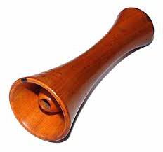
Auscultation of the fetus is the simplest and most effective method of determining its viability. To perform this simple operation, an obstetric stethoscope is required (photo below). If necessary, use a phonendoscope.
You can conditionally divide the entire pregnancy period into several periods (according to the rate of fetal heartbeats, as well as the nature of their fullness).
Interestingly, at the 6th week after conception, the child’s heart rate coincides with the mother’s. The difference can be 3 strokes up or down. Then the number of contractions begins to increase. If we take into account that the heart rate increases by 3 beats every day, then it is permissible to determine the age of the fetus histologically.
After two months of pregnancy, the heart itself is divided by partitions into 4 chambers - atria and ventricles. The adult organ also has a similar structure. At the beginning of the 9th week, the embryo's heart beats approximately 175 beats per minute. Further, the frequency decreases and, starting from the second trimester, 140-160 beats become the norm for the fetus. Any deviations from it indicate hypoxia, with tachycardia characterizing the initial degree of oxygen deficiency, and bradycardia characterizing a severe stage requiring immediate intervention.
Fetal palpation
By palpation in the second half of pregnancy, you can determine the position of the fetus and its individual parts in the uterus. In addition, the gestational age is determined by the height of the uterine fundus, as well as by the baby’s head: if it is pressed tightly to the entrance to the pelvis, these are the first harbingers of labor. In obstetrics, the Leopold method is used, which consists of four main techniques.
Auscultation and childbirth
Deafness of heartbeats can be both a manifestation of pathology and an elementary difficulty in listening. This happens when the mother’s abdominal wall is thickened (obesity), the fetus is abnormally positioned (for example, a posterior view of the occipital or breech presentation), polyhydramnios, etc. Muted heartbeat tone is especially common during the labor period. Diagnosis of the fetal body at this time is of paramount importance.
One of the methods of examining a pregnant woman is palpation. It helps determine the location of the fetus and its presentation. But the same result can be achieved when cardiac auscultation is used to diagnose intrauterine development. Listening points are characteristic. If the heartbeat is detected more clearly above the mother's navel, then the fetus has a breech presentation, if lower - a cephalic presentation. The baby may be hyperactive, turning over from side to side throughout the entire pregnancy. Listening to clear tones at the level of the navel indicates a transverse position.
Auscultation in the diagnosis of pulmonary diseases

Auscultation is a method that plays a decisive role in making a diagnosis of pulmonary diseases. There are correct (or vesicular) breathing and various forms of deviation from the norm. Also a characteristic sign of various diseases are dry or moist rales, which have certain listening characteristics. The auscultation points of the lungs are located symmetrically.
Physiologically altered vesicular respiration
If a person has well or, conversely, poorly developed muscle mass, or has hypertrophied adipose tissue, the change in breathing can be either in the direction of weakening or strengthening. Listening takes place using a phonendoscope.
Increased vesicular respiration is typical in childhood. Its other name, which can be heard in medical circles, is puerile. There is one characteristic feature - the same breathing in symmetrical areas on the right and left sides.
Diagnosis of bronchitis by auscultation
Auscultation for bronchitis is carried out in the usual way. When listening to the acute stage, the vesicular type is characteristic. This is the body's reaction to inflammation and narrowing of the bronchioles. Against the background of harsh breathing, dry wheezing is detected, and they can be different in tone, and also resemble a buzzing and whistling. This depends on the size of the bronchi and the degree to which they are filled with secretions. They are clearly audible in both phases of breathing.

As bronchitis progresses, mucus production in the respiratory tract increases, and mid-bubble rales are detected on auscultation.
It is best to listen to the lungs when the patient is standing. It is necessary to compare the sounds of breathing and wheezing at the same points on the right and left organs. There is a certain sequence of auscultation - auscultation points - lungs.
You need to start from the tops and then examine the front surface, then the side and back. With prolonged bronchitis, additional sounds may be added, for example, crepitus, which indicates the transition of inflammation to the lower respiratory tract.
Auscultation of the lungs is carried out in several stages: during normal and deep breathing and after coughing. The auscultation points that are most “suspicious” to the doctor are examined in particular detail.
Diagnosis of chronic bronchitis is also based on auscultation data and laboratory studies of biological materials. When listening to the lungs, breathing is determined to be of the vesicular type in the presence of a longer exhalation or hard, as in the acute stage. Sometimes chronic bronchitis provokes the development of a more severe disease - in this case, breathing becomes “cotton-like.” During an exacerbation, wheezing is heard over the entire surface of the lungs.
Percussion of the lungs

Percussion examination can be carried out in three ways: by tapping directly on the examined area, through a plate or with a finger on a finger. Currently, the last one is most applicable. This method does not require the doctor to have additional equipment, and it is quite informative in examining the lungs.
Percussion can be comparative or have a topographical focus. The most popular is the first option, which is used to identify pathological foci. They are compactions, so the percussion sound over them is duller than over healthy lung tissue.
There are a large number of shades and tones extracted when examining sounds. Normally, it should be loud, ringing and prolonged. If deafness, dullness of tone, a metallic tint, box or tympanitis appear, this indicates that the patient has inflammatory or other processes in the lungs that require medical intervention.
Auscultation in the diagnosis of diseases of the gastrointestinal tract
Listening is used as a method for diagnosing a number of diseases of the gastrointestinal tract. The examination is carried out by a doctor using a stethoscope or placing the ear to the abdominal wall. Using this method, the presence (absence) of peristalsis in the intestines or stomach is determined.
Auscultation is carried out on a comparative principle, that is, to obtain an adequate picture, it is necessary to listen at different points. The examination should be carried out in silence and, if possible, without pressing on the abdomen.
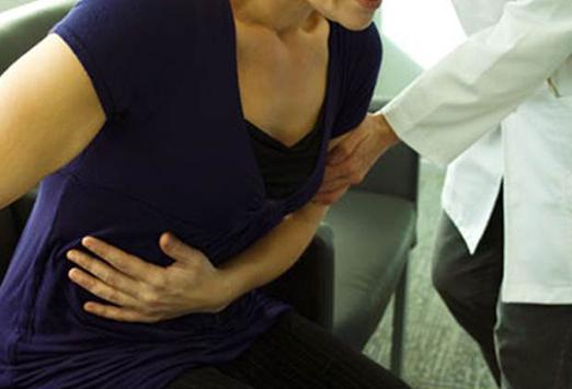
Palpation of the abdomen
In examining the abdominal organs, the palpation method provides maximum information. It is performed with gentle pressure on the abdomen. You need to start from the left groin area with warm hands so as not to cause discomfort to the patient. This is required to eliminate reflex tension of the abdominal wall.
The examination is carried out using the method of comparative analysis of the right and left halves from bottom to top. Pressure on the epigastric area is final. This is used to determine pain in various organs, tension in the abdominal wall, and the presence of fluid in the abdominal cavity (fluctuation syndrome).
Percussion of the abdomen
The percussion method also makes it possible to identify the spleens, since they have an absolutely dull sound (femoral). In addition, by comparing gastric and intestinal tympanitis, the doctor can make a diagnosis of obstruction of any of the sections.
Absolute hepatic dullness is normally determined on the right side in the 4th intercostal space at the level of the midline of the nipple. If, upon examination of this area, a tympanic sound is detected, this indicates perforation of the organs, that is, there is fluid in the cavity.
Percussion of the spleen has no practical significance: its lower edge can be easily palpated.