Uveitis - inflammation choroid eye, which is manifested by soreness, hypersensitivity to light, lacrimation, blurred vision.
The uveal tract has a complex structure, located between the sclera and the retina, outwardly resembling a bunch of grapes. It consists of vessels that supply the eyes with nutrients. The uveal tract is formed by the iris, the vitreous and ciliary bodies, and the choroid itself.
Disease classification
According to anatomical structure of the uveal tract, the following types of uveitis are distinguished:
- Front. The development of inflammation in the iris and vitreous is characteristic. This is the most common type of disease that can occur in the form of iritis, anterior cyclitis,;
- Intermediate. Inflammation affects the ciliary body, retina, vitreous body, choroid. Pathology proceeds in the form of posterior cyclitis, pars planitis;
- Rear. Characterized by damage to the choroid, retina, optic nerve. Depending on the localization of the pathological process, chorioretinitis, retinitis, choroiditis, neurouveitis may occur;
- Generalized. The inflammatory process affects all parts of the uveal tract. In such cases, they talk about the development of panuveitis.
Depending on the nature of inflammation, 4 forms of pathology are distinguished:
- Serous;
- Purulent;
- Fibrinous-plastic;
- Mixed.
According to etiological factors, uveitis is usually divided into:
- Endogenous. Infectious agents enter the eye with blood flow;
- Exogenous. Infection occurs as a result of injury to the choroid of the eye.
Uveitis can develop as a primary disease when it is not preceded by pathological processes. Secondary uveitis is distinguished when the pathology occurs against the background of other eye diseases.
According to the nature of the flow, there are:
- acute process, the duration of which does not exceed 3 months;
- chronic pathology that lasts more than 3-4 months;
- recurrent uveitis, when, after complete recovery, inflammation of the uveal tract develops again.
Etiological factors
The following causes of uveitis are distinguished:
- bacterial infection caused by streptococci, staphylococci, chlamydia, toxoplasma, tubercle bacillus, brucella, pale treponema, leptospira;
- viral infection: herpes virus (including the causative agent of chickenpox), cytomegalovirus, adenovirus, HIV;
- fungal infection;
- presence of foci chronic infection- tonsillitis, caries, sinusitis;
- development of sepsis;
- autoimmune diseases (rheumatism, systemic lupus erythematosus, spondyloarthritis, ulcerative colitis, Crohn's disease, polychondritis, interstitial nephritis, glomerulonephritis);
- eye injury, burns, foreign bodies;
- hormonal disorders;
- eye damage from chemicals;
- genetic predisposition;
- development of hay fever, food allergies;
- metabolic disorders.
The disease often develops in patients who have a history of other eye pathologies. In childhood and old age, infectious uveitis is mainly diagnosed, which occurs against the background of allergies or stressful situations.
Symptoms of the disease
The clinical picture depends on the location inflammatory process, states immune system the nature of the disease. In acute anterior uveitis, patients report the following symptoms:
- soreness and redness of the affected eye;
- pupil constriction;
- increased lacrimation;
- photophobia;
- decreased visual acuity and clarity;
- increased .
Chronic inflammation of the anterior uveal tract is characterized by an asymptomatic course. Only in some cases, patients note a slight reddening of the eyeballs, the appearance of dots before the eyes.
A characteristic sign of peripheral uveitis is the defeat of both eyes. Patients complain of decreased central vision, the appearance of "flies" before the eyes.
Posterior uveitis is characterized by the following symptoms:
- feeling of blurred vision;
- objects become distorted;
- the appearance of floating dots before the eyes;
- decrease in visual acuity.
It is also possible to develop macular edema, optic neuropathy, macular ischemia, retinal detachment.
Diagnostic measures
Diagnosis of uveitis is carried out by an ophthalmologist. As part of the initial appointment, the specialist must examine the eyes, check visual acuity, visual fields, conduct tonometry to determine the value of intraocular pressure.
Additionally, the following studies are carried out:
- ultrasound of the eye;
- study of pupillary reaction;
- biomicroscopy, which involves examining the eye using a slit lamp;
- gonioscopy to determine the angle of the anterior chamber;
- . The study is carried out in order to study the fundus;
- fluorescein angiography of the retina;
- tomography of various structures of the eye, if necessary;
- electroretinography;
- rheoophthalmography, which allows you to measure the speed of blood flow in the vessels of the eyes.
Features of treatment
Medical therapy for anterior and posterior uveitis involves the use of following groups drugs:
- Antibiotics a wide range actions (fluoroquinolones, macrolides, cephalosporins). The drugs can be administered subconjunctivally, intravitreally, parenterally. The choice of a suitable antibiotic depends on the type of pathogen, its sensitivity to drugs;
- Antiviral drugs are prescribed for the treatment of viral uveitis. Widely used:, against the background of taking Viferon or Cycloferon. Medicines prescribed in the form of intravitreal injections or taken orally;
- Non-steroidal anti-inflammatory drugs, glucocorticosteroids allow you to stop inflammation in a short time. Assign subconjunctival dexamethasone or prednisolone in drops, orally take Ibuprofen, Movalis or Butadion;
- Immunosuppressants are used when anti-inflammatory treatment is ineffective. Reception of Cyclosporine, Methotrexate which are capable to oppress immune reactions is shown;
To prevent the occurrence of adhesions, Cyclopentolate, Tropicamide, Atropine drops are recommended; - Fibrinolytics have a resolving effect. Widely used: Gemaza, Lidaza, Wobenzym;
- Complex multivitamins;
- Antihistamines: Claritin, Lorano, Cetrin, Clemastine, Suprastin.
If drug therapy has helped eliminate acute inflammation, then physiotherapy is indicated. Electrophoresis, infitotheraphy, laser blood irradiation, vacuum impulse massage, phototherapy, phonophoresis, have high efficiency. laser coagulation, cryotherapy.
Surgical intervention
The development of complications or severe course of uveitis requires surgical treatment. The operation may include the following steps:
- dissection of the adhesion between the iris and the lens;
- removal of the vitreous body, glaucoma or;
- retinal soldering with a laser;
- removal of the eyeball.
Surgery does not always have a favorable outcome. In some cases, the operation causes an exacerbation of the inflammatory process.
Traditional medicine methods
During the treatment of uveitis, some folk recipes. However, before any manipulation need to consult a doctor.
The following recipes will help to effectively eliminate inflammation:
- eye wash medicinal decoction. It is necessary to take in equal amounts the flowers of chamomile, calendula, sage. Grind the raw material. Take 3 tablespoons of the mixture, pour a glass of boiling water. The composition is insisted for 1 hour. Strain the resulting product, rinse the eyes with a decoction;
- aloe juice is bred cold boiled water in a ratio of 1:10. The resulting solution is dripped 1 drop no more than 3 times a day into the affected eye;
- lotions from the root of marshmallow. Raw materials should be crushed, pour 3-4 tablespoons of 200 ml cold water. The remedy is insisted for 8 hours, after which it is used for lotions.
Complications and prognosis
In the absence of effective treatment, uveitis can lead to the development of serious illnesses eyes:
- cataract, in which the lens becomes cloudy;
- damage to the retina up to it;
- developing due to impaired outflow of fluid inside the eye;
- persistent clouding of the vitreous body;
- damage to the optic nerve;
- pupillary infection, in which the pupil stops responding to light due to adhesion to the lens.
With timely and complex therapy acute inflammation the patient's eyes can be completely cured in 3-6 weeks. However, chronic uveitis is prone to recurrence during exacerbation of the underlying pathology, which significantly complicates therapy and worsens the prognosis.
Uveitis is an inflammatory pathology of the choroid of the eye, which can lead to complete loss of vision. Therefore, it is so important to diagnose and start treatment of the disease in time. Great value has the prevention of the disease, which involves timely therapy pathological processes in the body, exclusion of domestic eye injuries, allergization of the body.
The choroid of the eye has a complex structure and consists of three sections: the iris, the ciliary (ciliary) body and the choroid itself (choroid). Each of these departments, as already indicated in the lecture on the anatomy of the eye and its age characteristics, has an originality in structure and functions. The most important in the anatomy of the iris is the presence in it of a muscle that narrows the pupil, and a muscle that expands it, the first is innervated by the oculomotor parasympathetic, and the second by the sympathetic nerve. Sensitive nerve endings are "representatives" of the trigeminal nerve; due to the anterior ciliary vessels, anastomosing with the posterior long ciliary vessels of the ciliary body, its blood supply is carried out. The function of the iris is to regulate the amount of light entering the eye due to the "automatic" diaphragming of the pupil, depending on the level of illumination. The more light, the narrower the pupil, and vice versa. The iris is involved in ultrafiltration and outflow of aqueous humor, in thermoregulation, in maintaining ophthalmotonus, and in the act of accommodation.
The ciliary body is, as it were, a gland of intraocular secretion and is involved in the outflow of aqueous humor. It provides an act of accommodation due to the interweaving of the fibers of the zinn ligament into it, participates in the regulation of ophthalmotonus and thermoregulation. All these functions are due to the complexity of its glandular and muscular structure. It is innervated by both parasympathetic, sympathetic, and sensitive nerve endings, and vascularization is provided by the posterior long cilpar vessels, which have recurrent arteries (anastomoses) both to the iris, as already noted, and to the choroid. Each of the 70 processes of the glandular part of the ciliary body has "its" nerve branches and "its" vessels.
Thanks to the activity of the ciliary body, continuous nutrition of the avascular structures of the eye (cornea, lens, vitreous body) is ensured.
Particular attention should be paid to the fact that the choroid is richly vascularized due to the many branches of the posterior short arteries located in its choriocapillary layer, to which the pigment layer lies on the outside, and the retina on the inside. The choroid is involved in the nutrition of the retinal neuroepithelium, in the outflow intraocular fluid, in thermoregulation, in the regulation of ophthalmotonus, in the act of accommodation. The vessels of the choroid anastomose with the posterior long ciliary vessels of the ciliary body. Thus, all three sections of the choroid have a vascular relationship, and the iris and ciliary body have innervation. The choroid is very poorly innervated and essentially has only sympathetic nerve endings.
The rich sensitive innervation of the iris and ciliary body causes their pronounced pain during inflammation and damage.
Inflammation of the choroid of the eye
Inflammation of the choroid accounts for about 5% of cases among all eye pathology. Inflammation of the choroid of the eye can occur in the form of keratoiritis, which was mentioned in relation to keratitis.
Iritis, iridocyclitis (these are anterior uveitis), posterior cyclitis (hypercyclic crises), cyclochoroiditis, choroiditis, chorioretinitis, chorioneuroretinitis (these are posterior uveitis) can occur independently (isolated) or in combination.
In addition, in some cases, inflammation can be total in nature - these are panuveitis.
There are also so-called peripheral uveitis, although they can be classified as posterior cyclitis or cyclochoroiditis.
Uveitis
Before presenting information about some features clinical picture various uveitis, it is appropriate to point out that uveitis in children, regardless of their nature, has a certain originality. So, they often have an inconspicuous onset, a subacute course, the symptoms are slightly expressed, the corneal syndrome is weak, the pain is small, the precipitates are polymorphic, the exudate is often serous, the posterior synechiae are relatively weak and thin, the lens and vitreous body (opacities) are often involved in the process, reactive papillitis it is weakly expressed, frequent relapses, short remissions, there are no complaints of decreased vision, although it is reduced, the process is often bilateral. However, all parts of the choroid are more often involved in the inflammatory process.
As for the clinical picture of uveitis in adults, the disease is more severe than in children, and there are many complaints of significant discomfort in the eye (s).
Types of uveitis
By their nature, uveitis, regardless of their location, can be congenital and acquired, exogenous and endogenous, toxic-allergic and metastatic, granulomatous and non-granulomatous, generalized and local, prolonged and abortive, single and recurrent, acute, subacute and chronic, with concomitant general pathology and without it, with reverse development and with complications.
By the nature of exudation (transudation), uveitis can be serous, fibrinous, purulent, hemorrhagic, plastic and mixed.
To make a correct clinical diagnosis of uveitis, one should begin the examination of the patient with a brief, targeted history of the disease. Then it is necessary to sequentially check visual functions, examine each eye visually and with the help of instruments, examine other organs and systems (palpation, auscultation, using thermography, tonometry, etc.).
Further, a complex of targeted clinical and laboratory studies (X-ray, bacteriological, serological, immunological, virological, etc.) is prescribed. The main attention should be paid to identifying as many symptoms of the disease as possible, bearing in mind that the initiation of treatment is always symptomatic.
Anterior uveitis
What are possible symptoms anterior uveitis (iritis, iridocyclitis)? The first sign of inflammation of the choroid, which may attract attention, is a small and sometimes pronounced corneal syndrome, i.e. photophobia, lacrimation, blepharospasm, redness of the eye with a purple tint (pericorneal injection).
By checking the patient's vision immediately, you can make sure that it is somewhat reduced and does not improve when using weak plus or minus glasses. In the process of examining the eyes with lateral illumination or biomicroscopy, one can detect "fogging" (haze) of the corneal endothelium, as well as precipitates that are different in number, size, shape, tone (color), and exudate in the anterior chamber moisture of various types and quantities ( serous, purulent, etc.).
The iris is of a changed color, plethoric (edematous, hyperemic) with newly formed vessels, tuberous (granulomas).
The pupil may be narrowed, its reaction to light is slowed down. In the process of "playing" the pupil under illumination and darkening, and later when expanding it with mydriatics, posterior synechia (commissures of the pupillary edge of the iris with the anterior lens capsule) and exudate deposits on the lens can be detected.
Finally, with light palpation of the eyeball, its soreness is revealed. In addition, there may be a general depressed, restless, uncomfortable state of the patient.
All these symptoms indicate inflammation of the choroid. But to establish whether it is anterior uveitis or more common, ophthalmoscopy is performed. If at the same time the vitreous body is transparent, and there are no changes in the fundus, then the diagnosis of anterior uveitis is beyond doubt.
Diagnosis of posterior uveitis
It should be immediately noted that the diagnosis of isolated posterior uveitis, in contrast to the diagnosis of anterior uveitis, can be outward signs is difficult and the suspicion of the presence of posterior uveitis arises from such indirect symptoms as impaired visual function in the form of decreased visual acuity, defects in the field of vision (microscotomas, photopsy, etc.). In this case, the anterior segment, as a rule, is not changed.
Signs of inflammation of the posterior choroid are detected only ophthalmoscopically and biomicrocycloscopically, when inflammatory foci are found that are diverse in appearance, size, quantity and localization. Assessing the variety of these foci, i.e., the picture of the fundus, we can assume a possible etiology and activity (severity) of the inflammatory process in the choroid.
The cardinal signs of panuveitis include all of the listed possible symptoms characteristic of anterior and posterior uveitis, the diagnosis of panuveitis is relatively easy. In this disease, as a rule, changes are noted in all parts of the choroid, as well as in the lens, vitreous body, retina and optic nerve. There are also often violations of the regulation of ophthalmotonus (hypotension, hypertension).
Rheumatic uveitis
The most common rheumatic uveitis is characterized by the fact that it occurs against the background of acute course(attacks) of rheumatism.
Rheumatic uveitis is manifested by a sharp corneal syndrome and pain in the eye area. The mixed injection of an eye is expressed. On the endothelium of the cornea, multiple gray small precipitates are noted, there is abundant gelatinous exudate in the moisture of the anterior chamber, the iris is full-blooded, its vessels are dilated, multiple thin pigmented posterior synechiae are relatively easily torn after instillation of mydriatics (scopolamine, but not atropine). The lens and vitreous body are practically intact. On the fundus, more or less pronounced vasculitis is determined in the form of grayish "couplings" on the vessels.
All changes are reversed effective treatment and stabilization of rheumatism, the process recurs against the background of another attack of the disease.
Treatment of uveitis of this type is local, symptomatic.
Tuberculous uveitis
Tuberculous uveitis occurs more often against the background of active intrathoracic (pulmonary) or mesenteric, sometimes bone tuberculosis, and often against the background of a chronic course of the disease or remission.
The process in the choroid can first of all be suspected by decreased vision and corneal syndrome. Inflammation often occurs in one eye. Hyperemia of the eye in the form of a mixed injection is slightly expressed, the corneal syndrome is hardly noticeable. Very characteristic of tuberculous uveitis are "sebaceous" large precipitates on the endothelium of the cornea.
In addition, there are pathognomonic grayish-pinkish, surrounded by vessels (like infiltrates in tuberculous keratitis) nodules (granulomas-tuberculomas) in the iris and "guns" (snowflake-like deposits) on the pupillary edge of the iris. Synechia in this process are wide, powerful, planar, poorly torn under the action of mydriatics. A yellowish exudate is often found in the anterior chamber of the eye. New blood vessels form in the iris.
Exudate can often be deposited on the anterior lens capsule, germinate with newly formed vessels and regenerate (organize) connective tissue. Exudation can spread to the posterior chamber of the eye and the vitreous body, and as a result of this, clouding of the posterior capsule of the lens and vitreous body (golden rain) occurs. Posterior sequential cataract disrupts the nutrition of the lens, and its inner layers gradually become cloudy.
On the fundus can be found in different departments tuberculous foci of various sizes, without distinct contours, yellowish in color, protruding from the choroid into the retina. These foci do not merge and pigment is deposited on their periphery, and in the center they acquire a grayish tint. Naturally, the retina is also involved in the process, as a result of which, to varying degrees (depending on the location and size of the foci), visual functions (visual acuity, changes in the visual field, as well as color vision. Such a picture of tuberculous uveitis indicates that it develops according to the type of panuveitis, but there are cases when it is characterized by signs of anterior uveitis (iridocyclitis) or posterior uveitis (choroiditis).
Syphilitic uveitis
Syphilitic uveitis can occur with congenital and acquired syphilis. With congenital syphilis, inflammation of the choroid, as well as the cornea, may appear already in utero, which is detected in a newborn child.
Uveitis in acquired syphilis is characterized by mild corneal syndrome, mixed injection, serous exudate in the anterior chamber of the eye, and multiple polymorphic small precipitates.
In the altered iris, yellowish-reddish nodules-papules are revealed, to which newly formed vessels approach. Posterior synechiae are massive, wide, rupture after instillation of mydriatics, in their place on the anterior capsule of the lens remain pigmented polymorphic clumps. Small-pointed floating brownish opacities are possible in the vitreous body. Possible post-inflammatory changes in the fundus, reminiscent of "scattered salt and pepper." This picture is characteristic only for syphilis. Changes in the anterior and posterior sections of the eye in syphilitic uveitis can be observed both in combination and in isolation. In cases where uveitis occurs in the form of choroiditis, its diagnosis is childhood difficult, since the process is not accompanied by changes in the anterior part of the eye. Choroiditis is manifested only by disturbances in the visual field (discomfort), and children, as you know, do not pay attention to this and do not make any complaints. Inflammation of the posterior part of the eye is detected either by chance, for example, with eye injuries, or in connection with other manifestations of syphilis. As a rule, this pathology is bilateral.
Collagenous uveitis
Collagenous uveitis most often occurs against the background of nonspecific, the so-called rheumatoid arthritis, which appears and progresses uncontrollably mainly in preschool and school age. However, there are not isolated cases when uveitis appears long before the development of polyarthritis.
The eyes are affected in collagenoses in about 15% of cases. Eye disease begins gradually and, as a rule, on one, and then later different times and on the other eye. Uveitis proceeds mainly in the form of iridocyclitis, i.e., anterior uveitis. It is characteristic that most often, although not always, the eye during a normal visual examination is calm and there is no suspicion of an inflammatory process in it. This is especially dangerous in cases where there are no manifestations of polyarthritis, which could "give a signal" to the examination of the eyes. In the meantime, the inflammation almost "asymptomatically" progresses, and its initial stage is missed.
Early signs of uveitis can be detected only in cases where the disease has already been detected (albeit late) in one eye, and the other eye was still healthy. One of the first signs of collagenous uveitis is a gentle hyperemia of the iris and a slow reaction of the pupils to light. On closer biomicroscopic examination, rear surface of the cornea, predominantly in its lower segment, different-sized gray precipitates are found. After instillation of mydriatics, the pupil expands slowly and insufficiently, but its shape is rounded, i.e. there are no posterior synechia at this time yet. After weeks - months, the iris becomes pale, grayish, with clearly visible vessels and alternation of distinct gaps and crypts, which indicates degenerative changes in the structures of the iris.
The continuation of the inflammation process is evidenced by the occurrence of posterior synechia, which, when the pupil expands, seem massive (wide) planar, almost not torn after installations of strong mydriatics (scopolamine + dimexide + cocaine) and subsequent applications or subconjunctival injections of 0.1% adrenaline solution. The pupil at the same time acquires an irregular stellate shape. Gradually, the synechia completely "block" the connection between the anterior chamber and the posterior one. The pupillary margin and iris tissue are completely fused with the anterior lens capsule.
The inflammatory process in the eye proceeds according to the proliferative type, as a result of exudation, shaped cellular elements are deposited in the pupillary zone, they are regenerated by connective tissue, grow into newly formed vessels of the iris and, thus, not only the fusion of the iris with the anterior lens capsule occurs, but also complete infection of the pupil connective tissue. As a result, the anterior chamber first becomes uneven, and then, due to the lack of outflow of intraocular fluid from the posterior chamber to the anterior chamber, the iris acquires a funnel-shaped shape. This largely closes the angle of the anterior chamber, and as a result of the deterioration of the outflow of intraocular fluid, hypertension may occur, and then secondary glaucoma, which occurs in some protracted untreated cases.
As it appears from the picture drawn, collagenous anterior uveitis is characterized by great originality and severity of the course.
But, as studies show, the matter is not limited only to the defeat of the anterior and middle sections of the choroid. Simultaneously or some time after the onset of symptoms of uveitis, polymorphic small inclusions such as calcifications are found in the conjunctiva of the eyeball. Further, crescent-shaped grayish-whitish opacities are detected biomicroscopically in the surface layers at the border of the limbus and the cornea in the zone of 3 and 9 hours. Gradually, these opacities spread over the surface of the cornea in the area of the open palpebral fissure in the form of a ribbon with "enlightenment bays".
Thus, in collagenous uveitis, the inflammatory-dystrophic proliferative process is localized not only in the anterior choroid, but also extends to the lens, cornea, and conjunctiva. Such a picture of eye changes is commonly called the ocular triad of Still's disease - a combination of uveitis, sequential cataracts, and ribbon-shaped corneal dystrophy. As a rule, both in the initial and advanced stages of collagenous uveitis, there is no pronounced pathology in the choroid and other parts of the fundus.
Uveitis in other diseases
Uveitis can occur and practically (in 10-15% of cases) occur with almost all bacterial, viral, adenovirus and many systemic diseases. Therefore, in essence, in any general infectious and systemic disease, there should be a rigorous and urgent eye examination, followed by a careful examination of the eyeball and its auxiliary apparatus.
So, for example, the eyes of a patient with influenza, chickenpox, in the presence of herpes, in Behcet's disease (ophthalmostomatogenital syndrome), cytomegaly, in Reiter's disease (urethro-oculosynovial syndrome), in Besnier-Beck-Schaumann disease (sarcoidosis), in toxoplasmosis and in many other diseases and syndromes. With all these diseases, there may be keratitis and, more dangerously, uveitis, since both keratitis and uveitis almost always end in a decrease in visual functions.
Hypercyclic crisis
Especially, literally in a few words, it is necessary to say about the so-called hypercyclic crises. Hypercyclic crises occur, as a rule, in young and middle-aged women. These conditions appear unexpectedly in the daytime and manifest as a sharp pain in one eye, nausea, vomiting, headache up to fainting. Pulse increases significantly arterial pressure increases, the heartbeat appears. The eye at this time is almost calm, but there is a short-term decrease in visual functions. On palpation, the eyes are painful and hard (T+2). The attack lasts from several hours to 1-2 days and, as it appeared, suddenly disappears without any residual effects.
However, other local manifestations of this pathology are also possible. So, against the background of a general serious condition, a predominantly congestive injection may appear in the eye, the cornea swells, large gray precipitates are deposited on the corneal endothelium, the iris swells sharply, but the pupil does not expand (as in glaucoma), vision drops sharply. This picture of the crisis resembles an acute attack of primary glaucoma. The hypercyclic crisis continues for hours (days).
Similar attacks can be repeated. The etiology of this process has not yet been established.
Medical care during an attack is symptomatic and consists in taking antispasmodics, analgesics. An intravenous infusion of 5-10 ml of a 0.25% solution of novocaine in isotonic solution sodium chloride (introduce very slowly). Anesthetics (novocaine, trimecaine, pyromecaine), corticosteroids, dibazol, glucose, taufon, amidopyrine, adrenaline are prescribed locally hourly in the usual pharmacological dosages.
Treatment of uveitis
Due to the fact that the symptoms of uveitis, which are different both in etiology and in the course, have much in common, their treatment, especially before the etiology is clarified and specific agents are prescribed, should be symptomatic, as has been repeatedly indicated.
Treatment of uveitis should include the use of the following medications:
- anesthetics (novocaine, pyromecaine, trimecaine, dimexide, etc.);
- antihistamines (diphenhydramine, suprastin, pipolfen, tavegil, diazolin, etc.), calcium preparations;
- non-specific anti-inflammatory drugs (amidopyrine and other salicylates, corticosteroids, etc.);
- vasoconstrictor agents (rutin, ascorbic acid and etc.);
- antimicrobial agents (antibiotics, sulfonamides, etc.);
- antiviral drugs (kerecid, florenal, banafton, poludan, etc.);
- neurotropic agents (dibazole, taufon, vitamins of group B, etc.);
- absorbable preparations (potassium iodide, ethylmorphine hydrochloride, lecozyme, etc.);
- cycloplegics (scopolamine, homatropine hydrobromide, mezaton, etc.);
- specific drugs.
In addition, physiotherapy, laser treatment, surgical methods. Drug treatment of uveitis should be hourly (except for mydriatics, ethylmorphine hydrochloride, etc.).
All patients with suspected uveitis or with diagnosed uveitis are subject to treatment in the relevant hospital departments (dispensaries) and specialized sanatoriums.
Persons who have undergone uveitis are subject to dispensary care for at least 2 years after undergoing a treated local or general process.
Uveitis - inflammatory disease choroid of the eye. Its causes, manifestations are so diverse that even hundreds of pages may not be enough to describe them, there are even ophthalmologists who specialize only in the diagnosis and treatment of this pathology.The anterior and posterior parts of the choroid are supplied with blood from different sources, so isolated lesions of their structures are most common. Innervation is also different (iris and ciliary body - trigeminal nerve, and the choroid has no sensory innervation at all), which causes a significant difference in symptoms.
The disease can affect patients regardless of gender and age and is one of the leading causes of blindness (about 10% of all cases) in the world. According to various sources, the incidence is 17-52 cases per 100 thousand people per year, and the prevalence is 115-204 per 100 thousand people. Average age patients - 40 years.
What it is?
Uveitis is a general term for an inflammatory disease of the choroid of the eyeball. Translated from the Greek "uvea" - "grape", since in appearance the choroid of the eye resembles a bunch of grapes.
Causes
In most cases, uveitis is provoked by such a cause - an infection that enters the eye through the bloodstream, being transferred from another infected organ, or through trauma to the eye from environment. There can be a variety of bacteria and viruses. Basically, bacteria enter from the outside, while viruses and other microorganisms are carried along the bloodstream.
But we will not exclude other causes of uveitis:
- Hypothermia.
- Low immunity.
- Diseases of the blood.
- Reiter's syndrome.
- Allergic reaction to food or medicine.
- Metabolic or hormonal disruptions: diabetes, climax.
- Injuries to the eye when a foreign body, piercing objects or burns get into it.
- Infectious or chronic diseases:, psoriasis, rheumatism, etc.
- Other eye diseases: scleritis, retinal detachment, etc.
Classification
In medicine, there is a certain classification of the disease. It all depends on where it is located:
- Peripheral. With such a disease, inflammation affects the ciliary body, choroid, vitreous body, and also the retina.
- Front. A type of disease that occurs much more often than others. Accompanied by damage to the iris and ciliary body.
- Rear. The optic nerve, choroid, retina becomes inflamed.
- When there is inflammation throughout the choroid of the eyeball, this type of disease is called "panuveitis".
With regard to the duration of the process, they distinguish acute type disease when symptoms worsen. Chronic uveitis is diagnosed if the pathology disturbs the patient for more than 6 weeks.

Symptoms of uevitis
Depending on where the inflammatory process develops, the symptoms of uveitis are also determined (see photo). In addition, it matters how much the human body can resist pathogens, at what stage of development it is. Depending on these factors, the signs of the disease may be aggravated, have a certain sequence.
Peripheral uveitis occurs with the following symptoms:
- often both eyes are affected symmetrically,
- flies before eyes
- deterioration in visual acuity.
Posterior uveitis is characterized by a late onset of symptoms. They are characterized by:
- blurred vision,
- distortion of objects
- floating dots before the eyes,
- decrease in visual acuity.
Anterior uveitis is characterized by the following features:
- chronic lacrimation,
- pupil constriction,
- pain,
- eye redness,
- photophobia,
- decreased visual acuity,
- increase in intraocular pressure.
In the chronic course of anterior uveitis, symptoms are rare or mild: only slight redness and floating points before the eyes.
Diagnostics
In the diagnosis, an important role is played by the patient's history and information about his immunological status. With the help of an ophthalmological examination, the localization of inflammation in the choroid of the eye is specified.
The etiology of uveitis of the eyes is specified by skin tests for bacterial allergens (streptococcus, staphylococcus or toxoplasmin). In the diagnosis of a disease of tuberculous etiology, the decisive symptom of uveitis is a combined lesion of the conjunctiva of the eyes and the appearance of specific acne on the patient's skin - conflicts.
Systemic inflammatory processes in the body, as well as the presence of infections in the diagnosis of uveitis of the eyes, is confirmed using analyzes of the patient's blood serum.
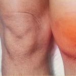
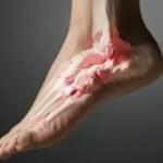
What does uveitis look like: photo
The photo below shows how the disease manifests itself in adults.

Complications
Serious complications of uveitis include profound and irreversible loss of vision, especially if the uveitis was unrecognized or the wrong therapy was prescribed.
Also, the most common complications include detachment of the retina, optic disc or iris and cystoid macular edema (the most common cause of visual impairment in patients).
Treatment of uveitis of the eye
The treatment of uveitis is complex, consisting in the use of systemic and local antimicrobial, vasodilating, immunostimulating, desensitizing drugs, enzymes, physiotherapy methods, hirudotherapy, traditional medicine. Usually, patients are prescribed drugs in the following dosage forms: eye drops, ointments, injections.
For drug treatment anterior and posterior uveitis use:
- vitamin therapy.
- Antihistamines - "Clemastin", "Claritin", "Suprastin".
- Viral uveitis is treated antiviral drugs- "Acyclovir", "Zovirax" in combination with "Cycloferon", "Viferon". They are prescribed for topical use in the form of intravitreal injections, as well as for oral administration.
- Broad-spectrum antibacterial agents from the group of macrolides, cephalosporins, fluoroquinolones. The drugs are administered subconjunctivally, intravenously, intramuscularly, intravitreally. The choice of drug depends on the type of pathogen. To do this, conduct a microbiological study of the detachable eyes on the microflora and determine the sensitivity of the isolated microbe to antibiotics.
- Immunosuppressants are prescribed when anti-inflammatory therapy is ineffective. The drugs of this group inhibit immune reactions - "Cyclosporin", "Methotrexate".
- Anti-inflammatory drugs from the group of NSAIDs, glucocorticoids, cytostatics. Patients are prescribed eye drops with prednisolone or dexamethasone, 2 drops in a sore eye every 4 hours - Prenacid, Dexoftan, Dexapos. Inside take "Indomethacin", "Ibuprofen", "Movalis", "Butadion".
- Fibrinolytic drugs have a resolving effect - Lidaza, Gemaza, Wobenzym.
- To prevent the formation of adhesions, eye drops "Tropicamide", "Cyclopentolate", "Irifrin", "Atropine" are used. Mydriatics relieve spasm of the ciliary muscle.
Treatment of uveitis is aimed at the speedy resorption of inflammatory infiltrates, especially in sluggish processes. If you miss the first symptoms of the disease, not only the color of the iris will change, its dystrophy will develop, but everything will end with decay.
Folk remedies
In the treatment of uveitis, you can use some methods of traditional medicine, after discussing the possibility of such treatment with your doctor:
- You can use crushed marshmallow root. To do this, pour 3-4 tablespoons of marshmallow root with a glass of water at room temperature. You need to insist it for 8 hours, and then use it for lotions.
- A decoction of chamomile, rosehip, calendula or sage helps with uveitis. To prepare it, you need 3 tablespoons of herbs and a glass of boiling water. The mixture should be infused for about an hour. Then you should strain it, and rinse your eyes with this decoction.
- Aloe can also help. You can use aloe juice for instillation into the eyes, diluting it in cold boiling water in a ratio of 1 to 10. You can make an infusion of dry aloe leaves.
Usually, folk remedies- These are additional treatment options that are used in a complex manner. Only timely adequate therapy of an acute inflammatory process in the eyeball gives a good prognosis, that is, it guarantees that the patient will recover. This will take a maximum of 6 weeks. But if it is a chronic form, then there is a risk of relapse, as well as exacerbation of uveitis as the underlying disease. Treatment in this case will be more difficult, and the prognosis is worse.

Surgery
Surgical intervention is required if the disease proceeds with serious complications. As a rule, the operation involves certain stages:
- the surgeon dissects the adhesions that connect the shell and the lens;
- removes the vitreous body, glaucoma or cataracts;
- removes the eyeball;
- using laser equipment, attaches the retina.
Every patient should know that surgical intervention doesn't always end a positive result. A specialist warns him about this. After surgery, there is a risk of exacerbation of the inflammatory process. Therefore, it is important to identify the disease in a timely manner, diagnose it, and prescribe effective therapy.
2965 09/18/2019 5 min.The eyes are an important part of the whole body. Sometimes, during diagnostics, the source of the problem is found not at all where it was previously searched for. The treatment of any health problem must be approached comprehensively. This is especially true for such eye disease like uveitis. It is important to treat not only the symptoms, but to identify the cause of the disease.
What is uveitis?
Uveitis is a general concept that refers to inflammation of various parts of the choroid (iris, ciliary body, choroid). This disease is quite common and dangerous. Often (in 25% of cases) uveitis leads to and even to blindness.
The appearance of this disease contributes to the high prevalence of the vascular network of the eye. At the same time, the blood flow in the uveal tract is slowed down, which can lead to the retention of microorganisms in the choroid. Under certain conditions, these microorganisms are activated and lead to inflammation.
Lacrimation as one of the signs of uveitis
The development of inflammation is also influenced by other features of the choroid, including different blood supply and innervation of its various structures:
- the anterior section (iris and ciliary body) is supplied with blood by the anterior ciliary and posterior long arteries, and is innervated by the ciliary fibers of the first branch of the trigeminal nerve;
- the posterior section (choroid) is supplied with blood through the posterior short ciliary arteries and is characterized by the absence of sensitive innervation.
These features determine the location of the uveal tract lesion. The anterior or posterior section may suffer.
Classification
The anatomy of the eye predisposes to the fact that the disease can be localized in different places of the uveal tract. Depending on this factor, there are:
- Anterior uveitis: iritis, anterior cyclitis. Inflammation develops in the iris and. This variety is the most common.
- Median (intermediate) uveitis: posterior cyclitis, pars-planitis. The ciliary or vitreous body, retina, choroid are affected.
- Posterior uveitis: choroiditis, retinitis, neurouveitis. The choroid, retina and are affected.
- Generalized uveitis - panuveitis. This type of disease develops if all parts of the choroid are affected.

Forms
The nature of inflammation in uveitis can be different, and therefore the following forms of the disease are distinguished:
- serous;
- hemorrhagic;
- fibrinous-plastic;
- mixed.
Depending on the duration of inflammation, acute and chronic (more than 6 weeks) form of uveitis is distinguished.
Causes of inflammation
Uveitis can develop due to a variety of reasons, the main of which are:
- infections;
- trauma;
- systemic and syndromic diseases;
- metabolic disorders and hormonal regulation.
The most common infectious uveitis: they occur in 43.5% of cases. Infectious agents in this case are mycobacterium tuberculosis, streptococci, toxoplasma, pale treponema, cytomegalovirus, herpesvirus, fungi. As a rule, such uveitis is associated with infection entering the vascular bed from any focus of infection and develops with sinusitis, tuberculosis, syphilis, viral diseases, tonsillitis, sepsis, dental caries, etc.
In the development of allergic uveitis, an increased specific sensitivity to environmental factors plays a role - drug and food allergy, hay fever, etc. Often, with the introduction of various sera and vaccines, serum uveitis develops.
Uveitis can occur against the background of systemic and syndromic diseases, such as:
- rheumatism;
- rheumatoid arthritis;
- psoriasis;
- spondyloarthritis;
- sarcoidosis;
- glomerulonephritis;
- autoimmune thyroiditis;
- multiple sclerosis;
- ulcerative colitis;
- syndromes of Reiter, Vogt-Koyanagi-Harada, etc.
Post-traumatic uveitis occurs due to penetrating or contusion damage to the eyeball, foreign bodies entering the eyes.
The following diseases also contribute to the development of uveitis:
- metabolic disorders and hormonal dysfunction (diabetes mellitus, menopause, etc.);
- diseases of the circulatory system;
- diseases of the organs of vision (, conjunctivitis, keratitis, blepharitis, scleritis, perforation of a corneal ulcer).
And this is not the whole list of diseases that can cause and develop uveitis.

Symptoms and Diagnosis
On initial stage disease, the color of the iris changes and adhesions appear. The lens of the eye becomes cloudy. Further, uveitis can manifest itself in different ways, depending on the type and form of inflammation. General symptoms are:
- photophobia;
- chronic lacrimation;
- aching or sharp pains;
- pain and discomfort;
- deformation, ;
- the appearance of a light "fog" before the eyes;
- deterioration of visual acuity, up to blindness;
- fuzzy perception;
- increased intraocular pressure (with a feeling of heaviness in the eye);
- transition of inflammation to the second eye.
Any violation normal operation the membranes of the eye will lead to serious changes for the entire organ of vision. That is why uveitis, like any other ophthalmic pathology, must be treated quickly. About what types of this disease are, what caused its development and how it should be treated, will be described in detail in this article.
Uveitis is the medical term for an inflammatory process that can occur in different parts of the choroid. It's pretty rare disease and in 25% of cases it leads to visual impairment, and sometimes even blindness.
In men, the pathology develops somewhat more often. This can be explained from an anatomical point of view. Uveal (vascular) pathways look like a branched vascular network with slow blood flow. This becomes the main reason that infectious agents linger here. With normal immunity, they do not affect human health in any way, but as a result of exposure to negative factors, they begin to activate and cause an inflammatory process.
Important: you need to contact an ophthalmologist when the very first signs of eye pathology appear. This will allow time to stop the development of the disease and cure it.
The uveal membrane has a rather complex structure. It occupies the space between the retina and sclera, looks like grapes. From here comes its name - "uvea", which in Russian means "grape".
It has 3 main sections:
- iris;
- ciliary body;
- choroid - the choroid itself (located directly under the retina, lines it from the outside).

To the number important functions, which are assigned to the choroid, include:
- Flow regulation sun rays. This protects the eyeball from excess light.
- Transport nutrients throughout the retina.
- Removal of decay products from the eye.
- Participation in the adaptation of the eyeball, i.e. change in refractive power optical system eyes for a clearer and clearer perception of various objects that are at different distances from it.
- Production of intraocular fluid.
- Normalization of pressure inside the eye.
- Thermoregulation.
The main function of this shell is to supply the organs of vision with blood. Thanks to the anterior, posterior short, as well as long ciliary arteries, blood is transported to all areas of the eye. However, due to the fact that each part of the eyeball is supplied with blood from its own source, the infection also occurs separately.
Etiology
Uveitis of the eye can occur due to infection,  the onset of allergies, due to poor metabolism, injury, severe hypothermia, or against the background of any general illness.
The most common is considered to be infectious uveitis, leading to the development of inflammation. The infection is caused by fungi, streptococci, mycobacterium tuberculosis, treponema, toxoplasma, herpes virus, etc.
Acute allergic uveitis can begin as a result of the use of any food products or medicines. Background diseases are rheumatism, rheumatoid arthritis, glomerulonephritis, ulcerative colitis, psoriasis or multiple sclerosis.
Injuries include eye burns of varying severity, foreign bodies and other penetrating injuries of the eyeball.
Hormonal dysfunction can also cause uveitis, the causes of this: menopause, disorders menstrual cycle etc.
Classification of the disease and its symptoms
The main morphological forms of pathology: anterior uveitis, median, posterior, peripheral and diffuse. The anterior, in turn, is divided into iritis, cyclitis and iridocyclitis. The posterior one is called choroiditis, and the diffuse one is called panuveitis or iridocyclochoroiditis.
Depending on the nature of the course, acute, chronic and recurrent uveitis are distinguished.
Each form of uveitis has a number of its features. Anterior uveitis is characterized by the following symptoms:
- redness of the pupils;
- fear of light;
- chronic lacrimation;
- constriction of the pupils;
- Pain in the eyes;
- increased intraocular pressure.
Peripheral uveitis of the eye, symptoms:
- eye damage, which is symmetrical;
- the appearance of "flies" before the eyes;
- marked deterioration in visual acuity and quality.

With posterior uveitis, symptoms appear later. A person will have the following symptoms:
- blurred vision;
- everything visible around is distorted;
- impaired perception of colors;
- the patient constantly sees "flies" floating before his eyes, there may often be peculiar flashes;
- sharply reduced visual acuity.
The severity of the inflammatory process also differs in different forms of the disease. It is most intense in anterior uveitis. The iris at the same time becomes greenish or rusty-brown, the pupil is greatly narrowed and almost does not react to light. Tiny plaques appear on the cornea, freely moving in the eye fluid. They arise as a result of a large number pigment proteins together with lymphocytes.
The acute form lasts up to 1.5-2 months. If left untreated, it passes into the chronic stage, which begins to recur with the onset of cold weather.
Peripheral uveitis is indolent and has the most vague symptoms, so it is difficult to diagnose. When it affects such structures of the eye, which are very difficult to examine. But if you don't accept necessary measures, the appearance of serious complications and the development of secondary eye diseases are possible.
Diagnosis of the disease
To make an accurate diagnosis, you need to full analysis organs of vision. Diagnostic tools include:
- examination by an ophthalmologist;
- determining how much the patient acute vision;
- microscopic examination retina;
- ultrasound diagnostics;
- angiography - examination of blood vessels and identification of the cause of blood flow;
- biopsy followed by examination of the sample taken.

Ways to treat uveitis
If the course of the disease has been started, then treatment should have an integrated approach. Drug therapy involves the use of external agents and decoctions.
traditional medicine
These drugs include:
- mydriatics - cyclopentol, atropine and others. These drugs eliminate muscle spasm and eliminate the effects of adhesions;
- steroids - prednisolone, dexamethasone and others. If there is no benefit from them, the doctor may prescribe immunosuppressive drugs;
- eye drops;
- antihistamines if an allergic reaction occurs;
- in the presence of infections, antimicrobial and antiviral drugs.

Traditional medicine
Various herbs help in the fight against uveitis, treatment is carried out using these recipes:
- a decoction of calendula, chamomile, birch buds and sage. To prepare it, you need to mix 1 tsp. crushed plants, brew in 100 ml of boiling water and rinse with a warm solution 2-3 times a day;
- aloe drops. They need to be bred in warm water in a ratio of 1:10, and then instill in each eye 3 times a day, 2-3 drops;
- grind fresh marshmallow root to a state of gruel, wrap in clean gauze and apply to the eyes for half an hour. After the procedure, they must be washed with herbal decoction.
Prevention of uveitis
Complete relief from uveitis will occur in a few weeks if treatment is started on time. If the course of the disease was started or the patient did not complete the course of treatment, then there is a high probability that uveitis will become chronic. To cure it, you will need a long and difficult therapy, so it is better to avoid the appearance of the disease.
To do this, you need to observe simple hygiene of the organs of vision, to avoid injuries and the penetration of bacteria. It is very important to start treatment immediately. allergic diseases, since some of them can provoke the development of uveitis.