One of the most pressing problems for modern women I developed a disease called fibroids.
In this article you can find out what it is, how to treat it, and whether it is dangerous. In accessible language Everything about uterine fibroids in women will be described in detail: symptoms and treatment, classification, causes, complications and consequences.
Attention: At the end of the article, photos of the formations in a living form will be provided, so viewing them is not recommended for impressionable and easily inspired people! Videos of actual operations will also be provided.
First, let's look at the definition of the disease and its varieties.
What is fibroid
Uterine fibroids are a disease of the female reproductive system, which is characterized by the formation and growth of a benign tumor in the uterus, namely in its muscle layer. Most often, this disease is found in women 30-40 years old or older. But there are more and more cases where this diagnosis is made at a younger age, including in virgins 20-25 years old.
Myoma nodes can be located inside the body of the uterus; this localization option is found in 95% of cases, and in the cervix in 5% of patients. If a tumor develops from connective tissue, it is called fibroma, and if it is muscle, it is called leiomyoma.
The growth rate of the tumor depends on hormonal levels, therefore, one of the treatment methods is hormone therapy, but a more effective option for eliminating the problem is surgery.
Kinds
Depending on the location relative to the myometrium, the classification of fibroids will be as follows:
- intermuscular (interstitial, intramural) – the node is located inside the muscle layer;
- subperitoneal (subserous) - the node is located near the peritoneum under the mucous membrane of the outer layer of the uterus;
- submucosal (submucosal) - the node is located in the uterine cavity under its internal mucous layer;
- interligamentous (intraligamentary) – the tumor is located between the wide uterine ligaments.

There are also different types knots, they can be on a leg or on a flat base. Based on the number of nodes, fibroids can be single or multiple, but most often the appearance of one node entails the appearance of other neoplasms.

By size
Gynecologists indicate the size of fibroids in centimeters, meaning the size of the node itself, or in weeks. If the diagnosis says “fibroids at 13 weeks,” it means that the uterus with the fibroid node has increased to the size that is typical for pregnancy at 13 weeks. Based on these criteria, the tumor can be classified into 3 types:
- small sizes– up to 2 cm (4 weeks);
- average– 2-6 cm (10-11 weeks);
- large sizes– more than 6 cm (12 or more weeks).

Why are uterine fibroids dangerous?
Any tumor poses a danger to human body, since the neoplasm is abnormal for it.
On initial stages some women still do not fully understand why a benign tumor is dangerous, but as it grows, they begin to feel compression of the organs in the pelvis. In the presence of fibroids, blood circulation in neighboring organs is disrupted, since the formation itself acquires its own vascular network. In addition, fibroids can be a sign of endocrine diseases, which in turn can cause additional health problems.
What will happen if left untreated?
Many women who have been diagnosed with fibroids are interested in what will happen if it is not treated. Let's list all the consequences of an indifferent attitude towards this disease:
- recurrent miscarriages, hypoxia, fetal malnutrition;
- infertility;
- uterine bleeding which provoke the development of anemia;
- the neoplasm can develop into sarcoma, from which the woman can ultimately die;
- rapid increase in tumor size;
- torsion of the leg of the myomatous node with disruption of nutrition in it;
- decreased uterine tone, which leads to postpartum blood loss;
- hyperplastic processes various types;
- hydronephrosis or pyelonephritis.
What provokes the appearance of formation
Modern medicine has not yet established the exact reason why fibroids appear, but there are a number of factors that can increase the likelihood of its development:
- genetic predisposition;
- hormonal imbalance;
- hyperplastic processes in the endometrium;
- pregnancy, childbirth and abortion;
- infectious and inflammatory diseases organs reproductive system;
- chronic stress;
- diabetes mellitus and diseases of the endocrine system, problems with excess weight;
- chronic diseases internal organs and systems, hypertension;
- low physical activity;
- irregular sex life and lack of orgasms.

Let's take a closer look at some of the reasons why fibroids are most likely to appear:
- excess estrogen, lack of progesterone. Since fibroids are a hormone-dependent neoplasm, they can develop against the background of hormonal imbalance. It is quite natural that it is most often diagnosed in women of childbearing age, usually due to an imbalance of estrogen and progesterone. Obesity only makes the situation worse, since adipose tissue is also capable of producing estrogen;
- pregnancy, childbirth and abortion. Abortion, diagnostic curettage, difficult childbirth and spontaneous abortions increase the likelihood of a neoplasm, and successful childbirth, especially with subsequent breastfeeding, on the contrary, reduces the risks;
- woman's nutrition. Unbalanced diet and unhealthy food can disrupt hormonal balance in female body. Refined foods, trans fats and fiber deficiency contribute to increased concentrations of female sex hormones. Unhealthy food can lead to obesity and, as a result, to the development of fibroids. A woman needs to eat a lot of vegetables, fruits, seafood, cereals, less fat and sugar;
- inferiority of intimate life. Due to irregular sexual intercourse and lack of orgasms, venous blood stagnates in the pelvic organs, which can lead to hormonal disorders and tumor development.

Oral contraception can also provoke the development of fibroids, mechanical injuries reproductive organs, prolonged exposure to ultraviolet radiation on a woman’s body.
Diagnostics
A doctor may suspect the presence of fibroids during a routine gynecological examination, since The size of the uterus with this disease will be increased. Diagnosis of pathology may include the following activities:
- transvaginal ultrasound of the pelvic organs. Ultrasound can assess the size and shape of the tumor;
- hysteroscopy. During the procedure, the doctor can simultaneously perform a biopsy of the affected area and send the resulting material to a histology laboratory;
- laparoscopy. Prescribed in case of controversial situations when it is impossible to distinguish uterine fibroids from ovarian tumors;
- CT and MRI. The studies, although informative, are expensive and therefore rarely used.
How does a tumor manifest itself?
On early stages the disease may not manifest itself at all, the first ones may appear when the size of the node reaches 2-6 cm:
- sharp pain in the lower abdomen that is not associated with the arrival of menstruation. The nature of the pain is cramping, pulling;
- painful periods, although they did not have this feature before;
- increased menstrual flow;
- heavy intermenstrual bleeding;
- disruption of the regularity of the menstrual cycle, both its lengthening and shortening;
- problems with conception.

Symptoms of this disease depends on its age, the age of the patient, the size and location of the tumor, the rate of its growth and the presence of concomitant chronic diseases.
Very often, fibroids do not make themselves felt in any way; they can only be detected at the next gynecological examination. By palpating the abdomen, the doctor will find an enlarged uterus and will necessarily send the woman for an ultrasound, where echoes of fibroids will confirm the preliminary diagnosis.
This disease has a number of characteristic symptoms, if you discover them, you should urgently consult a doctor:
- pain during the intermenstrual period, which are felt in the lower abdomen and radiate to the lower back and limbs;
- menstrual irregularities. The duration of the cycle changes, intensifies menstrual pain, the intensity of discharge increases during regulation, bleeding begins in the middle of the cycle;
- a woman can't long time get pregnant.
If the myomatous node quickly increases in size or already has an impressive volume, the abdominal girth also increases, while body weight remains almost unchanged. There is discomfort and aching pain in the lower abdomen, which increases during psycho-emotional and physical stress.
Large myomatous nodes can put pressure on neighboring organs, thereby causing constipation, frequent trips to the toilet “in small ways” and painful urination.
If the leg of the myomatous node is twisted, the body of the fibroid may become necrotic, and additional symptoms may appear “ acute abdomen", such as sharp pain in the lower abdomen, rapid heartbeat, cold sweat, fainting. In such cases, emergency removal of fibroids is performed.

Pathology can affect other organs and systems, which provokes the appearance of the following symptoms:
- at frequent bleeding anemia develops;
- may often feel sick and dizzy;
- pain behind the sternum;
- neuroses and neurotic states arise.
If any of the above symptoms appear, you should definitely seek medical help.
Is it curable or not?
Patients who have not previously encountered gynecological problems, having learned about the diagnosis of fibroids, do not have an accurate idea of whether the pathology can be cured or not.
Although uterine fibroids are a tumor-like disease with many characteristics of a tumor, they still have a number of distinctive features, which allow treating the disease not only surgical methods, but also conservative.
Most nice feature This neoplasm is its ability to decrease in size and completely disappear.
The choice of treatment method is carried out individually depending on the severity of the disease and the characteristics of the body; only in extreme cases is surgical intervention performed.
How to treat uterine fibroids
There are 2 ways to treat fibroids:
- conservative treatment, in which medications and non-invasive procedures are prescribed;
- surgery, in which the operation is performed.
What to do in each specific case can only be determined by a doctor.
How uterine fibroids are treated will depend on the severity of the pathology, clinical symptoms, size of the tumor, the patient’s age and intention to have offspring in the future.

Treatment of fibroids medicines will be effective only under certain conditions:
- with small node sizes, when the uterus does not exceed 12 weeks of pregnancy in size;
- if the disease is accompanied by a small number of symptoms;
- when the node has a wide base and is located subserosally or inertially.
Myoma can be treated with early stages, the longer the neoplasm is present in a woman’s body, the less opportunity there is to use conservatism in treatment. If there are serious contraindications to surgery drug treatment is the only way out.
Treatment without surgery includes the following set of measures:
- diet;
- use of immunomodulators;
- phytotherapy;
- physiotherapeutic procedures;
- hormonal drugs.
During drug treatment, a patient with fibroids goes through the following stages:
- inflammation and infections are eliminated;
- work intensifies immune system by using special drugs;
- the diet and daily routine are adjusted;
- the functioning of the endocrine system is normalized;
- an even psychological background is formed;
- bleeding is eliminated and the anemia caused by it is treated;
- the menstrual cycle is normalized.
Having discovered a fibroid in a patient, the doctor determines the rate of development of the pathology over a year. If it grows within a year to the size of a 4-week pregnancy, then it is considered fast-growing and goes on to surgical treatment.
Now let's look at each of the treatment methods in more detail.
Medicines
Conservative treatment is carried out using the following groups medications:
- androgen derivatives;
- gestagens;
- combined oral contraceptives;
- analogues of gonadotropin releasing hormone aGnRH.

One of the innovative medicines is the drug Esmya, the main active ingredient of which is ulipristal acetate. This medicine, after 3 months of treatment in patients who were prescribed surgery, significantly reduces the size of the tumor and reduces the intensity of symptoms, and in 50% of cases there is no need for surgery. There are no tablets side effects, and six months after their use, the tumor does not resume its growth.

Now let's look at medications from other groups in more detail.
Gonadotropin-releasing hormone agonists
These drugs put a woman into artificial menopause by suppressing ovarian function with hormones. Natural gonadotropin-releasing hormone agonist drugs suppress the secretion of sex hormones by the pituitary gland, which affect the activity of the ovaries. Drugs in this group include:
- Buserelin;
- Triptorelin;
- Leuprorelin;
- Goserelin.
Under the influence of drugs, the ovaries stop their activity, ovulation does not occur, the inner uterine layer does not change and menstruation stops coming. This is a reversible process; after discontinuation of the drug, the ovaries resume their work. The course of treatment is six months. During this time, the size of the tumor should decrease by 50%, and symptoms should decrease significantly.
These medications have a number of negative aspects:
- fibroids can fully recover in size after stopping medication;
- The drug should not be taken for more than six months, as the risk of developing osteoporosis and other complications due to estrogen deficiency increases.
Most often, agonist drugs are prescribed before surgery to reduce the size of the tumor.
Antigonadotropins
Most often in this series, Danazol and Nemestran with the active ingredient gestrinone are prescribed. These medications reduce the intensity of symptoms but do not reduce the size of fibroids. When taking them, a number of side effects are possible:
- hair on the face and body grows rapidly;
- the timbre of the voice changes;
- rashes appear.

These drugs are most often prescribed when there is no effect from hormonal therapy.
Gestagens
The drugs in this series include Duphaston, Norkolut and Utrozhestan. They normalize the growth of the endometrial layer in the presence of hyperplasia that occurs against the background of fibroids. Fibroids are poorly treated exclusively with gestagens; they are prescribed only when the neoplasm develops simultaneously with endometrial hyperplasia. The course of treatment is 8 months.
Another drug that contains a progestin (levonorgestrel) is the Mirena intrauterine device. It is given for 5 years and, in addition to blocking tumor growth, also has a contraceptive effect.
Antiprogestogens

Operations
Unfortunately, drug treatment cannot always give a good result.
If fibroids cannot be treated with conservative methods, surgery is indicated. First, let's look at the cases in which surgery is prescribed for fibroids.
Indications for surgery
The operation is prescribed in the following conditions:
- the size of the node exceeds the uterus of the 12th week of pregnancy;
- the tumor puts pressure on nearby vessels and organs, interfering with them normal operation;
- the neoplasm provokes heavy bleeding;
- there are very strong ones painful sensations;
- the leg of the node twisted and its death began;
- a submucosal myomatous node appeared;
- in addition to fibroids, endometriosis was diagnosed;
- there is a suspicion of malignant processes in the neoplasm;
- If the tumor grows very quickly, surgery is urgently needed.
Now we will consider in more detail the types of surgical interventions and situations in which their use is necessary.
Embolization

IN Lately many practicing surgeons use the method of uterine artery embolization. This is a minimally invasive intervention during which an embolus is injected into the uterine vessel to block the arterial lumen. The blood supply to the myomatous node stops, and its tissues die.
This is the most effective method for patients with fibroids who still plan to have children in the future.
Hysterectomy
The most radical method in which the organ is completely removed. There are 3 options for online access:
- abdominal- the most commonly used method, which involves supravaginal amputation of the uterus or its extirpation. Frequently negative consequence extirpation is urinary incontinence. Supravaginal amputation is possible provided that the cervix is healthy and the nodes between the cervix and uterus are not large;
- laparoscopic;
- vaginal– used for small myomatous nodes.

Before or during surgery, the surgeon determines the advisability of removing the uterus and ovaries. The decision is made individually in each case, taking into account the patient’s age and the presence or absence of tumors on the ovaries.
This method is prescribed in the following cases:
- fibroids are larger in size than 13 weeks of pregnancy;
- drug treatment is ineffective;
- the tumor is growing rapidly;
- the ovaries are affected by a tumor;
- Acute bleeding began.
Myomectomy
Women who are of reproductive age and still want to have children are initially given recommendations on how to treat fibroids with medication; if they do not help, then a conservative myomectomy may be prescribed. During this intervention, the myomatous node is peeled off to healthy tissue. The intervention is performed laparoscopically or abdominally.

Fuse ablation
This is a non-invasive method of treating pathology, which is carried out under MRI control. During the procedure, the cells of the myomatous node are heated by an ultrasonic pulse until they are completely destroyed.

The process is carried out in several stages. Initially, the doctor examines the tumor and plans surgery using MRI. At the second stage, under the control of an MRI machine, the doctor begins to heat the cells of the node to a certain temperature with ultrasound pulses. After the cells die, the specialist cools the tissue. Depending on the size of the tumor, there may be several such sessions. The procedure itself takes about 4 hours. A control MRI is done using contrast.
Since FUS ablation is a non-invasive technique, it has many positive aspects:
- there is no need for anesthesia and postoperative care;
- no complications and adverse reactions, type of bleeding, elevated temperature and intoxication;
- both the uterus and reproductive function women;
- rehabilitation takes place quickly;
- no relapses;
- the method is also effective in the treatment of large nodes;
- fibroids decrease in size immediately after the session;
- You can quickly get rid of uncomfortable symptoms.
During the procedure, the patient lies motionless on her stomach. If any discomfort she immediately reports this to the attending physician. The procedure should not cause burning, stabbing or shooting pain, so the occurrence of such symptoms should be immediately reported to the medical staff conducting the treatment.
Folk remedies to help
Traditional methods of treatment are reduced only to the use of tampons and douching herbal infusions and decoctions at home.
Not a single similar method will help get rid of the underlying internal causes that provoked the development of fibroids.
The use of any alternative methods of treating this disease should be discussed with your doctor.
Prevention If you belong to the fair half of humanity, then no are not able to 100% protect you from the occurrence of fibroids. The only thing that may be in your power is to reduce the factors that provoke the growth of fibroids. There are several basic recommendations for women who do not want to face this disease:
- make annual visits to the gynecologist regular, it is better to visit a specialist 2 times a year;
- do an annual ultrasound of the reproductive system;
- have sex regularly and achieve orgasm;
- to prevent abortions, and to protect against unwanted pregnancy, the use of hormonal contraceptives will help;
- control weight, lead an active lifestyle and play sports;
- take vitamin-mineral complexes with antioxidant effect, which include vitamins A, E, C, iron, zinc, iodine, selenium.
A few words about pregnancy

Among women reproductive age Those who have been diagnosed with fibroids always have a number of questions regarding the compatibility of this disease with the possibility of having children. We will provide answers to the most popular ones.
Is it possible to get pregnant?
You can get pregnant if you have fibroids in the uterus.
Everything will depend on the size of the tumor and its location. If it does not prevent the fertilized egg from passing through the fallopian tube and gaining a foothold in the uterine wall, then conception will occur. It is important that the entire process of bearing a child is under strict medical supervision.
In the first 2 trimesters, due to hormonal changes in a woman’s body, a slight growth of the myomatous node may occur, but in the last months of pregnancy the fibroid does not grow, but only undergoes destructive changes.
What are the risks to fetal health?
Fibroids can disrupt blood circulation and nutrition of the uterine walls, which affects the ability of the myometrium to contract properly, so the presence of a tumor in the uterus increases the risk of miscarriage.
Premature termination of pregnancy, spontaneously or according to indications, is the main threat to the fetus, but due to tumor growth, a delay may occur intrauterine development, fetal hypoxia, premature or protracted labor, which is also a serious risk for future offspring.
Photo
Finally, we will give you some photos of fibroids so that you can understand what they look like in real life.
Impressionable people should not watch!
A hormone-dependent tumor that often occurs in women of childbearing age is called uterine fibroids (myoma). Vivid symptoms and signs of uterine fibroids appear, as a rule, at later stages of development, and in the early stages this disease develops asymptomatically, and there are usually no complaints from patients.
What is it - fibroids?
Myoma (fibromyoma, leiomyoma) is female disease, which is a benign tumor that forms on the walls or in the uterine cavity from connective tissue. Myoma develops when elevated level female sex hormones - estrogens, because of this it is classified as hormone-dependent. The tumor ranges from a small nodule to a very large size (weighing about 1 kg). When large, it can be easily recognized by palpation. Symptoms of the disease often do not appear immediately; with advanced disease, fibroids are more difficult to treat and there is a greater likelihood of complications. Although the disease has signs of tumors, it is different from it, so it would be more correct to classify fibroids as tumor-like formations.
This neoplasm can cause a lot of trouble (despite the fact that it is a benign disease) - uterine bleeding and complications during pregnancy can occur, which is why treatment is mandatory.
Leading clinics in Israel
Note! Most often, the localization of such a neoplasm is the body of the uterus, but its location is also possible in the cervix. If the disease develops in muscle tissue, it is considered typical, and if it occurs in the neck or ligaments, it is considered atypical.
The size of the neoplasm ranges from several millimeters to several centimeters, often multiple fibroids (when several tumors form at once).
Due to the fact that the tumor is a hormone-dependent tumor, with the onset of menopause its growth may stop and its size may decrease; sometimes it resolves completely and simply disappears.
Remember! Although this disease is benign and quite rarely degenerates into a malignant form, tumor growth can lead to serious disruptions in the functioning of the patient’s reproductive organ.
 Most often, formation occurs in the uterus, but can be found in the mammary glands, vagina, sometimes it can form in the digestive organs: stomach, esophagus, intestines, kidneys, it is also diagnosed in the bladder, on the skin, transverse skeletal muscles, and heart muscle. Fibroids can affect the brain, bones, limbs - shins, legs, arms and other organs.
Who is at risk
This disease is diagnosed in almost 25% of women of fertile age, of which in 3% of cases this formation is first discovered during a routine examination; it can be felt. This disease can also occur in young and elderly nulliparous women, after childbirth, during pregnancy, menopause, and after gynecological operations. But women who have given birth have a much lower risk of getting fibroids.
The incidence of the disease among all women by the age of 35 reaches 35-45% of cases. The peak of the disease occurs at 35-50 years of age.
Remember! Myoma in a row gynecological diseases is in 2nd place. If the frequency of diagnosis in reproductive age is about 20%, then in premenopausal age it reaches 35% of cases.
Classification of the disease and stage of development
Uterine fibroids are classified by the number of nodes, according to this, it can be:
- multiple;
- single.
In accordance with the size, tumors are determined:
- large;
- average;
- small myomatous neoplasms.
Depending on the size of the nodes, they are compared with the stages of pregnancy and distinguished:
- small fibroids (5-6 weeks);
- medium size (7-11 weeks);
- large (more than 12 weeks).
Depending on the size and location of the nodes, the following varieties are distinguished:

Based on its location relative to the myometrium (muscular layer), the neoplasm is divided into the following types:
- interstitial fibroid. It is located in the center of the muscular layer of the uterine wall - it grows into the uterus (60% of all cases), usually of large size;
- submucosal (submucosal). This species grows towards the endometrium. When the node is located for the most part in the myometrium, it is called intermuscular with centripetal growth. Such a neoplasm can be pedunculated or even broad-based; pedunculated tumors can “fall out” of the cervical canal, become infected and twist;
- subserous (subperitoneal). Her node is located near the peritoneum, under the mucous membrane of the outer layer of the uterus. This type can be divided into the following types:
- type 0 – looks like: a knot on a wide base – 0-A, on a leg – 0-B;
- Type 1 – a large proportion of the node is located in the serous membrane;
- Type 2 – a large proportion is located in the thickness of the myometrium;
- diffuse. Doesn't show itself a certain type tumors, but has a diffusely unformed character.
If we consider the tumor by location, it can be located in the area:

If we consider the stages of formation, we get three stages of morphogenesis:
- development of the bud (active growth area) in the myometrium;
- development of an undifferentiated tumor;
- development and maturation of a tumor with differentiated cells.
Tumor growth does not always proceed in the same way, so they distinguish:
- simple myoma, when the tumor grows slowly and has small size, often single, mild symptoms;
- proliferating, this species is fast growing, has clinical manifestations. It is considered multiple uterine fibroids or single large ones.
Based on the nature of growth, the neoplasm can be divided into:
- true;
- false.
What does it come from?
There are many causes of the disease:
- hormonal disorders (sharp decrease or increase in the amount of progesterone or estrogens);
- Sex life is irregular (especially after 25 years). Due to the lack of sexual dissatisfaction, stagnation of blood flow in the pelvic area predominates, and this provokes the formation of fibroids;
- the presence of chronic inflammation;
- genetic predisposition;
- late birth (or no birth at all);
- mechanical damage (traumatic childbirth, abortion, consequences of gynecological operations, etc.);
- various stresses, hard physical work;
- various pathologies - endocrine diseases (thyroid problems, diabetes).
Indirect causes of fibroids can be noted, for example, smoking, physical inactivity, and external factors, like poor ecology and poor nutrition. In order to prevent the appearance of fibroids, it is necessary to eliminate these provoking factors.
The growth rate of nodes depends on various factors:

Remember! Myoma begins to develop due to a mutation of one cell; its subsequent development and division is influenced by changes in hormonal levels, imbalance of estrogen and progesterone.
Symptoms of the disease
The early stages of the disease are most often asymptomatic.
Later the following signs begin to appear:

But the presence of all these signs is not enough to diagnose fibroids; to confirm the suspicion of the presence of this neoplasm and exclude its erroneous diagnosis, you must undergo a thorough examination and do an ultrasound. Some of these signs may indicate other, more dangerous diseases (uterine cancer, ovarian cancer, endometriosis).
Some signs of fibroids may indicate its localization:
| Localization of fibroids | Symptoms |
|---|---|
| Submucosal | menstrual irregularities occur; |
| abundant and long periods, uterine bleeding, which causes anemia; | |
| there is no pain syndrome, but when the myomatous node from the submucosa enters the uterine cavity, there may be cramping, very severe pain. | |
| Intramural | accompanied by cycle disruption; |
| pain in the pelvic area. | |
| Subserosal | usually without any symptoms; |
| pain is minor, occurs rarely - in the lower back, back, lower spine; | |
| There may be urinary problems and constipation. |
Pregnancy
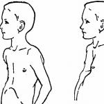
 Usually, with small nodes in the early stages of pregnancy, problems should not arise; an exception occurs when fibroids form near the placenta. The tumor produces substances that provoke contractions of the uterus and the pregnancy may be terminated. If this happens at a later date, then the risk of premature birth is high. Often, due to compression by the tumor, the fetus may develop deformation of the skull bones or torticollis, and the intake of sufficient quantities of nutrients and oxygen to the fetus.
If during gestation the neoplasm does not have a negative impact on the development of the fetus, then childbirth may be complicated by malpresentation of the fetus. In this case, a cesarean section is recommended, during which the tumor is also removed.
Also possible complications during childbirth there may be:
- early rupture of amniotic fluid (as the tone of the muscular layer of the uterus is increased);
- there is a high risk of heavy postpartum bleeding;
- there is a high probability of premature placental abruption (if the tumor is located behind it).
Would you like to receive an estimate for treatment?
*Only upon receipt of data on the patient’s disease, a representative of the clinic will be able to calculate an accurate estimate for treatment.
Uterine fibroids are diagnosed in approximately 8% of pregnant women; in most of them, the size of the tumor does not increase during pregnancy or, on the contrary, decreases.
Important! When fibroids are located near the embryo, the likelihood of miscarriage increases several times.
Complications of the disease
Uterine fibroids can pose a threat to a woman’s health due to the development of complications of the disease:
- miscarriage or infertility;
- postpartum hemorrhage;
- degeneration of a tumor into malignant oncology;
- formation of a submucosal node;
- necrosis of myomatous node;
- posthemorrhagic anemia;
- hyperplastic processes of the endometrium.
Diagnosis of the disease
Due to the fact that the symptoms of the disease are similar to a number of other diseases, a series of diagnostic measures for a correct diagnosis.
Diagnostic methods include:

How to treat
If the size of the tumor is small, the tumor is located deep in the muscles of the uterus or subserous, and it does not grow, then conservative treatment is chosen.
Assign medications such groups:
- androgen derivatives (gestrinone), which interfere with the formation of ovarian hormones, prevent tumor growth in the future;
- gestans (norkolut, utrozhestan), they stop the growth of the endometrium without affecting the fibroid;
- gonadotropic hormone agonists (zoladex, buserelin) – reduce the size of fibroids and its symptoms.
Surgical removal is prescribed for tumors big size, if there is bleeding, pain, a tendency to rapid growth, miscarriage or infertility.
The following types of surgical medical intervention are used:

Treatment tactics are selected taking into account the location of the tumor, its type, the patient’s age, and her state of health. Although fibroids are not a dangerous disease, self-medication at home is fraught with complications.
Nutrition for uterine fibroids
There is no special diet for this disease, but it is recommended to adhere to some general nutritional recommendations:
- it must be balanced, include a sufficient amount of vitamins and microelements;
- You should eat 5 times a day, do not overeat and do not take long breaks between meals;
- Avoid cooking methods such as frying and smoking. Replace them with stewing, boiling, baking;
- Fatty meat, lard, sausages, fatty cheeses, butter, sweets;
- Your diet should include more vegetables, fruits, herbs, dark breads, and fish.
Traditional methods of treatment
 The use of various folk remedies is possible only with the permission of a doctor and in conjunction with the main treatment
You can also use tampons soaked in sea buckthorn oil. The course of treatment is two weeks.
An infusion of boron uterus is prepared as follows: pour 50 g of herb into 0.5 liters of vodka and leave in a dark place for 10 days, shaking regularly. After this, take a teaspoon once a day for 10 days, a tablespoon for the next 10 days, then take a break and repeat the course.
Disease prognosis
With timely detection of fibroids and its proper treatment the prognosis is favorable. After organ-conserving surgery, pregnancy may occur. If fibroids grow rapidly, radical surgery with complete removal of the uterus may be required.
Disease prevention
Preventive measures boil down to:
- regular sexual intercourse with one partner;
- taking vitamins and microelements;
- active lifestyle;
- preventive annual examinations.
If the diagnosis is confirmed and you are diagnosed with uterine fibroids, then the following recommendations should be followed:
- avoid lifting heavy objects;
- eliminate stress;
- increase the amount of fruits, vegetables, and seafood in the diet;
- walk more often;
- refuse to engage in sports that are aimed at heavy loads on the pelvic area;
- do not be exposed to thermal influences - sunbathe, visit saunas, baths.
Video on the topic
Uterine fibroids are a gynecological disease characterized by the appearance benign neoplasms in the uterine muscle layer.
The size of fibroids can vary: from small, the size of a pea, to huge ones, when uterine fibroids reach a weight of 3 kg and correspond in volume to a full-term pregnancy. Moreover, it can be one tumor or several tumors, small in size, the so-called multiple uterine fibroids.
In terms of frequency of occurrence, this disease continues to occupy a leading position among all gynecological pathologies. According to statistics, such neoplasms are found in more than 28% of women during their lifetime. The majority of patients are women over 30 years of age.
In this article we will look at what uterine fibroids are, what signs precede this disease, and what symptoms you should pay attention to when fibroids have already formed. We will not ignore the methods of treating uterine fibroids, including talking about treatment without surgery. Reviews of women with this problem can be read in the comments.
Causes
Why does fibroid develop, and what is it? Uterine fibroids are a benign tumor that develops from the muscle tissue of the uterus. In medicine, this disease is also called fibromyoma, leiomyoma.
As a benign tumor, fibroids has a number of characteristic features:
- is the most common tumor in women 35-55 years old;
- capable of regression (reduction in size) and even complete disappearance during the postmenopausal period;
- can maintain its size for a long time and not grow, or grow very quickly;
- may either not manifest itself in any way and be an accidental finding during an ultrasound examination, or be accompanied by certain symptoms.
The main reason for the development of fibroids is considered to be spontaneous division of uterine cells; this can occur as a result of some changes in the woman’s body:
- frequent inflammatory processes,
- abortions,
- operations on the uterus,
- use of an IUD,
- improper use of hormonal drugs,
- other factors.
In other words, almost all abnormalities affecting the reproductive organs can lead to the subsequent development of a tumor. Sometimes the reasons for the development of fibroids are not clear, but, nevertheless, the disease must be treated, otherwise the nodular formations can increase in size and cause a number of symptoms.

Classification
There are several types of this tumor depending on the type of tissue from which it originates:
- Intramural. It is the most common form of fibroids. Comes from the middle muscle layer. With this type of disease, an increase in fibroid size also means an increase in the size of the uterus itself. Expressed by the following symptoms: the occurrence of pain, the appearance of a feeling of heaviness and pressing sensations in the pelvic area, menstrual irregularities often occur.
- Subserous. At this type disease, the tumor develops in the outer muscle layer. It is actually located outside the uterus, outside, but at the same time it grows into the pelvic cavity. With this type of disease, the menstrual cycle is not disrupted, but if the tumor is large enough, the woman may experience some discomfort and discomfort.
- Submucosal or submucosal. This is one of the least common uterine fibroids. This neoplasm develops under the thin mucous membrane of the inner layer of the uterine wall. That is why submucous fibroids give the most pronounced clinical symptoms. IN in this case Myomatous nodes can have a rather long stalk, which allows them to descend into the cervix and even the vaginal cavity. In such situations, clinicians talk about a “nascent” tumor node.
Depending on the size of the myomatous nodes, which are compared with the duration of pregnancy, distinguish:
- small fibroids (5-6 weeks),
- medium (7-11 weeks),
- large sizes (over 12 weeks).
Myoma nodes can be located in groups or occur as single formations. Their size varies depending on the severity of the fibroids, ranging from a couple of millimeters to tens of centimeters. In 95% of cases, nodes are located directly in the body of the uterus, less often in the area where the ligamentous apparatus of the organ is located or in the cervix.
First signs
In the case of uterine fibroids, the signs of the disease are as follows:
- painful, heavy and prolonged menstruation;
- output of large blood clots during menstruation;
- acyclic intermenstrual bleeding and discharge mixed with blood;
- periodic pain;
- pressing and pulling, as well as constant heaviness.
The larger the fibroid and the extent of its spread, the more intense its manifestations will be.

Symptoms of uterine fibroids
Often, uterine fibroids are an accidental discovery during a routine examination by a gynecologist. Many women with uterine fibroids do not complain or do not attach importance to the signs of the disease.
In cases where uterine fibroids do manifest themselves, their symptoms may include:
- Unusually heavy periods (you have to change more than 3 pads in 1 hour);
- Unusually long periods (more than 7 days);
- A sharp change in the nature of menstruation. For example, if your already irregular periods have become even more unpredictable;
- Irregular, scanty, spotting from the vagina in the period between menstruation;
- Long-lasting, nagging pain lower abdomen;
- Pain during sexual intercourse;
- Increase in abdominal circumference, without significant increase in body weight;
- Feeling of pressure in the lower abdomen.
With large sizes of uterine fibroids, compression syndrome of neighboring organs will occur. In this case, frequent urination or chronic constipation is often observed. In the presence of submucosal fibroids, difficulties with conception and pregnancy develop.
The symptoms that appear are not always fibroids; a similar clinical picture is observed in other gynecological diseases, namely genital cancer, endometriosis, etc. In order to accurately determine the disease, a timely examination by a specialist is necessary.
Uterine fibroids: photo
What uterine fibroids look like, a photo of benign neoplasms is presented below.

Diagnostics
Diagnosing uterine fibroids is quite simple. Already at the first gynecological examination, the doctor can make a diagnosis with 90-100% confidence. Palpation reveals an enlarged uterus with one or more dense nodes. Her mobility is not limited.
Additional examination methods include:
- Ultrasound of the pelvic organs. It is carried out using a vaginal sensor. To improve visualization, the study is performed with a filled bladder. This method highly informative, allows you to identify the exact size and shape of the tumor;
- Hysteroscopy. This method is informative for recognizing fibroids, the growth of which deforms the cavity. During this procedure, the gynecologist takes a biopsy (a piece of tissue) from the uterine cavity for further analysis;
- Laparoscopy. This method is used only in cases where a specialist cannot distinguish between uterine fibroids and an ovarian tumor.
Diagnostic curettage of the uterine cavity is indicated for all identified uterine fibroids in order to establish endometrial pathology and exclude uterine cancer.
Uterine fibroids: treatment without surgery, reviews
If uterine fibroids are detected, treatment depends on many factors: the presence or absence of symptoms, the patient’s age, concomitant diseases, and the size of the formation. If the size of the tumor is not large, there are no symptoms or little concern, then the doctor may prescribe drug treatment for uterine fibroids, that is, without surgery, as well as periodic examination. This will help monitor the growth of the tumor and the course of the disease.
Conservative therapy consists of prescribing special medications. Medicines from different groups are used:
- gestagens (norkolut, duphaston, premolut) for a course of 4-6 months;
- androgen derivatives (danazol, gestrinone) for a course of 6-8 months;
- gonadotropin releasing hormone agonists (buserilin, zoladex) for 3-6 months;
- oral hormonal contraceptives(Yarina, Janine, Regulon);
- intrauterine device Mirena (contains gestagen - levonorgestrel) for 5 years.
Main directions Conservative treatment of uterine fibroids is as follows:
- sanitation of sexually transmitted infections;
- stimulation, activation of the immune system with the help of herbal medicine and other medicines;
- correction of nutrition, food intake;
- normalization of metabolism;
- normalization of psycho-emotional state;
- , normalization of the menstrual cycle, elimination of bleeding.
Also, treatment of fibroids without surgery includes a special diet, certain immunomodulatory drugs, herbal medicine, homeopathic medicine, and special physiotherapeutic procedures.

Surgical intervention
When deciding on the nature of the surgical intervention and its volume, the patient’s age, state of general and reproductive health, and the degree of expected risk are taken into account. Depending on the objective data obtained, surgical intervention can be conservative, with preservation of the uterus, or radical, with complete removal of the uterus. For young, nulliparous women with uterine fibroids, whenever possible, conservative surgical treatment tactics are chosen to preserve reproductive function.
Exist indications for surgical treatment of the disease:
- size for surgery of a tumor greater than 12 weeks of pregnancy;
- submucosal fibroids, in which the nodes grow in the direction of the internal os of the uterus;
- fast growth tumors despite conservative treatment;
- the presence of other diseases of the female genital area;
- menorrhagia and metrorrhagia (acyclic uterine bleeding), leading to anemia;
- miscarriage and infertility.
How to treat? Based on the type, size and location of the tumor, the doctor decides which operation to remove the fibroid:
- Laparoscopy – performed through small holes in the abdomen;
- Hysteroscopy – work with the uterus through the vagina using a special instrument;
- Abdominal surgery– an incision in the lower abdomen provides access to fibroids (an extremely rare operation);
- Hysterectomy – complete removal uterus, is prescribed to patients without hope of stopping the growth of fibroids with the help of gentle operations.
Laparoscopy and hysteroscopy are the most popular operations because they have a number of advantages: almost complete absence of traces from the operation, preserving the woman’s ability to give birth to a child in the future, very fast recovery after surgery.
Uterine artery embolization
A modern method of treating uterine fibroids, the principle of which is to stop blood flow through the uterine arteries and replace the fibroid nodes with connective tissue.
The method involves passing a catheter through the femoral artery into the uterine artery and blocking blood flow there using embolization material. The procedure is performed in a cath lab, is a minimally invasive procedure and does not require anesthesia. Typically, hospitalization for one day is necessary.
Some doctors claim that after such an operation a woman can become pregnant and carry to term. healthy child, others believe that this can be problematic, everything is very individual and depends on the type of fibroid, its size, and the successful operation.
Folk remedies
Many women are looking for some effective folk remedies for the treatment of uterine fibroids. Almost all treatment methods at home come down to inserting tampons and douching with herbal medicinal solutions.
However, there is not a single effective folk remedy that can change the deep internal causes of fibroids. In any case, before trying any traditional methods treatment of fibroids, you should definitely tell your doctor and discuss this method.
The use of any alternative methods of treating this disease should be discussed with your doctor.
There are no special measures to prevent uterine fibroids. At the same time, if you suspect uterine fibroids, the most important thing is timely diagnosis and timely treatment. Thus, the only prevention is periodic examinations by a gynecologist at least 1-2 times a year
Forecast
With timely detection and proper treatment of uterine fibroids, the further prognosis is favorable. After organ-preserving operations in women in reproductive period pregnancy is likely.
However, the rapid growth of uterine fibroids may require radical surgery to exclude reproductive function, even in women young. Sometimes even small uterine fibroids can cause infertility.
Uterine fibroids are one of the most common gynecological diseases. It affects women of reproductive age, most often diagnosed at 30–40 years of age, but there are cases of earlier formation of nodes (at 25 years of age). During the postmenopausal period, uterine fibroids occur extremely rarely, while taking hormonal medications containing estrogen.
Causes of the disease
Gynecologists call fibroids a hormone-dependent benign tumor. This means that its development occurs due to the influence of female sex hormones. But some scientists are confident that an increase in estrogen levels is only a trigger for the disease. The causes of myomatous node are the incorrect division of a single myometrial cell ( muscular body uterus). Why does a cell begin to divide incorrectly? this moment not installed. There are 2 main theories:
- a genetic defect in which the cell is formed in the prenatal period of development, and begins to divide when the girl reaches the age of her first menstruation;
- the cell divides incorrectly due to the small number of pregnancies, when several cycles in a row the body of the uterus prepares for conception, but it does not occur.
 But in both cases, tumor growth is affected by an increase in estrogen in the woman’s blood. That is why it is called hormone-dependent.
In the terminology of doctors, the concept of benignity is associated with how the tumor behaves in relation to other tissues and organs. Uterine fibroids are not cancer; unlike malignant neoplasms, they do not spread beyond the body of the uterus. Nevertheless, fibroids must be treated: at a certain location, they interfere with normal gestation during pregnancy, and a large node begins to put pressure on neighboring pelvic organs.
The development of a myomatous node can be provoked by the following factors:
- inflammatory gynecological diseases;
- endometriosis;
- hormonal changes (puberty, pregnancy, etc.);
- injury to the mucous membrane and muscular layer of the uterus associated with surgical interventions (abortion, caesarean section, etc.);
- endocrinological diseases;
- overweight and sedentary lifestyle.
Provoking factors are unable to cause fibroids without its main prerequisite - the presence of a defective cell. But they can speed up the growth process of the myomatous node.
Forms of the disease
There are non-nodular and nodular fibroids. In the first case, the tumor does not form a pronounced node localized in any part of the myometrium; a large area of the uterine body is affected. This is such a rare form of the disease that this condition is considered only in the specialized literature. Most often we are talking about.

With nodular fibroids, the tumor has clearly defined boundaries, as can be seen in the photo. Depending on the number of detected formations, single and plural forms. With multiple, each myomatous node is formed from its own cell, the process has nothing to do with metastases in malignant neoplasms, and existing nodes do not provoke the formation of new tumors.
The main classification is carried out according to the location of nodes with single or multiple fibroids. The following types are distinguished:

When forming a node, only muscle cells(uterine leiomyoma), connective and muscle(fibromyoma), only connective (fibroma).
Symptoms of the disease
 With a small size of a single node, uterine fibroids can for a long time don't show yourself. Some women experience dull cramping or constant pain during menstruation. As the tumor grows, the symptoms increase and appear as follows:
- long and painful periods;
- increased discharge during menstruation and the appearance of intermenstrual bleeding;
- pressure or heaviness in the lower abdomen (occurs with a subperitoneal tumor);
- disturbances in the functioning of the bladder and rectum (constipation, urinary incontinence, etc.), which are caused by the pressure of the growing node on the organs;
- infertility or problems with bearing a fetus;
- abdominal pain radiating to the legs or lower back;
- anemia caused by constant blood loss and general malaise in the form of weakness, dizziness, etc.
In cases of fibroids with the formation of a pedunculated node, symptoms may include sharp pain in a stomach. It occurs when the leg is twisted and requires immediate medical care, because in a node deprived of blood flow, the necrotic process begins.

The first signs of fibroids may appear when the size of the node is 2–6 cm. They are often expressed in a change in the amount of discharge during menstruation, the appearance of pain that did not occur before. Characteristic sign - bloody issues between menstruation, they can be a symptom of both fibroids and cancer, so you should not ignore such phenomena.
Why are uterine fibroids dangerous?
The development and growth of a node can provoke many complications that are directly or indirectly related to fibroids. That is why, when the first signs of illness or suspicions about this appear, you should contact antenatal clinic. If a small tumor is detected in time, conservative treatment and prevention of severe conditions associated with the growth of the myomatous node will be possible.
Among the complications are:

You should not neglect your doctor’s advice about undergoing surgery if treatment of fibroids by other means is impossible.
If you have fibroids, it is unacceptable to self-medicate or try to use any folk remedies. They can serve as part of a treatment regimen prescribed by a doctor, but are unable to resolve or “remove” fibroid nodes from the body. Such activities will only waste time and may lead to unexpected side effects.
Diagnosis and treatment of fibroids
 The best way is ultrasonography(ultrasound). To determine the need for this method, the doctor must examine the patient and listen to her complaints. If the node is large, it can be easily determined even at the stage of examination and taking smears and blood for tests. If the node is small and located deep in the muscle layer, then an ultrasound may be prescribed based on suspicion.
In addition, other examinations are prescribed if differentiated diagnostics are needed:
- biopsy;
- magnetic resonance imaging;
- transvaginal ultrasound, etc.
As soon as the presence of myomatous nodes, their location, multiplicity and size is established, specialists will select the appropriate treatment:

In treatment, extracts of medicinal plants are used, the action of which is aimed at eliminating or alleviating the symptoms of the disease when conservative therapy or helps restore a woman’s health after surgical intervention. Plant extracts are not used for self-therapy of fibroids.
How to prevent the development of fibroids?
Prevention of uterine fibroids involves eliminating provoking factors. Considering that abortions and gynecological inflammations can provoke the growth of nodes, a woman should be more attentive to her health and protect herself from unwanted pregnancies. You need to visit a gynecologist for a preventive examination 2 times a year. Detected problems must be treated in a timely manner without triggering them.
To avoid the risks associated with inactivity (stagnation of blood in the pelvic organs), you need to devote sufficient time to classes, dancing, and try to walk more. Swimming is a good means of prevention. To regulate metabolism and reduce the risk of recruitment overweight a balance should be maintained between consumed and expended kilocalories, and this is also largely due to an active lifestyle.
In case of endocrinological disorders, the help of a doctor is required. But only the woman herself can contact a specialist on time. Therefore, it is important to pay attention to your health and undergo preventive examinations.