Complete collection and description: kidney hydronephrosis, treatment in infants and other information for human treatment.
Hydronephrosis is a disease in which the ureter is blocked by a stone, tumor, blood clot, or the flow of urine from the kidneys is impaired as a result of an abnormal development of the urinary organs.
Usually occurs in newborns congenital pathology, which affects one of the kidneys (most often the left). But there is also a bilateral lesion (in 11% of cases of the total number of detected diseases). With this option, it is necessary to apply urgent surgical intervention, otherwise the baby may die as a result of the development of insufficiency. The combination of dilation of the renal pelvis and ureter is called ureterohydronephrosis.
With a diagnosis such as hydronephrosis in newborns, treatment is carried out primarily surgically; all conservative methods are used only for some relief of the condition and in order to prepare for surgical intervention. Medicines are also used in postoperative period, to reduce the likelihood of complications.
Boys develop this disease three times more often than girls. The cause of the development of pathology is an unhealthy lifestyle, which leads future mom during pregnancy, as well as illnesses suffered during this period.
Manifestations of hydronephrosis
This disease is dangerous because it most often begins to manifest itself only in the third stage. And timely detection of hydronephrosis in a newborn lies entirely with the baby’s parents. You should pay attention if the child becomes capricious, eats poorly, or has trouble sleeping. As abdominal pain develops, he may twist his legs and pull them up.
It is especially important to pay attention to the quality and condition of urine. You should immediately go to the doctor if you see bleeding.
The main symptom of this disease is an enlarged kidney, which can be easily felt by bimanual palpation or by ultrasound. A strong enlargement of the baby's kidneys can be seen by the enlarged abdomen. The addition of an infectious complication, which often accompanies hydronephrosis, causes an increase in temperature. In this case, an increased content of leukocytes is detected in the urine.
Causes
In newborns, hydronephrosis can develop for several reasons:
- Narrowing of the junction of the pelvis and the ureter.
- Narrowing of the ureteric junction bladder. In this case, not only hydronephrosis develops, but also fluid accumulation in the urethra (megalureter).
- Development of vesicoureteral reflux. In this condition, fluid backflows from the bladder into the ureter. Occurs as a result of congenital underdevelopment or absence of valves that prevent the reverse flow of urine.
- Non-obstructive hydronephrosis. This pathology is rare. With it, there is a violation of urine excretion by the kidneys without visible mechanical obstacles.
- Polycystic renal dysplasia.
- Presence of a posterior urethral valve.
- Ureterocele.
- Traumatic injury or tumor process.
Types of treatment depending on the degree of the disease
In a newborn baby, there are three degrees of the disease:
- The first degree is called pyelectasis. It develops as a result of a slight disturbance in the excretion of urine from the kidney, while its functional abilities are completely preserved. There may be some enlargement of the cavities.
- The second stage is called hydrocalycosis. Several months usually pass from the beginning of the first stage. In the tubules and pelvis there is a significant accumulation of fluid, which puts pressure on the parenchyma and causes dysfunction of the kidney.
- The final, or terminal third stage leads to irreversible changes in the renal parenchyma. The function of the organ is significantly changed or stops completely
Indications for surgery
Hydronephrosis of the kidneys in newborns can go away on its own. For example, pyeelectasis is not an indication for surgery. In some cases, it is physiological in nature and can go away on its own. Such a child must be registered with a doctor and undergo regular examinations. The question of a radical solution to the problem may arise when the first stage begins to progress and moves into the second and third. These variants of the disease are considered a pathology and require urgent action.
Modern treatment methods make it possible to provide significant assistance with minimal aftercare. surgical complications. The following deviations may be indications for surgery:
- expansion of the kidney cavities;
- the presence of stones in the kidneys;
- significant narrowing of the ureter;
- tumor formation in the pelvis area.
Laparoscopic intervention
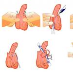
The most gentle and modern method is laparoscopic plastic surgery. It consists of introducing a laparoscope in the form of a tube through small incisions, at the end of which there is a screen. Such an intervention can be performed at any stage of the disease, and regardless of the patient’s age. It is contraindicated only for premature newborns and if they have other developmental defects. The child's stay in the hospital after surgery lasts for one week.
It is very important that the baby must then register with a urologist, whom he must visit with his parents at least 3-4 times a year. To prevent complications, he takes uroseptics for some time. The duration of this course is up to two weeks, sometimes the doctor can extend it, if necessary. In addition, you should take a urine test during the first year after surgery, twice a month.
The rehabilitation period may be extended depending on the availability concomitant pathology, other diseases, and the degree of likelihood of complications. After urine begins to flow freely into the bladder, the size of the kidney becomes normal, and the damaged tissues restore their structure and function. The normalization of blood circulation in the periphery of the organ can be determined using Dopplerography.
With a diagnosis such as renal hydronephrosis in newborns, treatment using the laparoscopic method sometimes gives some complications, including infection or bleeding. It should be noted that the likelihood of their development during surgery in a newborn is slightly higher than in an adult.
In some cases, surgical treatment may be offered before the baby is born. However, such manipulation can lead to premature birth, so the woman is usually warned about the possibility of such an outcome from the operation.
The success of the operation depends on the degree of renal dysfunction. The likelihood of an unfavorable outcome remains high if treatment is carried out on a child who is less than six months old.
The disease can be diagnosed in the fetus at 14-20 weeks. intrauterine development. The detection rate of prenatal hydronephrosis is one case per hundred pregnancies. The study proved that the incidence of this pathology is about two percent; mainly boys suffer from prenatal disease. In this case, surgery is also possible. But this may result in premature birth, which the doctor must inform the mother about.
Often this condition can be transient and disappear without a trace after childbirth, or after some time.
Forecast
Modern methods of surgical intervention guarantee a high probability of recovery. Minimally invasive techniques do not require a long hospital stay and help minimize the development of complications. The chances of successful treatment are somewhat reduced if there is concomitant diseases kidneys (for example, polycystic disease).
Such a serious illness as hydronephrosis is characterized primarily by the fact that, due to the most various reasons the outflow of fluid from the renal pelvis and its calyces is impaired.
As a result of this defect, the cavity system of this organ expands, the pressure in the kidney increases, which ultimately can lead to a slowdown in blood circulation and even atrophy of the parenchyma itself. In other words, kidney function is impaired, sometimes irreversibly.
This disease can be acquired or congenital, and sometimes doctors can diagnose hydronephrosis in the fetus. Unfortunately, developmental defects are quite common today genitourinary system still at the embryonic stage, including such as congenital hydronephrosis.
It is very important to diagnose it on time, that is, during pregnancy. Since if this pathology is detected in utero, after birth, the child may well recover and lead a normal, full life.
According to statistics, renal hydronephrosis in the fetus is detected in 5% of cases of this disease.
Interestingly, boys suffer from this disease much more often. And in a quarter of children with hydronephrosis, the lesion is bilateral.
Causes of congenital pathology
A 4-month-old embryo's kidneys are almost the same as those of a newborn - there is an excretory system, parenchyma, pelvis, and calyces. The fluid is already leaving, the fetus empties its bladder several times a day.
Congenital hydronephrosis in an unborn baby develops due to defects of the genitourinary system that arose at the intrauterine stage. Most often, the lumen of the ureter is blocked in one way or another, so the excretory function is impaired.
Basically, pathology occurs for the following reasons:
- abnormal development of the kidney in the form of a horseshoe;
- multicystic disease (usually the left kidney);
- the presence of an additional vessel in the kidney;
- incorrect origin (location) of the ureter.
If we talk about the specific causes of hydronephrosis, then it occurs as a result of:
- narrowing of the internal lumen of the ureter;
- squeezing it from the outside with a vessel, inflamed tissue or tumor;
- obstruction of urine flow due to reflux, the presence of a stone, or the presence of injury;
- special pathological structure of the ureteral mucosa.
Why exactly certain defects in the fetus develop during pregnancy is not exactly clear. However, experts name factors that can lead to a certain risk, including:
- pollution environment and other environmental problems;
- ionizing radiation;
- burdened heredity.
Thus, gynecologists refer families planning a new pregnancy to a geneticist who have previously given birth to a baby with a defect, in order to rule out various congenital pathologies of the unborn child.
Classification
Hydronephrosis in children is divided into congenital (it is called primary) and acquired (this is the so-called secondary hydronephrosis). Naturally, in a fetus it can only be of the first type.
The disease can be one-sided, this occurs in most cases and affects either the right or left kidney; and there are also cases of bilateral hydronephrosis, when changes affect both organs. This is diagnosed in approximately 5% or 9% of all cases of this disease.
Congenital hydronephrosis, like acquired hydronephrosis, occurs in three stages (degrees):
- The first (pyelectasia). Urine puts pressure on the kidney cavities, accumulating due to impaired outflow. As a result, the organ stretches slightly, becomes larger, but continues to function normally.
- The second (hydrocalicosis). This stage begins after a few months. Urine accumulates in the tubules of the parenchyma, putting pressure on it, disrupting the functioning of one or both kidneys. The organ is enlarged, its walls are thinned.
- Third (terminal). Here, irreversible atrophy of the parenchyma itself already occurs. The kidney is very large, as is the pelvis with the cups, the organ ceases to function. A child with such a problem may even lose a kidney.
Manifestations of hydronephrosis in the fetus
As a rule, hydronephrosis, even in newborns and even more so in the fetus, can only be detected during special diagnostics. It is impossible to determine from a pregnant woman that a child may suffer from a similar illness. However, sometimes hydronephrosis leads to oligohydramnios, and the woman experiences pain when the child moves.
But the fact that the disease does not clinical signs, does not mean that you can ignore it. Due to hydronephrosis, other disturbances in the development of the embryo may occur.
For example, the placenta is poorly supplied with blood, fetal hypoxia occurs, and the child is born weakened, with numerous dysfunctions, including respiratory ones.
There is also a risk of infection in utero. That is why women expecting a child are constantly monitored by a doctor, conducting routine examinations, including ultrasound.
Diagnosis in utero
Typically, a pregnant woman undergoes three screenings during the process of bearing a child - one ultrasound in each trimester. However, if there is a suspicion of a defect, the doctor may prescribe additional examinations.
Already from the 16th week, the embryo’s kidneys begin to work, so at the second ultrasound (which is performed from the 18th to the 20th week inclusive), the doctor checks whether the embryo has congenital pathologies of the kidneys, as well as the bladder and ureters. If the screening shows only the presence of fluid, but neither the cups nor the pelvis are enlarged, such a deviation is not considered hydronephrosis.
Also, the diagnosis is not made if the pelvis has increased by only 5-8 millimeters. But when the expansion is greater, we can talk about hydronephrosis. Most often in the fetus it is one-sided.
There are also cases when a child in the womb already has a severe and advanced stage of hydronephrosis. In this case, when performing sonography, it is clear that the kidney tissues are significantly thinned, which subsequently leads to renal failure.
Embryo treatment
The tactics of a doctor who discovers hydronephrosis in a fetus is based on the fact that the pregnant woman is examined very often, and in most cases even hospitalized. However, treatment of the disease in an unborn baby is not carried out using conservative methods.
In the most extreme case, surgery may be prescribed. Usually they go for it when there is a serious risk that the kidney will rupture. However, such intervention is fraught with premature birth.
A common practice for this pathology is to place a catheter in the fetal bladder to drain urine from its kidneys. This procedure helps to avoid various complications.
If a pregnant woman gets to an experienced doctor on time, who carries out competent treatment, the prognosis for her child’s renal hydronephrosis is quite favorable.
Hydronephrosis in newborns
Peculiarities of this disease in infants is that it can develop differently. As of today, not yet exact method, allowing doctors to make a reliable prognosis. Therefore, the urologist, as a rule, never prescribes surgery right away, even if the child was diagnosed with hydronephrosis in the prenatal stage.
They observe the baby and watch the dynamics. Sometimes the genitourinary system can develop, tissues and organs can mature, pathologies can disappear on their own. It happens that due to unstable water metabolism at this age, the functioning of the kidneys changes; the size of the pelvis returns to normal 3-4 weeks after birth.
On the other hand, serious deterioration may occur in just a couple of months, so surgical intervention may be late. Therefore, it is so important to constantly monitor the course of renal hydronephrosis in newborns.
In most cases, the pathology is caused by congenital abnormalities and anatomical features both external and internal. Internal causes include, for example, congenital underdevelopment of the lumen of the ureter, leading to its narrowing.
An external cause may be the formation of an additional vessel that compresses the ureter, coupled with an abnormal discharge of the latter from the pelvis.
Symptoms
Pyeelectasia (enlargement of the renal pelvis) is one of the main symptoms, which is detected by ultrasound diagnostics. In addition, hydronephrosis may manifest as hematuria (blood in the urine) or difficulty urinating, sharp pains in the abdominal area, fever, concomitant infections of the genitourinary system, as well as the presence of abdominal cavity formations of impressive size.
Diagnosis in children and infants
Hydronephrosis in newborns, whether it was detected earlier or after the birth of the child, in any case requires a series of studies. Unfortunately, children of this age are not yet able to talk about what symptoms they feel. An older child may complain of aching pain in the side, nausea; the baby just sometimes becomes extremely restless.
That is why, in addition to standard palpation, as well as urine and blood tests, which will show the presence of inflammation, blood in the urine or other hidden signs of complications, the diagnosis is also made on the basis of:
- Ultrasound. The specialist examines the bladder and kidneys – when they are full and after urination. Screening allows us to identify pathologies of the parenchyma, obstruction of the pelvis and ureter. If the results are in doubt, diuretics are used during the study, and the patient is given plenty of water to drink.
- Excretory urography. Contrast is injected into the newborn's vein and then observed as the substance is eliminated. This is how a conclusion is drawn about the ability of the kidneys and the entire system to filter, about the degree of obstruction.
- Computed tomography and MRI. Both of these studies varying degrees allow you to see a three-dimensional image of all urinary structures and draw conclusions about whether there is hydronephrosis in newborns, and if so, to what stage and extent. If they are made infant, then only under anesthesia.
- Nephroscintigraphy. This research is carried out using radioisotopes. During this procedure, a specialist evaluates how the kidneys work and how impaired the outflow of fluid is.
- Vaccine cystography. This study is done if there is reason to suspect the presence of vesicoureteral reflux (reflux of urine), or if there is simply poor outflow of fluid from the bladder. The child is also injected with contrast, but not into a vein, but into the urethra. The bladder fills, and when urination occurs, the doctor takes an x-ray. The image shows if, as a result of pathology, the contrast returns to the ureters.
Treatment
It depends on the stage of the disease. If the child has hydronephrosis of the first degree, then conservative therapy. It consists, first of all, in stimulating the outflow of fluid.
In addition, symptomatic treatment is necessary. So, in case of infection, antibiotics and anti-inflammatory drugs are prescribed. If necessary, medications that reduce pressure and relieve swelling of the ureters.
As already mentioned, the disease often goes away on its own, but in order not to miss its progression, children undergo regular ultrasound examinations. For children under 3 years of age - once every 3 or 6 months, for older patients - once a year.
Both positive and negative dynamics can be observed in the second and third degrees of the disease. In this case, screening is carried out once every 2-3 months. If the condition of the organ worsens, and as observation progresses, the pelvis enlarges and expands, surgical intervention is indicated.
Indications for surgery
More severe stages of hydronephrosis - starting from the second - do not in themselves serve as an indication for surgical intervention. An operation is necessary if, during the examination, both dilatation of the pelvis and narrowing of the ureter, both in the pelvis segment, were discovered in the child.
Fortunately, nephrectomy—removal of the kidney—has to be resorted to only in the most severe cases. They occur quite rarely, since urologists observe the child and do not allow hydronephrosis to progress sharply.
The most common type of surgery is pyeloplasty. It is carried out using different methods, the most effective of which is the Hines-Andersen method. It consists in the fact that during the intervention the narrowed section of the ureter is excised, and at the same time its normal connection with the pelvis is formed.
A blank catheter with a drainage tube brought out or a thin stent with an internal fluid outlet is installed in the area of the new connection. In the first case, the small patient spends an average of three weeks in the hospital after surgery. In the second case - no more than 9 days.
The surgeon decides whether to install a catheter or a stent, depending on the situation, the degree of damage and other factors.
Pyeloplasty is considered to be quite effective. It leads to significant improvements in 92-95% of all cases. Typically, the function of a diseased kidney is restored within six months to a year after pyeloplasty.
- Date: 02/17/2015
- Rating: 11
- Occurrence of disease
- Stages of development of hydronephrosis
- Symptoms of the disease and diagnostic methods
- Therapeutic measures
Hydronephrosis of the kidneys is quite unpleasant disease, which prevents the removal of urine from the collecting system of the kidneys. This entails serious consequences, including disruption of the proper functioning of these organs. This disease affects all age categories of the population. Both the right and left kidneys are affected.
Occurrence of disease
In a newborn, this disease is most often caused by congenital rather than acquired factors.
The main causes of pathology:
- incorrect position of the ureter;
- urinary tract dyskinesia;
- incorrect location of the arteries in the kidneys, due to which they put pressure on the ureter;
- the ureter does not extend correctly from the pelvis;
- the existence of an additional vessel that puts pressure on the ureter;
- congenital narrow ureter in a child due to underdeveloped lumen.
I would like to note the fact that the last of the above reasons is much more common in medical practice than others.
Hydronephrosis is a disease in which the ureter is blocked by a stone, tumor, blood clot, or the flow of urine from the kidneys is impaired as a result of an abnormal development of the urinary organs.
Newborns usually have a congenital pathology that affects one of the kidneys (most often the left). But there is also a bilateral lesion (in 11% of cases of the total number of detected diseases). With this option, it is necessary to apply urgent surgical intervention, otherwise the baby may die as a result of the development of insufficiency. The combination of dilation of the renal pelvis and ureter is called ureterohydronephrosis.
With a diagnosis such as hydronephrosis in newborns, treatment is carried out primarily surgically; all conservative methods are used only for some relief of the condition and in order to prepare for surgical intervention. Medicines are also used in the postoperative period to reduce the likelihood of complications.
Boys develop this disease three times more often than girls. The cause of the development of pathology is the unhealthy lifestyle that the expectant mother leads during pregnancy, as well as illnesses suffered during this period.
Manifestations of hydronephrosis
This disease is dangerous because it most often begins to manifest itself only in the third stage. And timely detection of hydronephrosis in a newborn lies entirely with the baby’s parents. You should pay attention if the child becomes capricious, eats poorly, or has trouble sleeping. As abdominal pain develops, he may twist his legs and pull them up.
It is especially important to pay attention to the quality and condition of urine. You should immediately go to the doctor if blood discharge appears on the diapers.
The main symptom of this disease is an enlarged kidney, which is easy to palpate during bimanual palpation examination, or with. A strong enlargement of the baby's kidneys can be seen by the enlarged abdomen. The addition of an infectious complication, which often accompanies hydronephrosis, causes an increase in temperature. In this case, an increased content of leukocytes is detected in the urine.
Causes
In newborns, hydronephrosis can develop for several reasons:
- Narrowing of the junction of the pelvis and the ureter.
- Narrowing of the junction of the ureter into the bladder. In this case, not only hydronephrosis develops, but also fluid accumulation in the urethra (megalureter).
- Development of vesicoureteral reflux. In this condition, fluid backflows from the bladder into the ureter. Occurs as a result of congenital underdevelopment or absence of valves that prevent the reverse flow of urine.
- Non-obstructive hydronephrosis. This pathology is rare. With it, there is a violation of urine excretion by the kidneys without visible mechanical obstacles.
- Polycystic renal dysplasia.
- Presence of a posterior urethral valve.
- Ureterocele.
- Traumatic injury or tumor process.
Types of treatment depending on the degree of the disease
In a newborn baby, there are three degrees of the disease:
- The first degree is called pyelectasis. It develops as a result of a slight disturbance in the excretion of urine from the kidney, while its functional abilities are completely preserved. There may be some enlargement of the cavities.
- The second stage is called hydrocalycosis. Several months usually pass from the beginning of the first stage. In the tubules and pelvis there is a significant accumulation of fluid, which puts pressure on the parenchyma and causes dysfunction of the kidney.
- The final, or terminal third stage leads to irreversible changes in the renal parenchyma. The function of the organ is significantly changed or stops completely
Indications for surgery
Hydronephrosis of the kidneys in newborns can go away on its own. For example, pyeelectasis is not an indication for surgery. In some cases, it is physiological in nature and can go away on its own. Such a child must be registered with a doctor and undergo regular examinations. The question of a radical solution to the problem may arise when the first stage begins to progress and moves into the second and third. These variants of the disease are considered a pathology and require urgent action.
Modern treatment methods make it possible to provide significant assistance with minimal postoperative complications. The following deviations may be indications for surgery:
- expansion of the kidney cavities;
- the presence of stones in the kidneys;
- significant narrowing of the ureter;
- tumor formation in the pelvis area.
Laparoscopic intervention
The most gentle and modern method is laparoscopic plastic surgery. It consists of introducing a laparoscope in the form of a tube through small incisions, at the end of which there is a screen. Such an intervention can be performed at any stage of the disease, and regardless of the patient’s age. It is contraindicated only for premature newborns and if they have other developmental defects. The child's stay in the hospital after surgery lasts for one week.
It is very important that the baby must then register with a urologist, whom he must visit with his parents at least 3-4 times a year. To prevent complications, he takes uroseptics for some time. The duration of this course is up to two weeks, sometimes the doctor can extend it, if necessary. In addition, you should take a urine test during the first year after surgery, twice a month.
The rehabilitation period may be lengthened depending on the presence of concomitant pathology, other diseases, and the degree of likelihood of complications. After urine begins to flow freely into the bladder, the size of the kidney becomes normal, and the damaged tissues restore their structure and function. The normalization of blood circulation in the periphery of the organ can be determined using Dopplerography.
With a diagnosis such as renal hydronephrosis in newborns, treatment using the laparoscopic method sometimes gives some complications, including infection or bleeding. It should be noted that the likelihood of their development during surgery in a newborn is slightly higher than in an adult.
In some cases, surgical treatment may be offered before the baby is born. However, such manipulation can lead to premature birth, so the woman is usually warned about the possibility of such an outcome from the operation.
The success of the operation depends on the degree of renal dysfunction. The likelihood of an unfavorable outcome remains high if treatment is carried out on a child who is less than six months old.
The disease can be diagnosed in the fetus at 14-20 weeks of intrauterine development. The detection rate of prenatal hydronephrosis is one case per hundred pregnancies. The study proved that the incidence of this pathology is about two percent; mainly boys suffer from prenatal disease. In this case, surgery is also possible. But this may result in premature birth, which the doctor must inform the mother about.
Often this condition can be transient and disappear without a trace after childbirth, or after some time.
Forecast
Modern methods of surgical intervention guarantee a high probability of recovery. Minimally invasive techniques do not require a long hospital stay and help minimize the development of complications. The chances of successful treatment are somewhat reduced in the presence of concomitant kidney diseases (for example, polycystic disease).
Hydronephrosis is a renal pathology in which the outflow of urine from the organ is disrupted, expansion of the calyces and pelvis occurs, and atrophy of the parenchyma occurs. Pyelocalyceal system It is a kind of funnel in which urine accumulates. If the body is healthy, fluid is freely excreted, and there are no problems with its accumulation.
In newborns, hydronephrosis is usually congenital. If you do not get rid of this problem in a timely manner, the gradual progression of the disease can lead to significant impairment of kidney function and renal failure. You need to pay attention to any disturbances in the child’s behavior and condition and immediately seek medical help.
Causes of pathology

The exact cause of hydronephrosis in newborns still cannot be determined. Most experts are confident that the beginnings of the disease can be laid in intrauterine development. Failure of a woman to comply with basic requirements during pregnancy (smoking, drinking alcohol, taking medications) significantly increases the risk of developing pathology in the child.
The development of hydronephrosis in infants is associated with physiological abnormalities of the urinary system:
- underdeveloped lumen of the ureter;
- incorrect structure of the pelvic region;
- narrowing of the bladder wall;
- violation of the innervation of the kidneys from the central nervous system;
- reflux (backflow of urine into the kidneys);
- stones in the ureter (occurs in rare cases in newborns).
The acquired form of hydronephrosis in newborns can develop against the background of other kidney diseases as a complication.
Symptoms of the disease
Pathology in newborns can be unilateral (one kidney is affected) and less often - bilateral. With hydronephrosis of 1 kidney, symptoms of the disease may not even appear, since the second kidney performs a compensatory function for the outflow of urine. Bilateral kidney damage can be life-threatening for the baby and cause uremia. Most often, hydronephrosis in newborns is diagnosed at the stage of development of kidney inflammation (for example,).
The child has the following symptoms of renal hydronephrosis:
- enlarged tummy;
- heat;
- lethargy and drowsiness;
- due to paroxysmal pain, the child screams, cries, and is very restless;
- refusal to eat;
- itching - occurs due to the accumulation of toxins in tissues due to impaired outflow of urine, the baby constantly tries to scratch himself, scratches the skin;
- There are streaks of blood in the urine.
When examined by a doctor, a tumor may be detected in the area of the affected kidney by palpation.
Degrees of renal hydronephrosis in children

In newborns, there are 3 degrees of kidney damage due to hydronephrosis:
- 1st degree (pyelectasia)- the pelvis expands from pressure and accumulation of urine, a slight enlargement of the kidney occurs, the parenchyma is not damaged, the functionality of the organ is not impaired.
- 2nd degree (hydrocalicosis)- the fluid begins to compress the parenchyma and accumulate in the tubules, the calyx expands even more, the organ functions only at 40%.
- Stage 3 (terminal)- the parenchyma irreversibly atrophies, the kidney increases significantly in size, and may gradually lose its function completely.
Possible complications
If hydronephrosis is not detected and treated in a newborn in time, complications will inevitably arise as it progresses:
- organ atrophy;
- bacterial pyelonephritis;
- renal failure.
To avoid this, it is necessary to conduct an examination during the period of intrauterine development for the timely detection of pathology.
Diagnostics

- general urine analysis;
- Ultrasound of the kidneys and bladder;
- excretory urography.
A good specialist can suspect hydronephrosis by palpation and detect a characteristic tumor.
Reference! Timely intrauterine diagnosis of a pregnant woman makes it possible to detect hydronephrosis in the fetus even before birth at 16-20 weeks. For every 100 pregnancies, 1 case of hydronephrosis is detected in the fetus.
General rules and methods of treatment
The choice of treatment regimen for kidney hydronephrosis in children is influenced by several factors:
- degree of pathology;
- duration of the inflammatory process;
- the presence of concomitant pathologies.
There are times when initial stage the disease may go away on its own. A child up to 3 years of age needs to undergo regular kidney ultrasound (every 3-6 months) to monitor the dynamics of the organ. Sometimes it takes time for organs to begin to fully perform their functions. During this period they can be used conservative methods which help stimulate the flow of urine.
Surgical intervention

If the dynamics are negative and the condition of the kidney worsens, surgery cannot be avoided. Usually, in case of hydronephrosis in newborns, they resort to laparoscopic plastic surgery, which today is the most gentle method. The operation should not be performed on premature babies with low birth weight.
No incisions are required during the operation. A laparoscope (a tube with a camera at the end) is inserted through small incisions. A narrow section of the ureter is excised. A new connection between the pelvis and the ureter is formed. The child may have an internal drainage stent installed, which will be removed after 2-3 months, or a catheter with a drainage tube. The doctor will determine the method of urine diversion based on the characteristics and results of laparoscopy.
Postoperative period
After the operation, the baby must remain in the hospital for about a week. In some cases, his stay there may be longer (up to 3 weeks). During this period, constant monitoring of the condition is important.
After discharge, the newborn is registered at the dispensary. Once every 1-2 months you need to undergo a follow-up examination with a urologist. The doctor may prescribe uroseptic drugs as a maintenance course for 1-2 weeks.
Around 6 months, urine tests may show increased white blood cells, protein, and hematuria. This is considered normal after surgery. Tests must be taken 2 times a month.
To determine the microcirculation of the periphery of the kidneys, a Doppler study is performed. When urinary excretion is restored, the kidney returns to its normal size and tissue regeneration occurs.
Find out the instructions for using stamen bud tea.
The rules for use and indications for use of the drug Cyston are described on the page.
Go to the address and read about what calcium oxalate in urine means and methods for correcting the indicators.
Consequences
Although 95% of surgeries are successful, young children have a higher risk of complications than adult patients. After laparoscopy, the child still requires surgical correction and constant monitoring by a urologist and neonatologist.
After surgery you may experience:
- bleeding;
- inflammatory processes of an infectious nature;
- if the operation is performed during the intrauterine period, termination of pregnancy and premature birth are possible.
Thanks to minimally invasive surgical techniques, the use of modern absorbable sutures, internal insertion of drainage tubes and the use of antibacterial agents, number of complications in Lately decreased significantly.
Prevention measures

Prevention of hydronephrosis in newborns should be taken care of during pregnancy. In infants, this is a congenital pathology, but its likelihood can be reduced if the pregnant woman adheres to certain recommendations.
- do not take medications without a doctor’s prescription;
- do not drink alcohol;
- no smoking;
- carry out all necessary examinations on time;
- eat well.
The earlier hydronephrosis is detected in a newborn, the greater the chance of restoring the kidneys to a normal state and returning their functionality. Parents need to be attentive to the manifestation of any disturbances in the functioning of the child’s body. Babies cannot describe their complaints themselves, so you should always monitor their condition and regularly conduct necessary diagnostics to be able to record any deviations from the norm.
In the next video, a specialist from the Moscow Doctor clinic will tell you more about the methods and features of the treatment of hydronephrosis in children:
Today, various kidney disorders in children are not considered uncommon. In this article you will become familiar with the specifics of the disease, disease factors and varieties, symptoms of the disease in infants, and also the main methods of diagnosing early stages pregnancy, as well as treatment of illness.
This disease is a violation of the outflow of fluid from the renal pelvis and its calyces:
- As a result, the organ cavity expands and the pressure increases. All this can cause circulatory problems. In other words, the functioning of the kidneys is impaired.
- The disease can be acquired or congenital, and sometimes a doctor can diagnose the disease while the fetus is developing. Unfortunately, today quite often we come across defects in the formation of the genitourinary system in the embryonic period.
- If this pathology is identified earlier, even before birth, the child can be completely cured. According to statistics, kidney disease in the fetus occurs in 5% of situations. Boys suffer from this pathology much more often.
- Every year, up to 50 children with hydronephrosis from 1 month to 17 years old receive help.
To understand what the disease is, you need to become familiar with the structure of urination. The pelvis and calyces have the shape of a funnel with accumulated liquid. During a normal stay, there are no disturbances in the transmission of urine, but when the cavities expand, this process is disrupted.
There are two types of disease:
- disease affecting one kidney;
- a disease when both kidneys function poorly.
In the first case, the patient, as a rule, does not feel any signs of symptoms. With a bilateral manifestation, the disease is most dangerous, since a severe, fatal condition can occur as a result of the accumulation of urea in the blood.
Based on its origin, the disease is divided into:
- congenital;
- acquired formation.
The innate configuration occurs much more often than the acquired form. Therefore, the study of organs using ultrasound after birth has already been introduced into the mandatory diagnostic procedure by doctors.
The congenital disease, like the acquired one, has a III period:
- A disorder that is accompanied by poor urine flow;
- Enlargement or stretching of the renal calyx;
- Reduction of parenchyma. The kidney and pelvis with calyces are large, the organ stops working. For what reason these or other abnormalities occur in the fetus during pregnancy has not been specifically established. But experts present conditions that can cause the disease, including:
- pollution of the surrounding environment;
- the effect of x-rays;
- The baby's parents are carriers of the disease.
Causes
In the unborn
This type of disease in an unborn baby is formed due to emerging disorders of the genitourinary organization.
Violation appears under the following factors:
- improper formation of the organ;
- a rare disease in which the renal parenchyma is gradually replaced by cysts;
- the obviousness of an extra vessel in the organ;
- incorrect placement of the tubular organ;
- compression of the ureter by a vessel, inflamed tissue or tumor;
- pathologies of fluid patency as a result of the presence of injury or stone;
- specific structure of the mucous connective tissue tube.
In children
Peculiarities congenital hydronephrosis These are structural features of the baby’s body. At the moment, it is impossible to note exactly why this disorder appears, however, women, from the moment they learn of pregnancy, continue to harm the body with alcohol and cigarettes, use medical supplies without the doctor’s permission, they risk the baby’s well-being.

The key stages of hydronephrosis include 3 levels:
- Level 1 hydronephrosis
Changes in the pelvis with intact parenchyma and preservation of the basic functions of the kidney.
- Level 2 hydronephrosis
An increase in the volume of the kidney, a defect in the parenchyma, a huge enlargement of the pelvis, a decrease in the functioning of the organ up to 40%.
- For third level hydronephrosis
There is an increase in the kidneys, a decrease in the parenchyma, as well as a decrease in the activity of the organ by about 40% or a lack of activity of the organ.
Intrauterine examination at the time of pregnancy can show the presence of this disease in the child before birth.
By medical indicators, impaired renal function is a consequence of:
- stenosis;
- discharge of the ureter;
- problems with the structure of the pelvis;
- stone formation;
- CNS disorders;
- narrowing of the bladder neck;
- decrease in liquid.
The consequences of the disease lead to the child’s inability to go to the toilet normally.
Symptoms
The course of the disease is usually asymptomatic. Painful sensations in most cases, they begin with the appearance of microlith in the organ, as well as with the appearance of inflammatory processes.
Symptoms when inflammatory process in the kidneys in children looks like this:
- temperature increase;
- pain when urinating;
- lumbar pain;
- appearance bloody discharge in urine.
The insidiousness of hydronephrosis in children may not be noticed. But the first signs are the following symptoms:
- enlarged belly;
- when attacking infectious diseases on the body;
- increase in temperature;
- weakness and itching occurs;
The disease prevents the normal removal of fluid, and toxic substances remain in the baby’s body. Toxic substances are irritating skin child, which causes itching. A doctor, examining such a child, is able to identify a neoplasm by palpation.
There are cases when the first sign of the onset of the disease is the formation of bloody clots during urination.
Diagnosis
Diagnostics include:
- urine and blood tests;
- Ultrasound of the organ.
- The main way to diagnose hydronephrosis in a child is the X-ray method. Thanks to this method it is possible to establish the ratio of the volumes of a particular kidney in relation to the parameters of a healthy organ.
- To clarify the diagnosis, computed tomography of the kidneys, radioisotope studies and screening are performed.

If the child’s illness is already quite mature for a long time, the disease can cause severe complication- . Therefore, it is necessary to monitor the child’s condition and follow all the doctor’s instructions. Today, medicine makes it possible to detect the disease at the stage of intrauterine formation of a child - from the 20th week of pregnancy.
In order not to miss the further course of the disease, children are constantly undergoing ultrasound examinations. A child under three years of age should be examined at least once every six months; older patients should be examined once every 12 months.
Diagnostics also includes:
- Ultrasound examination method. The expert examines the bladder and kidneys and determines the functioning of the organ in a filled state and after fluid has been removed from the body. Diagnostics helps to detect disorders of the parenchyma, pelvis and ureter. If the results are in doubt, drugs that promote a diuretic effect are used during the research. At the same time, the patient is given a lot of water to drink.
- Excretory urography. A substance is injected into the baby's vein, while observing how the substance is excreted. Thus, a conclusion is made about the functioning of the kidneys.
- Radionuclide research carried out in the case of assessing the location, formation, size of the organ in order to recognize the main cause of the organ disorder. This method determines the activity of the renal parenchyma.
- X-ray method kidney studies and urinary tract during the act of urination using radiopaque agents. This study is carried out if there are reasons to suspect the presence of urine reflux.
Intrauterine diagnosis
Typically, a pregnant woman undergoes 3 examinations - once during the trimester:
- Already from the 16th week, the embryo’s kidneys gradually begin to function, for this reason a second study is carried out.
- If during screening only the formation of fluid is noticeable, but neither the cups nor the pelvis have increased in volume, this disorder does not apply to hydronephrosis.
- There are situations when an advanced stage of this disease is observed in the baby in the womb. During ultrasound scanning, it is noticeable that the kidney tissue is very thin. This leads to a syndrome of dysfunction of all kidney functions, leading to disorders of water, electrolyte, nitrogen and other types of metabolism. Unfortunately, babies can't explain what they feel. An older child may complain of pain in the side.
The child is also injected with contrast, but into the urethra.
The bladder fills, and if urination occurs, the doctor takes an X-ray. The image shows that due to the violation, the substance returns to the ureter.
Treatment
In the first stage of the disease, it is better to use conservative treatment methods. This therapy is aimed at stimulating the flow of urine. In most cases this is enough for effective treatment. The second stage of the disease implies the presence of two dynamics, positive and negative, in the following cases:
- the kidney condition is worsening, so surgical intervention is necessary;
- positive dynamics imply continuation of the prescribed treatment and systematic supervision.
Embryo treatment
The position of the doctor who has identified hydronephrosis in the fetus is based on the fact that a woman in the position is constantly examined, and in many situations even hospitalized, as well as:
- Surgical intervention consists of eliminating the narrowing in the area of the transition of the renal pelvis to the ureter, carried out using various methods.
- The Anderson-Hines method is considered the most productive. This method is used during surgery, when the narrowed area of the ureter is cut out, which leads to normal integration with the pelvis.
- A tube or a very thin prosthesis with internal fluid outlet is placed in the area of the new union. In the very last case, a surgical intervention may be prescribed.
When a pregnant woman sees a qualified doctor in a timely manner, he will conduct competent therapy, which will only lead to a positive result in treatment. The postoperative period is not the same for all children. The child may have a drainage tube placed and will be monitored in the hospital for a couple of weeks. Also, once the prosthesis is installed, the baby will be discharged within 9 days. After a couple of months, the prosthesis will be removed using special equipment. If the defect is diagnosed on time and surgery is performed, the probability of a positive outcome is 96 - 98%.
You can also learn about the expansion of the renal pelvis from this video.
Sometimes at birth a diagnosis such as “hydronephrosis of the kidney” is made in a newborn. a disease characterized by blockage of the ureter by a tumor, stones, blood clots or there is a violation of urine discharge due to improper development of the organs that remove it. In newborns, this pathology usually affects one of the kidneys, mainly the left one. If the lesion is bilateral, urgent surgery is required, otherwise the baby may die.
This disease is mainly treated surgically, and conservative methods are used to slightly alleviate the patient’s condition and to prepare for surgery. Medicines used in the postoperative period to reduce the likelihood of complications.
How does the disease manifest itself?
Hydronephrosis of the kidneys in newborns is dangerous because its symptoms appear only at the third stage. Therefore, parents should notice the first symptoms. They should be alarmed that the baby has become lethargic, capricious, has begun to eat poorly, and has had trouble sleeping. When there is pain in the abdomen, the child twists his legs and pulls them up.
You should pay attention to the condition and quality of urine. If you suddenly start bleeding on your diapers, don’t delay your visit to the doctor.
The main symptom of the disease is an enlarged kidney, which can be easily palpated during bimanual palpation and can also be detected on ultrasound. A strong enlargement of the organ is manifested by a large volume of the abdomen. Hydronephrosis of the kidneys in newborns is often accompanied by infectious complication with increasing temperature. At the same time, urine contains a large number of leukocytes.
Causes
Hydronephrosis can be congenital or acquired. the most common congenital form is characterized by abnormal development of the kidneys and their vascular system during intrauterine development. A baby is born with an abnormal ureter that may be narrowed or incorrect location, extra arterial vessel. It, in turn, puts great pressure on this organ, resulting in the obstruction of urine flow.
A newborn may have a violation of the outflow of urine either from one kidney or from both at the same time. In this case, a diagnosis is made - unilateral or bilateral hydronephrosis.

It is still not completely clear what causes the abnormal intrauterine formation of the kidneys, leading to the occurrence of this disease. One thing is obvious - to provoke possible deviations In the fetus, a pregnant woman may abuse alcoholic beverages. In addition, smoking and taking certain medications negatively affect the health of the unborn baby.
The acquired form of renal hydronephrosis occurs due to complications that are caused by other kidney diseases.
Kinds
Kidney hydronephrosis in newborns, a photo of which clearly demonstrates the essence of the problem, is most often unilateral. Mostly boys are affected by this pathology. This disease is divided into three types.
Pyeelectasia is the first stage of pathology, characterized by enlargement of the pelvis due to high blood pressure. At this stage it is not yet affected, the main functions of the organ are preserved, but it itself is already slightly enlarged. With timely treatment, serious consequences can be avoided.
The second stage of hydronephrosis is hydrocalycosis. At the same time, the pelvis of the organ increases even more along with the calyces of the kidneys. Excess urine accumulating in the renal tubules begins to compress the parenchyma, which is why its functioning is almost completely disrupted.
The third stage of hydronephrosis is accompanied by active atrophy of renal tissue. In this case, the work of the kidney completely stops. At this stage, only surgical treatment is performed.
Diagnostics
Hydronephrosis of the kidneys in newborns is diagnosed using ultrasound examination, computed tomography, laboratory research, voiding cystography.

An experienced doctor is able to diagnose this pathology during the first examination. Using palpation, he determines whether the kidney is enlarged or not, and then sends the child for examination. With the help of modern equipment, it is possible not only to confirm the fact of the disease, but also to establish the extent of damage to the organ, and also to find out possible reasons leading to hydronephrosis.
Excretory pyelography is performed to assess excretory function, and with the help of voiding cystography, reflux can be detected, which in some cases provokes hydronephrosis.
If renal hydronephrosis is diagnosed in newborns, reviews from mothers recommend ultrasound diagnostics every three months to monitor changes in dynamics. In this case, complications can be prevented.
Treatment
If the kidney is slightly enlarged, then conservative treatment is carried out to normalize the outflow of urine. With timely, competent treatment, it is possible to achieve positive result, restoring kidney function.

When diagnosing the second stage of a disease such as renal hydronephrosis in newborns, treatment is also carried out mainly by medication with mandatory tracking of dynamics. If it is positive, then conservative treatment is continued according to the established regimen. Negative dynamics suggest mandatory surgical intervention.
At the third stage surgical intervention carried out in 100% of cases.
Indications for surgery

Thanks to modern methods treatment is provided effective help with minimal surgical complications. If renal hydronephrosis is diagnosed in a newborn, surgery may be prescribed in the following cases:
- expansion of organ cavities;
- strong;
- the presence of stones in the kidneys;
- the appearance of a tumor in the pelvis area.
Carrying out surgery
The most gentle method of surgical intervention is laparoscopic plastic surgery. The idea behind it is that a laparoscope, which is a tube with a screen at the end, is inserted through small incisions. With the help of such a device, a narrow section of the ureter is replaced by a new wide connection between this organ and the renal pelvis created by surgeons. This contributes to the normal restoration of urine outflow.

This type of surgery is performed at any stage of the disease. After this, the baby spends one week in the hospital.
The child must be registered with a urologist. For preventive purposes, the baby should take uroseptics for two weeks. In addition, during the first year after surgery, it is recommended to regularly take urine tests.
Treatment with folk remedies
In the presence of a disease such as renal hydronephrosis in newborns, treatment folk remedies involves eliminating the obvious symptoms of this pathology.

- 50 g each of Adonis grass, oat grain, horsetail, bearberry, nettle leaves and 150 g of birch leaves;
- 100 g each of birch buds, sedum grass, horsetail, adonis, oat grain, bedstraw and hop cones;
- 250 g each of bearberry and birch buds, 50 g each of hoofed grass, horsetail and knotweed, 75 g each of bean leaves and corn silks;
- 150 g each of juniper fruits, birch leaves and dandelion roots.
To prepare the tincture, 100 g of any collection is poured into a liter of boiling water, boiled for 10 minutes, then poured into a thermos along with the green mass and left overnight. Strain and drink 100 g of tincture before meals. The course of treatment lasts 4 months, with a break of 2 weeks, after which it is continued.
For children, it is necessary to prepare the tincture in such a daily dose herbal collection:
- up to 1 year - 0.5 tsp;
- up to 3 years - 1 tsp;
- 3-6 years - 1 decade. l.;
- 6-10 years - 1 tbsp. l.;
- over 10 years old - 2 tbsp. l.
It should be remembered that some herbs may have contraindications.
Conclusion
Thus, if you have been diagnosed with renal hydronephrosis in newborns, you should not despair. The main thing is if the degree mild illness, then it is necessary to monitor the dynamics of the development of this pathology and treat the child with medication. Surgical intervention is carried out in the most severe cases, but thanks to modern techniques a high probability of recovery is guaranteed.